

 PDF
PDF Citation
 Print
Print


INTRODUCTION
The importance of the anatomical features and position of the supraorbital foramen/notch has been widely focused on in maxillofacial surgery [1,2]. This is due to the possibility of damage to the supraorbital nerve or blood vessels among the supraorbital nerve territories. In addition, the anatomical features and position of the supraorbital foramen/notch are important in supraorbital nerve blocks because nerve damage may occur even in the nerve block. Supraorbital nerve block is a frequently used method for nerve blocks in clinical settings because it is used to treat migraines for those without an adequate response to medications [3], and it is also known to be effective in pain relief from supraorbital nerve pain [4]. The supraorbital foramen/notch form the upper boundary of the orbit and act as channels for the supraorbital nerve and blood vessels. The variations in these structures have been reported several times in studies with cadavers and skulls, but the results varied according to race and the measurement method [5,6]. A study investigated the variations in the supraorbital foramen/notch usingthe photos of 124 Korean skulls [7]; however, there were some limitations inaccurately analyzing the variations in the supraorbital foramen/notch of Korean because the study mainly reported on the relationship between the positions of the supraorbital foramen, infraorbital foramen and mental foramen.
Recently, multidetector CT (MDCT) has been frequently used in anatomical studies of each organ system because the measurement of blood vessels and bone structures can be done in live patients not in cadavers. Especially for bone anatomy images with three-dimensional computed tomography (3D-CT) using MDCT, the reliability of the measured values has been confirmed in various studies [8,9]. This reliability is due to the possibility of 3-dimensional reformulation of images, easy comprehension of bone structures and observation of more precise structures. The aims of this study were to investigate the variations in the supraorbital foramen/notch in Koreans using 3D-CT models from MDCT and to compare the results to those of other races.
Go to : 
MATERIALS AND METHODS
1. Study population
The images of 395 people who were referred for three-dimensional computed tomography (3D-CT) at Seoul National University Bundang Hospital were evaluated during the period from December 2007 to April 2012. The study population consisted of 232 men and 163 women who were 20 years or older. A total of 395 images were selected for this study, and patients with fractures or congenital abnormalities in the orbital region and frontal bone or patients who had surgery in the cranial area were excluded from the study.
2. Measurements
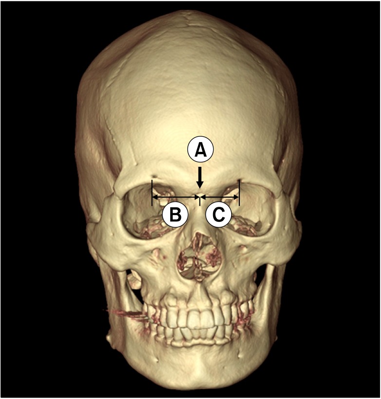
The images were taken with a 256-slice MDCT (Brilliance iCT, Philips Medical Systems, Eindhoven, Netherlands). The images were reconstructed into 3D with a 3D image reconstruction system (Rapidia, Infinite Co Ltd, Seoul, Korea) and they were transmitted and accessed through the Picture Archiving and Communication System (PACS). From the obtained images, first, the presence and features of the supraorbital foramen/notch were confirmed and second, the distance and diameter were measured from the nasion, which is the central point (Fig. 1), and the difference in sex and the left and right position were compared. The anatomical features were classified into the left and right hand side as follows: single foramen, single notch, double foramen, double notch, presence of both foramen and notch or absence of both foramen and notch (Fig. 2).

3. Statistical analysis
To compare the characteristics according to sex between the groups, one-way analysis Of variance (ANOVA) was done with the SAS 9.2 program. The mean value was compared with the paired sample comparison, and a P value < 0.05 was considered as significant in all the analyses.
Go to :
RESULTS
The presence and features of the supraorbital foramen/notch are shown in Table 1. On the right hand side, a single notch was most commonly observed (39.5%) followed by a single foramen which was observed in 37.0% of the images. On the left hand side, a single foramen was most commonly observed (42.3%) followed by a single notch which was observed in 35.4% of the images. Cases in which the supraorbital foramen or notch was absent were observed in 39 people (9.9%) and 46 people (11.7%) on the right and left hand sides, respectively. The presence of both the foramen and the notch was observed in 25 people on the right hand side and in 20 people on the left hand side.

The distance and diameter from the central nasion point are presented in Table 2. Without considering the sex and whether it was the left or right hand side, the mean diameter of the supraorbital foramen was 2.34 ± 0.78 mm (min. 0.65 mm - max. 5.71 mm), and the mean distance was 27.19 ± 4.03 mm (min. 11.4 mm - max. 40.67 mm). The mean size of the supraorbital notch was 3.37 ± 1.71 mm (min. 0.49 mm - max. 10.01 mm), and the mean distance from the nasion was 23.42 ± 2.45 mm (min. 16.34 mm - max. 30.5 mm)

When the measured values were compared between the sexes, in men, the distance to the right hand side foramen and notch was 28.11 ± 4.29 mm and 24.29 ± 2.45 mm, respectively, which were located more on the lateral side relative to women (the right hand side foramen, P = 0.0352; the right hand side notch, P = 0.0004). The same feature was observed on the left hand side in that the distance from the nasion to the foramen or notch was significantly greater in men (the left hand side foramen, P = 0.0330; the left hand side notch, P < 0.001).
Go to :
DISCUSSION
The supraorbital nerve is the terminal branch of the frontal nerve which branches out from the first branch of the trigeminal nerve. The nerve passes through the supraorbital foramen or notch and it even is distributed to the forehead and the middle of the frontal region. Therefore, in cases where the facial procedure including forehead and eyebrow lifts, damage to the supraorbital nerve and neurovascular bundle could occur [10], and it could cause supraorbital neuritis after the procedure. In addition, the location of the nerve is positioned between the skin and cranium not deep inside, which often is responsible for supraorbital neuritis or headaches when the nerve is under the pressure [4]. A supraorbital nerve block may help with supraorbital neuritis or headaches [11], but the anatomical features and location of the supraorbital foramen or notch need to be well understood to prevent nerve damage from occurring during the procedure.
When comparing the results of existing studies on the supraorbital foramen or notch, differences in anatomical features, size and location do exist along with differences due to race. In a study that examined the anatomical features of 110 adult Asian skulls, the frequency of both the supraorbital foramen and notch being absent was 5.5% on the right hand side and 10.0% on the left hand side [6]; however, in this study, the frequency was higher at 9.9% on the right hand side and 11.7% on the left hand side. In a study with 399 Caucasian skulls using MDCT, the frequency of a single notch on the right hand side was 69.4% and the frequency of a single notch on the left hand side was 68.2% [9]. However, the frequency of a single notch in the Korean population was considerably lower and the frequency of a single foramen was relatively high. In addition, differences were observed in the size and location. In a study with 507 cadaver skulls from an adult Western population, the mean distance to the supraorbital foramen or notch was 31 mm (min., 20 mm - max., 49 mm) [2]. In a study consisting mostly of 111 Indian skulls to determine the features, diameter and distance of the supraorbital foramen or notch, the mean distance from the nasion to the supraorbital foramen or notch was 32.02 mm (min. 15 mm - max. 38 mm), and the mean size of the notch was 5.70 mm, and the mean diameter of the foramen was 3.78 mm [5]. In comparison with the results of this study with those of the above 2 studies, the size of the supraorbital foramen and notch was smaller and the distance from the nasion (the mean distance to the foramen, 27.19 ± 4.03 mm; the mean distance to the notch, 23.42 ± 2.45 mm) was also shorter in the Korean population. In previous cadaver studies, the smallest supraorbital foramen was reported as 1.5 mm [5], but in a study conducted with MDCT, the diameter of the supraorbital foramen was as small as 0.8 mm, and the diameter or notch was 0.7 mm in size, and the analysis with CT was considered to be superior [9]. In comparison to a study that analyzed 124 Korean skull photos in which the mean distance from nasion to the supraorbital foramen or notch was 22.7 mm and the mean diameter was 4.7 mm, and the frequency of the notch (69.9%) was found to be higher than that of the foramen (28.9%). However, in this study, the frequency of a single notch and single foramen was similar on both sides; the mean diameter was smaller and the mean depth was longer for the supraorbital foramen or notch, and the minimum diameter of the supraorbital foramen or notch ranged from 0.65 mm to 0.49 mm in size.
This study has revealed the variation and characteristics of the supraorbital foramen or notch in a Korean population based on 3D-CT images using MDCT for the first time. In comparison with other races, the Korean population had a higher frequency of the foramen being present than the notch between the supraorbital foramen and notch, had a shorter distance from the nasion, and had a smaller diameter for the notch and foramen. It tells us that in the case of a nerve block, the location of the notch can be easily found by digital exploration of the top region of the supraorbital, but the location of the foramen can possibly be incorrect. In addition, familiarization of the distance from the nasion and the diameter of the supraorbital foramen and notch will contribute towards a decrease in complications such as nerve damage or vascular puncture during a supraorbital nerve block or maxillofacial procedure.
Go to :
 XML Download