

 PDF
PDF Citation
 Print
Print


Calcific tendinitis of the shoulder is a well-known source of the shoulder pain and characterized by a reactive calcification that affects the rotator cuff tendons. Calcium hydroxyapatite crystal deposits, the major contributor to calcific tendinitis, cause adjacent tissue inflammation and pain [1]. The conservative management of shoulder pain due to calcific tendinitis of the rotator cuff consisted of oral nonsteroidal anti-inflammatory drugs, physical rehabilitation to prevent loss of joint mobility, subacromial steroid injections, and intra-articular steroid injections [1]. If conservative medical treatment fails, open surgical or arthroscopic removal of the deposits can be done. Recently, other therapeutic methods such as extracorporeal shock wave therapy (ESWT) [2,3] and ultrasound guided fine needle technique (lavage or barbotage) [4] are considered as possible alternative to surgery which may lead to complications such as prolonged postsurgical disability.
Pulsed radiofrequency (PRF) lesioning is a non-destructive, minimal invasive procedure and this procedure is widely used to reduce pain from various causes [5,6]. The proposed mechanism of the PRF is that the rapid change of electronic fields produced by PRF can lead the alteration of pain signal [7].
Here, we report a patient who suffered from a persistent shoulder pain even after conservative management including ESWT, ultrasonography (US)-guided fine needle technique and intra-articular steroid injection. The patient got PRF treatment at supraclavicular nerve and axillary nerve and then her pain was relieved significantly with the size reduction of the calcification.
CASE REPORT
A 45-year-female presented with pain at right shoulder and right upper arm. The character of pain was dull and aching. The severity of pain was 5-6/10 on visual analogue scale (VAS). The pain was developed 3 years before coming to our hospital. Her shoulder pain was aggravated during internal rotation and abduction of right shoulder (range of motion was; flexion 160°, and abduction 140°). Through US, plain radiograph, and magnetic resonance imaging to evaluate the pain at orthopedics clinic before visiting our hospital, she was diagnosed with calcific tendinitis of the rotator cuff and treated with US-guided barbotage, ESWT and intra-articular injection therapy. After those treatments, the pain was relieved for about 10 days but was aggravated again. Also ESWT did not effective to her pain during all treatment.
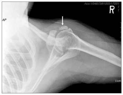
The physical examination informed us that there was tenderness on right acromio-clavicular joint, greater and lessor tuberosity of humerus and right coracoids, but Spurling's test and Jackson compression test were negative and therefore we could rule out cervical radiculopathy. The axillary lateral view of right shoulder showed calcified lesion (19 × 7 mm) at subscapularis tendon (Fig. 1).
For improving her pain, we performed subacromial-subdeltoid (SASD) bursa injection and suprascapular nerve block (SSNB) with 0.5% mepivacaine 5 cc and triamcinolone 5 mg at each site. After 2 weeks, her pain was aggravated, so acromio-clavicular joint injection, SASD bursa injection and SSNB were done again. She experienced pain relief to some degree following injection therapy but suffered the recurrence of shoulder pain several weeks later. After about 6 months from first visit, we performed SSNB and axillary nerve block (ANB) with 0.5% mepivacaine 5ml including 5mg triamcinolone at each site. She had complete pain relief after two nerves block but the pain returned 2 weeks later. Because the SSNB and ANB were very effective temporarily, we decided to perform PRF lesioning on the suprascapular and axillary nerve.
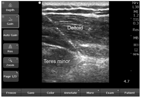
After placing the patient in the sitting position, skin preparation with betadine and draping were done. The operator was positioned behind the patient. The US transducer 10-5 MHz, multi-frequency linear (SonoSite, Inc., Washington, USA) was inserted into a sterile sheath containing ultrasound gel. To find suprascapular nerve (SSN), the US transducer in a transverse orientation was placed over the scapular spine at the most medial part. While imaging the supraspinatus muscle and the bony fossa underneath, the US transducer was moved laterally to locate the suprascapular notch. At this region, SSN is seen as a round hyperechoic structure. To find axillary nerve (AN), using US transducer parallel to the longitudinal axis of the shaft of the humerus, we could identify the surgical neck and the shaft of the humerus and the cross section of the posterior circumflex humeral artery (PCHA), using ultrasound Doppler. We could see other important ultrasonographic landmarks such as the teres minor muscle, which lies cranial to the PCHA, the posterior part of the deltoid muscle closest to the probe and the lateral and long head of the brachial triceps muscle in longitudinal section below the detoid muscle. The AN was located cranially in close relation to the PCHA in the neurovascular space between the teres minor muscle cranially, the deltoid muscle posteriorly, the triceps muscle caudally and the shaft of the humerus anteriorly. After infiltrating 1% lidocaine for local anesthesia at needle entry point of right shoulder, 10 cm radiofrequency needles, insulated with a 5-mm active tip (22 G, Radionics SMK-C10; Radionics Inc, Burlington, MA, USA), were inserted near right supraclavicular nerve and axillary nerve under ultrasound guidance using in plane technique. We checked proper needle position through sensory stimulation with 50 Hz, 0.5 V and motor stimulation with 2 Hz, 1 V. At SSN stimulation, the patient felt paresthesia over the shoulder area and contractions of the supraspinatus or infraspinatus muscles for sensory and motor stimulation, respectively. At AN stimulation, she felt paresthesia and contractions of the anterior, medial and posterior parts of the deltoid muscle. Following confirming the needle position, we performed PRF lesioning with 42℃ for 120 seconds at each time, and repeated this procedure for 3 times (Fig. 2).
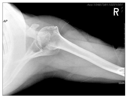
She experienced gradual alleviation of shoulder pain. Her pain score 1 month later, first follow up after PRF, was 3/10 on VAS. After 2 month, X-ray showed the calcification nodule was disappeared (Fig. 3) and she needed no more pain management. Three months later from PRF, the pain level was VAS 1/10 and the range of motion for right shoulder was nearly full. She complained no more pain when performing the internal rotation and abduction. She subsequently experienced pain relief during six months of follow up. There was no conservative management during that period.
Go to : 
DISCUSSION
The calcific tendinitis mainly affects individuals between 30 and 50 years of age and its clinical symptoms occur in from 34% to 45% of patients in whom calcifications are found [8,9]. A tissue hypoxia due to hypovascularization in the so-called 'critical zone' in the rotator cuff that becomes vulnerable to calcification, have been suggested as possible causes, however, the exact mechanism for calcific tendinitis is still a matter of controversy and speculation [1,10,11]. In most cases, this zone is located 1 to 2 cm from the insertion of the supraspinatus tendon on the greater tuberosity [1]. Tissue hypoxia causes fibrocartilaginous transformation and necrosis of the tendon tissue, which is then followed by a tendency to calcification deposits. There is a cyclic course of the disease with spontaneous resorption and reconstitution of the tendon. However, morbidity due to chronic pain may remain although the disease is self-limiting in some cases without therapy.
In our case, the patient in this case was treated for shoulder pain by various methods; however her pain was not decreased noteworthily for 3 years. The reason was not clear; however, the PRF procedure seemed to have played effective role in reducing the pain. We could postulate that the pain reducing effect of PRF on both nerves help the patient to exercise more, which enhanced the absorption of the calcium deposit.
Although the exact mechanism of action of PRF is unknown, there are several opinions about that of action of PRF. That may include inhibition of excitatory C-fiber responses by repetitive burst-like stimulation of A-delta fibers [7], the enhancement of noradrenergic and serotonergic descending pain inhibitory pathway [12].
The mechanism of the calcium deposit absorption could be the result of increased range of motion (ROM) due to the prolonged pain reduction after PRF. It had been advocated that active and passive ROM exercise is helpful to muscle-relaxing effect and consequent improvement in muscle capillary filling [13,14]. The improvement in muscle capillary filling and peripheral circulation is the similar effect from the deep heat application [13]. In our case, the ROM improvement results from the prolonged pain reduction after PRF lead to improvement of local circulation, which could help the absorption of calcium deposit.
We performed the PRF on axillary nerve as well as suprascapular nerve. The reason may be found in the innervations of both nerves. The suprascapular nerve is a mixed motor and sensory nerve originating from the ventral rami of C4, C5 and C6 spinal nerves and runs through the upper trunk of the brachial plexus. After passing underneath the transverse scapular ligament and through the suprascapular notch, it innervates the supraspinatus and infraspinatus muscles as the glenohumeral joint. It provides sensory input for much of the posterior shoulder capsule and joins with the lateral pectoral nerve in supplying sensory innervations to the AC joint, coracoclavicular ligament, and the subacromial bursa [15,16]. The axillary nerve derived from the C5 and C6 spinal nerves, is also mixed motor and sensory nerve. Its motor function affects deltoid, teres minor and the long head of the triceps brachii [17]. Its sensory branch together with subscapular and lateral pectoral nerve branches innervate the anterior shoulder joint capsule [18].
We thought that the patient's pain did not decrease significantly before combined with axillary nerve block, because the suprascapular nerve is responsible for 70% of the sensory innervation to the shoulder joint [19]. Therefore, we could deduce from ANB as well as SSNB that part of pain originated from area which other nerves innervated beside suprascapular nerve. In a study by Price and coworkers [20], axillary nerve block combined with suprascapular nerve block completely reduced pain following total shoulder joint replacement.
In conclusion, combination PRF lesioning of suprascapular and axillary nerve showed a contentable result in pain control and function in patients undergoing intractable shoulder pain. However, larger prospective randomized studies and long-term assessment are required before this procedure becomes popular for the treatment of calcific shoulder tendinitis.
Go to :
 XML Download