Journal List > Korean J Orthod > v.52(1) > 1159410

Barone, Cosentini, Bennardo, Antonelli, and Giudice: Incidence and management of condylar resorption after orthognathic surgery: An overview

Abstract

Objective

Condylar resorption (CR) is one of the major post-surgical complications of orthognathic surgery. This systematic review (SR) aimed to evaluate epidemiological data, risk factors, and therapeutical management of CR.

Methods

Six databases were screened by two investigators until September 2020 to obtain all SRs. After reading the titles and abstracts, eligible SRs were determined and data extraction was performed. Using the latest version of A Measurement Tool to Assess Systematic Reviews, the methodological quality of the included SRs was determined.

Results

Ten SRs with low or critically-low methodological quality were included in this review. Mandibular hypoplasia on the sagittal plane and hyperdivergent growth pattern on the vertical plane were the most common skeletal alterations in which CR could occur after orthognathic surgery. Post-operative condylar changes were analyzed both on two-dimensional and three-dimensional (3D) radiographic examinations. The incidence of CR was not related to the fixation method. Based on the severity of the pathological conditions, management of CR can include conservative or surgical therapy.

Conclusions

Despite the limited evidence in literature, CR is considered a consequence of orthognathic surgery. However, an accurate diagnosis of CR and a better orthognathic surgical planning must include 3D radiographic examinations to improve pre- and post-surgical comparison. Well-designed studies with long-term follow-up and 3D data are needed to clarify the findings of this analysis..

INTRODUCTION

For patients undergoing orthognathic surgery, several surgical risks with different severity rates must be considered.1 Condylar resorption (CR) is an uncommon but well-known clinical condition that can occur after surgical repositioning of the jaws.2,3 It represents the advanced stage of condylar remodeling, irreversibly affecting the temporomandibular joint (TMJ).3-5 As a result of mechanical load, the adaptive mechanisms that normally allow to balance bone resorption and bone formation on the condylar surface are completely lost, resulting in morphological alterations of the condylar structure.5,6 Microstructural changes include a significantly reduced condylar volume and a pathological deformed profile on the condylar surface.7-9 External changes have a significant influence on aesthetics and may include reduced posterior facial height, mandibular retrusion, and anterior open bite, especially if bilateral CRs occur.7,10
Based on the etiology, CR can be distinguished into primary and secondary CR.8 Primary CR is diagnosed when CR exists without a well-known cause.8,11 Conversely, both local (trauma, inflammation, infection, etc.) and systemic (rheumatic immune pathologies, steroid therapy, etc.) agents can be etiological factors of secondary CR.6 Referring to orthognathic surgery, all osteotomies can lead to CR, which can occur in both mono-maxillary and bi-maxillary surgery.2,12 Bilateral sagittal split osteotomy (BSSO) and/or Le Fort I osteotomy can modify the mechanical forces on the condylar surface, resulting in CR.12
Several studies have focused on condylar alterations after orthognathic surgery, and some systematic reviews (SRs) aimed to determine the most crucial risk factors for CR. However, the different methodological approaches do not allow to draw firm deductions. The rationale of this overview was to summarize the most recent knowledge on CR after orthognathic surgery (CROS), emphasizing the limitations of previous studies in order to critically evaluate their conclusions and identify the gaps in knowledge that should be filled by future researches with adequate protocols.
This study aimed to systematically review all SRs and meta-analyses (MAs) to analyze CROS in order to provide evidence-based information about epidemiological data, risk factors, and management of CR, evaluating each outcome in correlation with the qualitative methodological analysis of the included reviews.

MATERIALS AND METHODS

All SRs were systematically reviewed according to the Population, Intervention, Comparison, Outcomes, and Study criteria to assess CR in patients who underwent orthognathic surgery.

Search strategy

Six databases were investigated: PubMed, Cochrane Library, Google Scholar, Scopus, LILACS, and Web of Science. The electronic search was conducted until September 2020, using keywords and MeSH terms connected by the Boolean operator “AND.” The PubMed search combined the following term sequence: “orthognathic surgical procedures”[Mesh] AND “condylar resorption” AND “systematic review” and “condylar resorption” AND “orthognathic surgery” AND “systematic review.” The other search databases combined the following term sequence: “orthognathic surgery” AND “condylar resorption” AND “systematic review.” Additionally, a manual search was conducted in the reference lists of the selected SRs. No restriction of language or publication date was imposed.

Review selection

The electronic search was independently conducted by two investigators (SB and GC), screening titles and abstracts in parallel to evaluate the reviews for eligibility. In case of missing information, full-text reading was necessary for a final decision. A third author (AG) discussed and resolved any discrepancies between the two authors.SRs and MAs were included, which allowed for the extraction of data on CROS. Narrative reviews, overview of reviews, duplicate articles, studies with no evaluation of condylar morphology, and studies on orthognathic surgery of syndromic patients or cleft lip and palate patients were excluded. The Cohen's kappa coefficient (κ) was calculated to determine the inter-rater agreement between the two investigators (SB and GC).

Data extraction

The data extraction from the eligible reviews was independently performed by the same two investigators (SB and GC), recording the following information: author, publication date, study design (SR or MA), number of included studies, number of included patients, dentoskeletal malocclusion, type of intervention, methodological data, quality assessment of primary studies, outcomes, results of reviews, and author’s conclusion.

Assessment of methodological quality

The methodological quality of each SR was independently assessed by the two investigators (SB and GC) using the latest version of A Measurement Tool to Assess Systematic Reviews (AMSTAR-2).13 AMSTAR-2 includes 16 domains, 9 non-critical items, and 7 critical items that strongly influence the final score. The quality assessment can range from high to critically low.13

RESULTS

Search results and review selection

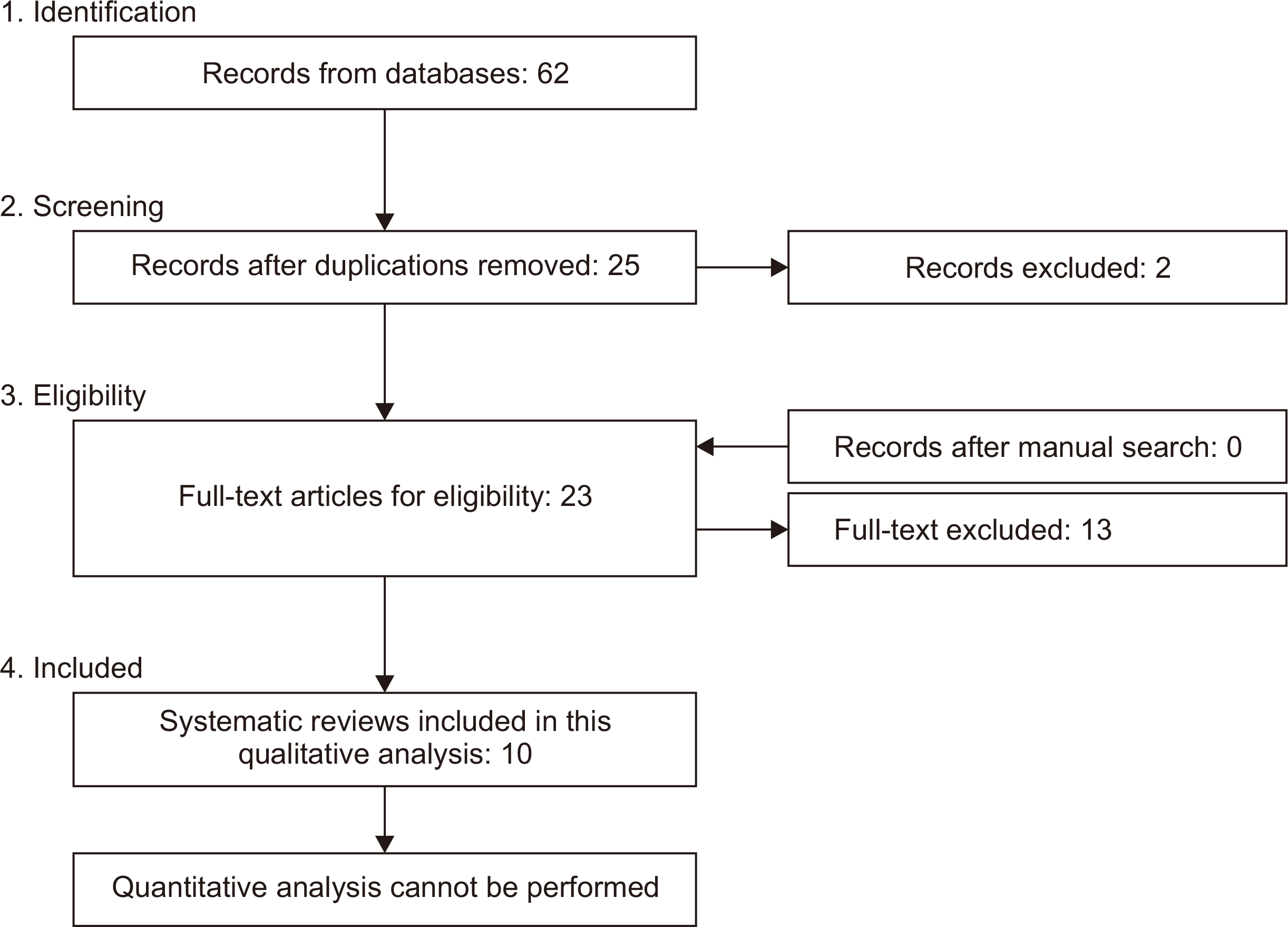
A total of 62 records were selected from the electronic search of six different databases (PubMed, n = 9; Cochrane Library, n = 0; Google Scholar, n = 25; Scopus, n = 13; LILACS, n = 1; Web of Science, n = 14), and no studies were added by manual search. After excluding the duplicates, 25 potentially significant studies were found. After screening titles and abstracts, 23 full-text articles were screened for eligibility, and 2 studies were excluded.5,14 After full-text reading, 13 reviews were excluded because they did not meet the inclusion criteria (Table 1). Figure 1 shows the flow diagram of the search strategy and SR selection: 10 SRs were included for the qualitative analysis.7,8,15-22 Quantitative analysis could not be conducted because no MAs were performed. The inter-rater agreement coefficient was κ = 0.93.

Data extraction

Table 2 summarizes all data extracted from the SRs. Each SR included a different number of studies (ranging from 6 to 76), and none performed an MA. The publication year ranged from 2008 to 2019. Regarding the study design, most of the SRs included observational retrospective and prospective studies. Non-randomized controlled trial (non-RCT) was the most common study type, with the others being case control study, cohort study, case series, and case report.7,8,15-22 Only two SRs described the results of RCTs.19,20 Dentoskeletal Class II was the most frequent malocclusion analyzed in the included SRs.7,8,15-22 Other studies evaluated patients with dentofacial malformations such as skeletal Class III, skeletal asymmetry, and skeletal open bite.7,17,20,21 The most common surgical procedure included BSSO with or without Le Fort I osteotomy.7,16-22 Five reviews reported the outcomes after Le Fort I osteotomy alone, unilateral sagittal split osteotomy, and intraoral vertical ramus osteotomy.7,16-18,22 Post-surgical changes of condylar morphology were analyzed on two-dimensional (2D) radiographic examinations (orthopantomography and/or lateral cephalogram), three-dimensional (3D) radiographic examinations (computed tomography, cone beam computed tomography, and magnetic resonance imaging), or combined 2D and 3D examinations.7,8,15-22 The follow-up period ranged between 12 and 60 months, with a minimum and a maximum follow-up period of 3 and 192 months, respectively.7,8,15-22 The number of patients included in the SRs ranged between 180 and 3,777, with a mean age of 26.6 years.7,8,15-22 Post-orthognathic CR was the primary outcome in the included studies evaluating the morphological changes of the mandibular condyle after surgery.7,15-22 Two SRs reported the effectiveness of CR management.8,16

Analysis of methodological quality

The methodological quality of each SR was determined using AMSTAR-2.13 Most of the included SRs had a low methodological quality.7,8,15,19-22 Three SRs had a critically low methodological quality.16-18 The most critical items of the AMSTAR-2 checklist were the absence of details for excluded studies and the absence of a comprehensive search strategy.

Condylar resorption

Nine of the included SRs aimed to describe the main characteristics and risk factors of CROS (Table 3).7,15-22 Jędrzejewski et al.19 reported that CR manifestations usually ranged from 6 months to 2 years after surgical treatment, but Catherine et al.16 extended this time to 6 years post-operatively. Although with different diagnostic methods, CR could be identified by both radiographic and clinical signs.8,16,20 Further, CR was recognized by 2D radiological examinations if there was a reduction of ramus or condylar height, with a minimum vertical decrease of 2 mm.20 A 3D analysis of the condylar profile assessed that a reduction of at least 17% of the condylar volume was considered as the cut-off value for the diagnosis of CR.8 Clinical signs of bilateral CR included an anterior open bite or a Class II malocclusion with retrognathia.16 On the contrary, an active process of unilateral CR could identify mandibular asymmetry with an ipsilateral Class II malocclusion and a contralateral open bite.16
There is no agreement among the included SRs about the role of sex or age in CROS.7,15-22 Five SRs reported that females showed a higher risk for CR than males, with an approximate female-to-male ratio of 5:1.16,17,19-21 Two SRs stated that young patients may be more prone to CR, while de Moraes et al.17 concluded that age could not be correlated with post-operative CR.16,17,21 In terms of pre-surgical evaluation, it was mandatory to exclude an active process of condylar atrophy, the presence of severe temporomandibular disorders, and an intra-articular inflammatory damage.8,16-18
Risk factors for CR were evaluated both in the sagittal and vertical planes.7,15-22 Angle Class II with mandibular hypoplasia was the most common malocclusion in which CR occurred after a significant mandibular advancement (incidence rate: 1.4–31%).7,15-22 An orthognathic plan with advancements greater than 10 mm may be a risk factor for CR.16 Furthermore, in Angle Class II malocclusions, micrognathia often involved condylar morphology characterized by a small volume, a reduced adaptive capacity to external load, and an increased risk of CR.8 Only one SR reported a more prone CR in Angle Class III malocclusion after a surgical mandibular set-back greater than 6 mm.8 On the vertical plane, a hyperdivergent skeletal pattern with an anterior open bite, a low posterior-to-anterior facial height, and an increased mandibular plane angle (MPA) could cause CR.7,15-22 However, the entity to define the critical clockwise rotation of the mandible was not unanimously accepted, but it could be described when the MPA was greater than 40°.16,18 In patients with a dolichofacial profile, a pre-surgical posterior condylar inclination was exposed to CR because the antero-superior surface and the latero-superior area were subjected to an excessive load on the sagittal and coronal planes, respectively.7,15-22

Fixation method

Six of the included SRs evaluated the type of fixation in relation to CR.8,16-19,21 Wire fixation, rigid fixation (bicortical miniscrew and miniplates), and intermaxillary fixation (IMF) are reported as the possible options.16,17 The incidence of CROS or surgical relapse was not affected by the fixation method in 6.3–13% of the cases.16,17,21 However, a majority of the authors recorded a high risk of CROS after a prolonged IMF rather than a rigid fixation, while no difference was found between wire and rigid fixation.8,18,19 Although no studies have compared the two types of rigid fixation, Jędrzejewski et al.19 reported that bicortical miniscrews could determine inferior alveolar nerve damage.

Management of condylar resorption

Two included SRs reported the main therapeutic approaches for CR.8,16 Despite the fact that there are no precise guidelines on the management of CR to date, several suggestions were made, differentiating the pre- and post-surgical phases of evaluation.8 In the pre-operative phase, the aim was to annul or limit the risk factors.8 In the post-surgical phase, the objectives included the treatment of inflammation and pain, as well as the correction of skeletal deformities and occlusal instability.8,16 Conservative or surgical treatment were proposed.8,16 Conservative therapy managed to avoid CR progression, stabilizing the pathologic conditions without improving dentofacial deformities.8,16 It included anti-inflammatory drugs, occlusal splints, physiotherapy, and in some cases, orthodontic or restorative treatments.8,16 Surgical treatment should be performed at least 6 months after orthognathic surgery, considering the severity of CR, the surgeon’s experience, and the patient’s intentions.8 Disc repositioning, condylectomy, chondro-costal graft, or complete prosthetic TMJ reconstruction were more invasive therapeutic options that were reported as possible treatments for CR.8,16

DISCUSSION

The purpose of this review was to examine the current evidence of CROS, relating the methodological analysis of each SR included in this study. CR has been recognized as one of the major post-surgical complications occurring in orthognathic patients.1 It can be considered an irreversible correspondence of the physiological condylar remodeling, characterized by a severe impairment of condylar morphology with or without clinical symptoms.5 In this overview, a population of patients with skeletal deformities who had undergone orthognathic surgery was analyzed, excluding the SRs of syndromic or cleft lip and palate patients in whom different heterogeneous comorbidities can be associated with skeletal malformations.23 This SR aimed to answer the following questions: “Can CR be considered a severe complication of orthognathic surgery?;” “What are the main risk factors for CROS?;” and “How can CROS be managed better?”
Ten SRs were included in this overview.7,8,15-22 Quantitative analysis could be reported because no MAs were performed.7,8,15-22 For each SR, the qualitative analysis recorded the final score of AMSTAR-2 obtained by the evaluation of its 16 items.13 Although the absence of MA precluded the evaluation of some critical domains, the methodological assessment of the included SRs ranged between low and critically-low quality.7,8,15-22 A comprehensive search strategy (item 4) and adequate details for the excluded studies (item 7) were the most common missing data in the AMSTAR-2 checklist.
The qualitative evaluation of the included SRs allowed to analyze the main characteristics of CROS, summarizing the most significant risk factors and better management reported in literature. CR related to orthognathic surgery can occur 6 months to 6 years after the surgical treatment, and both clinical and radiological signs should be examined. It was not unanimously accepted if sex or age could influence this pathological condition. However, young female patients seem to be more prone to CROS.7,15-22 Despite the limited evidence in literature, altered estrogen levels could interfere with the post-operative morphological changes occurring on the condylar surface.24 An increased inflammatory process negatively involves the synovial tissue and fibrocartilage synthesis, predisposing to TMJ instability and bone resorption.25 CR may expose the patient to skeletal relapse, resulting in an anterior open bite and a Class II dental relationship for mandibular clockwise rotation if it occurs bilaterally.16 Similarly, unilateral CR could be recognized by a contralateral open bite and an ipsilateral Class II malocclusion, emphasizing an asymmetric profile of the mandible.16 These clinical manifestations are consequent to CR because suitable joint relationships are required to allow mandibular functions, even with altered condylar morphology.21
Despite the low quality of evidence, all SRs identified Angle Class II malocclusion as the most frequent skeletal deformity in which CROS occurs.7,15-22 Surgical treatment of mandibular hypoplasia aimed to obtain a stable advancement of the jaw without post-operative relapse. An orthognathic plan with a mandibular advancement greater than 10 mm should be considered with caution because it represents a risk factor for CR.16 After surgery, backward suprahyoid muscle forces and reduced condylar volume are the most common problems in Class II patients, which decrease the adaptive capacity to external load, favor irreversible condylar alterations, and predispose to relapse.21,25,26
Despite the low qualitative assessment of the included SRs, hyperdivergent facial type was recorded as a significant risk factor for CROS on the vertical plane.7,15-22 Concerning the clockwise rotation of the mandible, the specific cut-off for CR was not unanimously accepted, but an MPA greater than 40° was reported as a critical value.16,18 Major condylar changes were emphasized in patients with a lower posterior-to-anterior facial height ratio due to a significant increase in perimandibular tissue stretching after surgery.27 Furthermore, hyperdivergent facial type is often associated with a pre-operative posterior inclination of the condyle.7,15-22 A large surgical advancement of the mandible with an important counter-clockwise rotation causes excessive forces on the antero-superior and latero-superior areas of the condylar head, predisposing to CROS because unbalanced load occurs on less dense surfaces.8,20
Referring to the management of CR, two SRs described the main treatment options for CROS.8,16 A conservative therapeutic approach aimed to stop the pathologic progression, stabilizing the clinical conditions with anti-inflammatory drugs, occlusal splint, physiotherapy, and orthodontic or restorative treatments.8,16,27 Surgical therapy is reserved for severe cases of CROS, and it should be performed no sooner than 6 months after orthognathic surgery.8,28 The goal of the surgical options is to improve the clinical situation, correct the post-operative relapse, and restore an adequate condylar morphology.8,16,25 However, a reoperation should be considered with caution, evaluating the pathological progression, and should be meticulously planned to avoid worsening of the disease.8
In this overview, the main limitation of the conclusions relates to the presence of SRs with low or critically-low methodological quality. All reported results should be interpreted considering an imprecise search strategy of the studies and a limited methodological assessment of CROS. Most of the SRs included studies that evaluated CR on 2D radiographs, but 3D analysis of condylar profile is fundamental to obtain more specific information about the trend and severity of CR.7,20,29,30 As reported in literature, a reduction of at least 17% of the condylar volume was considered a significant radiological sign of active CR.7,8,30 To date, orthognathic surgery has been a precise surgical procedure with a pre-operative 3D planning.14 An accurate pre-surgical 3D morphometric analysis of the condyles could improve the treatment plan and avoid post-operative relapses, allowing to maximize safe movements and minimize risky ones. The extent of the jaws’ movements, direction, and rotational component could be influenced by the pre-surgical condition of the condyle by evaluating the possible degree of compromise. For this reason, the pre-operative assessment of condylar morphology could also be performed on 3D scans, excluding an active process of condylar atrophy or intra-articular inflammation. Because a post-surgical 3D examination was often required, a pre-operative and post-operative comparison of the condylar surface could be achieved, allowing to recognize important details for the diagnosis and entity of CROS.7,30

CONCLUSION

CR is a possible consequence of orthognathic surgery, with an incidence rate of 1–31%. Mandibular deficiency, female hyperdivergent patients, and pre-surgical condylar alterations were the most reported risk factors for CROS. To limit inflammatory processes and improve clinical conditions, an adequate therapeutic approach involves conservative or surgical treatment, considering the severity of CR and disease progression. Although many advancements have been made with the application of 3D technologies, it is still difficult to find a standardized method for intraoperative condylar repositioning. To date, scientific evidence is scarce and limited in order to draw certain conclusions. Future perspectives must include improvements in both the diagnostic and prognostic phases. An accurate diagnosis must be based on 3D data after an adequate standardization of the radiological characteristics of CR has been defined. Additionally, well-designed RCTs with long-term follow-up and pre- and post-operative 3D comparisons of condylar surfaces should be implemented to confirm the findings of this review.

Notes

CONFLICTS OF INTEREST

No potential conflict of interest relevant to this article was reported.

REFERENCES

1. Pelo S, Saponaro G, Patini R, Staderini E, Giordano A, Gasparini G, et al. 2017; Risks in surgery-first orthognathic approach: complications of segmental osteotomies of the jaws. A systematic review. Eur Rev Med Pharmacol Sci. 21:4–12. PMID: 28121363.
2. Jung S, Choi Y, Park JH, Jung YS, Baik HS. 2020; Positional changes in the mandibular proximal segment after intraoral vertical ramus osteotomy: surgery-first approach versus conventional approach. Korean J Orthod. 50:324–35. DOI: 10.4041/kjod.2020.50.5.324. PMID: 32938825. PMCID: PMC7500571.
crossref
3. Ma RH, Li G, Yin S, Sun Y, Li ZL, Ma XC. 2020; Quantitative assessment of condyle positional changes before and after orthognathic surgery based on fused 3D images from cone beam computed tomography. Clin Oral Investig. 24:2663–72. DOI: 10.1007/s00784-019-03128-z. PMID: 31728734.
crossref
4. Hsu LF, Liu YJ, Kok SH, Chen YJ, Chen YJ, Chen MH, et al. 2021; Differences of condylar changes after orthognathic surgery among Class II and Class III patients. J Formos Med Assoc. doi: 10.1016/j.jfma.2021.01.018. [Epub ahead of print]. DOI: 10.1016/j.jfma.2021.01.018. PMID: 33583702.
crossref
5. Francisco I, Guimarães A, Lopes M, Lucas A, Caramelo F, Vale F. 2020; Condylar form alteration on skeletal class II patients that underwent orthognathic surgery: an overview of systematic reviews. J Clin Exp Dent. 12:e695–703. DOI: 10.4317/jced.56947. PMID: 32904976. PMCID: PMC7462380.
crossref
6. Catherine Z, Breton P, Bouletreau P. 2016; Management of dentoskeletal deformity due to condylar resorption: literature review. Oral Surg Oral Med Oral Pathol Oral Radiol. 121:126–32. DOI: 10.1016/j.oooo.2015.08.013. PMID: 26460272.
crossref
7. Vandeput AS, Verhelst PJ, Jacobs R, Shaheen E, Swennen G, Politis C. 2019; Condylar changes after orthognathic surgery for class III dentofacial deformity: a systematic review. Int J Oral Maxillofac Surg. 48:193–202. DOI: 10.1016/j.ijom.2018.06.008. PMID: 30007835.
crossref
8. He Z, Ji H, Du W, Xu C, Luo E. 2019; Management of condylar resorption before or after orthognathic surgery: a systematic review. J Craniomaxillofac Surg. 47:1007–14. DOI: 10.1016/j.jcms.2019.03.012. PMID: 30929993.
crossref
9. Claus JDP, Koerich L, Weissheimer A, Almeida MS. Belle de Oliveira R. 2019; Assessment of condylar changes after orthognathic surgery using computed tomography regional superimposition. Int J Oral Maxillofac Surg. 48:1201–8. DOI: 10.1016/j.ijom.2019.02.009. PMID: 30871846.
crossref
10. Lee JY, Lee SM, Kim SH, Kim YI. 2020; Long-term follow-up of intersegmental displacement after orthognathic surgery using cone-beam computed tomographic superimposition. Angle Orthod. 90:548–55. DOI: 10.2319/061119-398. PMID: 33378504. PMCID: PMC8028461.
crossref
11. Ji YD, Resnick CM, Peacock ZS. 2020; Idiopathic condylar resorption: a systematic review of etiology and management. Oral Surg Oral Med Oral Pathol Oral Radiol. 130:632–9. DOI: 10.1016/j.oooo.2020.07.008. PMID: 32807713.
crossref
12. NiÑo-Sandoval TC, Almeida RAC, Vasconcelos BCDE. 2021; Incidence of condylar resorption after bimaxillary, Lefort I, and mandibular surgery: an overview. Braz Oral Res. 35:e27. DOI: 10.1590/1807-3107bor-2021.vol35.0027. PMID: 33605357.
crossref
13. Shea BJ, Reeves BC, Wells G, Thuku M, Hamel C, Moran J, et al. 2017; AMSTAR 2: a critical appraisal tool for systematic reviews that include randomised or non-randomised studies of healthcare interventions, or both. BMJ. 358:j4008. DOI: 10.1136/bmj.j4008. PMID: 28935701. PMCID: PMC5833365.
crossref
14. Haas Junior OL, Guijarro-Martínez R, de Sousa Gil AP, da Silva Meirelles L, Scolari N, Muñoz-Pereira ME, et al. 2019; Hierarchy of surgical stability in orthognathic surgery: overview of systematic reviews. Int J Oral Maxillofac Surg. 48:1415–33. DOI: 10.1016/j.ijom.2019.03.003. PMID: 30910409.
crossref
15. Bermell-Baviera A, Bellot-Arcís C, Montiel-Company JM, Almerich-Silla JM. 2016; Effects of mandibular advancement surgery on the temporomandibular joint and muscular and articular adaptive changes--a systematic review. Int J Oral Maxillofac Surg. 45:545–52. DOI: 10.1016/j.ijom.2015.10.016. PMID: 26644217.
crossref
16. Catherine Z, Breton P, Bouletreau P. 2016; Condylar resorption after orthognathic surgery: a systematic review. Rev Stomatol Chir Maxillofac Chir Orale. 117:3–10. DOI: 10.1016/j.revsto.2015.11.002. PMID: 26682945.
crossref
17. de Moraes PH, Rizzati-Barbosa CM, Olate S, Moreira RW, de Moraes M. 2012; Condylar resorption after orthognathic surgery: a systematic review. Int J Morphol. 30:1023–8. DOI: 10.4067/S0717-95022012000300042. PMID: 28066126.
crossref
18. Gill DS, El Maaytah M, Naini FB. 2008; Risk factors for post-orthognathic condylar resorption: a review. World J Orthod. 9:21–5. PMID: 18426101.
19. Jędrzejewski M, Smektała T, Sporniak-Tutak K, Olszewski R. 2015; Preoperative, intraoperative, and postoperative complications in orthognathic surgery: a systematic review. Clin Oral Investig. 19:969–77. DOI: 10.1007/s00784-015-1452-1. PMID: 25804886. PMCID: PMC4434857.
crossref
20. Mousoulea S, Kloukos D, Sampaziotis D, Vogiatzi T, Eliades T. 2017; Condylar resorption in orthognathic patients after mandibular bilateral sagittal split osteotomy: a systematic review. Eur J Orthod. 39:294–309. DOI: 10.1093/ejo/cjw045. PMID: 27334905.
crossref
21. Nunes de Lima V, Faverani LP, Santiago JF Jr, Palmieri C Jr, Magro Filho O, Pellizzer EP. 2018; Evaluation of condylar resorption rates after orthognathic surgery in class II and III dentofacial deformities: a systematic review. J Craniomaxillofac Surg. 46:668–73. DOI: 10.1016/j.jcms.2018.02.002. PMID: 29534912.
crossref
22. Te Veldhuis EC, Te Veldhuis AH, Bramer WM, Wolvius EB, Koudstaal MJ. 2017; The effect of orthognathic surgery on the temporomandibular joint and oral function: a systematic review. Int J Oral Maxillofac Surg. 46:554–63. DOI: 10.1016/j.ijom.2017.01.004. PMID: 28174061.
crossref
23. Giudice A, Barone S, Belhous K, Morice A, Soupre V, Bennardo F, et al. 2018; Pierre Robin sequence: a comprehensive narrative review of the literature over time. J Stomatol Oral Maxillofac Surg. 119:419–28. DOI: 10.1016/j.jormas.2018.05.002. PMID: 29777780.
crossref
24. Nicolielo LFP, Jacobs R, Ali Albdour E, Hoste X, Abeloos J, Politis C, et al. 2017; Is oestrogen associated with mandibular condylar resorption? A systematic review. Int J Oral Maxillofac Surg. 46:1394–402. DOI: 10.1016/j.ijom.2017.06.012. PMID: 28684303.
crossref
25. Wolford LM. 2001; Idiopathic condylar resorption of the temporomandibular joint in teenage girls (cheerleaders syndrome). Proc (Bayl Univ Med Cent). 14:246–52. DOI: 10.1080/08998280.2001.11927772. PMID: 16369629. PMCID: PMC1305829.
crossref
26. Aneja V, Raval R, Aneja P, Rai KK, Agarwal S, Chuadhary S. 2017; Evaluation of mandibular condylar changes in patients following orthognathic surgery: a retrospective study. Niger J Surg. 23:37–41. DOI: 10.4103/1117-6806.199961. PMID: 28584510. PMCID: PMC5441214.
crossref
27. Gunson MJ, Arnett GW, Milam SB. 2012; Pathophysiology and pharmacologic control of osseous mandibular condylar resorption. J Oral Maxillofac Surg. 70:1918–34. DOI: 10.1016/j.joms.2011.07.018. PMID: 22014941.
crossref
28. Amarista FJ, Mercuri LG, Perez D. 2020; Temporomandibular joint prosthesis revision and/or replacement survey and review of the literature. J Oral Maxillofac Surg. 78:1692–703. DOI: 10.1016/j.joms.2020.05.021. PMID: 32610046.
crossref
29. Ha MH, Kim YI, Park SB, Kim SS, Son WS. 2013; Cone-beam computed tomographic evaluation of the condylar remodeling occurring after mandibular set-back by bilateral sagittal split ramus osteotomy and rigid fixation. Korean J Orthod. 43:263–70. DOI: 10.4041/kjod.2013.43.6.263. PMID: 24396735. PMCID: PMC3879282.
crossref
30. Xi T, Schreurs R, van Loon B, de Koning M, Bergé S, Hoppenreijs T, et al. 2015; 3D analysis of condylar remodelling and skeletal relapse following bilateral sagittal split advancement osteotomies. J Craniomaxillofac Surg. 43:462–8. DOI: 10.1016/j.jcms.2015.02.006. PMID: 25779607.
crossref

Figure 1
A flowchart of the search strategy and selection of systematic reviews.
kjod-52-1-29-f1.tif
Table 1
List of excluded studies
Study Reason of exclusion
Al-Moraissi, 2016 Incoerent topic
Al-Moraissi, 2017 Incoerent topic
Al-Ryhami, 2009 part 1 Incoerent topic
Al-Ryhami, 2009 part 2 Incoerent topic
Catherine, 2016 Incoerent topic
Francisco, 2020 Overview
Haas Junior, 2019 Overview
Ji, 2020 Incoerent topic
Kersey, 2003 Incoerent topic
Merhaban, 2020 Incoerent topic
Nicolielo, 2017 Incoerent topic
Romero, 2019 Incoerent topic
Sansare, 2015 Incoerent topic
Sonego, 2014 Incoerent topic
Verlinden, 2015 Incoerent topic
Table 2
List of included systematic reviews
Study Study design/
Search period
Searchdata-bases Language
restriction
Total subjects (T)/Population with CR (P) Dentoskeletal malocclusions (M)/
Intervention performed in patients with CR (I)
Included
studies (I)/
Quality assessment reported (Q)
Outcomes Radiological method of evaluation (OPG, CEPH, CBCT, CT, NMR, other) Qualitative analysis (AMSTAR-2) Results Conclusion Discussion of the
quality of studies
Bermell-Baviera, et al.15(2016) SR;from2002 to 2014 PubMed,Scopus, Embase, Cochrane Library Norestriction T: 790Mean age: 29.3 years M: Angle Class II malocclusion I: 22 (4 SRs, 5 prospective studies, 13 retrospective studies) Assessment of anatomical modifications in the condyle after surgical mandibular advancement CEPH: 7 Low Higher incidence of CROS occurred in dolichofacial patients with mandibular retrognathia, or in patients with preoperative alteration of condylar morphology The authors concluded that CROS could be accelerated after surgical mandibular advancement but it is not a contraindication to this treatment Heterogeneity of the included study was reported
P: 623 I: surgical mandibular advancement Q: CONSORT criteria (7 high-quality studies; 11 medium quality studies) CBCT: 3
CT: 3
3D photography: 1
Catherine et al.16(2016) SR;from 1970 to 2014 PubMed; manual search English and French language T: 2,994Mean age: 31.6 years M: NR I: 17 (8 cross-sectional studies, 4 observational descriptive studies, 1 cohort study; 1 case control study; 1 prospective study; 1 retrospective study; 1 case series) Examination of physiopathology, mechanism, and risk factors of CROS NR Critically low Most of the studies reported that incidence of CROS ranged from 1.2 to 20.2%. It occurred mainly in female, Class II patients treated after bimaxillary surgical approach The authors concluded that higher incidence of CROS occurred in female with pre-operative TMJ dysfunction, estrogen deficiency, Class II malocclusion with a high mandibular plane angle, and a posterior condylar inclination. Mandibular advancement > 10 mm and a counter-clockwise mandibular rotation were risk factors of CROS NR
P: 224 I: bimaxillary surgery (68%), BSSO (25%), LF I (7%) Q: NR
de Moraes et al.17(2012) SR;from January1978 to August 2010 Cochrane Database, PubMed, Medline, Ovid; manual search English language T: 2,567Age ranged from 14 to 46 years M: Class III and Class II malocclusion I: 8 (human clinical trials; randomized, clinical trials, prospective multicenter studies,or prospective clinical trial; retrospective studies) Assessment of risk factors of CROS NR Critically low Incidence of CROS ranged from 3% to 15%. Female patients had great predisposition to CROS. It occurred mainly in Class II patients with high mandibular plane angle The authors concluded that risk factors were sex (female patients), type of skeletal deformity (absolute mandibular deficiency and high mandibular plane angle) The authors recognized the heterogeneity of the studies and the lack of randomized clinical trials and prospectivestudies
P: 137 I: bimaxillary surgery (75.2%), BSSO (15.3%), LF I (9.5%) Q: NR
Gill et al.18(2008) SR;from January 1980 toAugust 2006 PubMed; Medline, Embase; PsycInfo; DARE; Cochrane Library; manual search NR T: 3,059Mean age 22.6 years
P: 155
M: Class II malocclusion and other not-specified surgical patients
I: bimaxillary surgery (104), BSSO (13), LF I (252)
I: 9 (7 retrospectivestudies, 2 prospective studies)
Q: NR
Assessment of the risk factors of CROS OPG: 8
CEPH: 9
CT: 1
Critically low Higher incidence of CROS occurred in female patients (68.4%), young patients (age < 20 years old), Class II malocclusion with mandibular retrognathia associated with high mandible plan angle. A posterior condylar neck inclination and a pre-surgical condylar atrophy predisposed to CROS. A mandibular advancement greater than 10 mm showed major risk for CROS than surgical advancement of 5 mm The authors concluded that risk factors for CROS included: sex (female), mandibular retrognathia with high mandibular plane angle, magnitude of surgical mandibular advancement, and pre-operative condylar atrophy The authors stated that prospective 3D studies with matched samples are required to better evaluate CROS
He et al.8(2019) SR;up to October 2018 PubMed; Embase; Cochrane Library; manual search No restriction T: 180Mean age 22 years M: NR I: 10 (1 prospective study, 9 retrospective studies) Assessment of therapeutic options for CROS NR Low CR could occur from 6 months to 2 years after OS. Conservative treatment (medication, occlusal splint, orthodontic therapy) is used to control the progress of CROS. Surgical therapy (disc reposition, condylectomy, condylar reconstruction) aims to correct dentofacial deformity The authors concluded that the management of CROS should be based on the severity of the condition, considering risks and benefits of conservative treatment or re-operation The authors recognized that few studies with small samples and few randomized studies were conducted. High-quality research is required to obtain more information on the treatment of CROS
P: 38 I: NR Q: Cochrane risk of bias for RCT showed a moderate risk of bias; MINORS for 9 non-randomized studies showed a moderate risk of bias
Jędrzejewski et al.19(2015) SR; up to February 2015 PubMed; Medline, ISI Web of Knowledge, Ovid, Cochrane Library; Embase Library; Google Scholar; manual search Articles publishedin English, German, French, orPolish T: NR M: NR I: 44 (5 RCTs; 39 non-randomized control trials) Examination of the post-operative complications after OS OPG Low CROS could develop from 6 months to 2 years after surgery. It occurred mainly in female patients with severe Class II malocclusion, a high mandibular plane angle, and a posteriorly condylar inclination The authors concluded that both intraoperative (surgical condylar reposition, incomplete green-stick split) and post-operative (intra-articular hemorrhage or edema, muscular forces) factors could influence CROS The authors declared that lack of high quality-studies precluded a reliable evidence on these results
P: NR I: NR Q: Cochrane Collaboration Tool (RCTs showed high risk of bias; 3 CTs showed low risk of bias; 33 CTs showed high risk of bias)
Mousoulea et al.20(2017) SR;articles publishedfrom 1946 to 2015 Medline; Ovid; PubMed; Embase; Cochrane Oral Health Group’s Trials Register; CENTRAL; Unpublished literature: ClinicalTrials.gov, the National Research Register, and Pro-Quest Dissertation Abstracts and Thesis database; manual search No restriction T: 862 Mean age 27.2 years M: Class I, Class II, and Class III malocclusion I: 14 (1 RCT; 3 prospective studies; 10 retrospective studies) Assessment of incidence and quantification of CR after BSSO OPG: 8 Low Incidence of CROS ranged between 1.4% and 31% after bimaxillary surgery and between 3.6% and 10% after BSSO. Vertical decrease of condylar height ranged between 2 mm and 8 mm. CROS occurred mainly in female young patients with mandibular retrognathia, a high mandibular plane angle, and a posteriorly condylar inclination. Considering the surgical procedure, higher incidence of CROS occurred after bimaxillary surgery and IMF The authors concluded that CR could be a post-surgical complication of orthognathic surgery with higher incidence in female retrognathic patients. 3D radiologic exams could improve the diagnosis of CROS The authors declared that the methodological heterogeneity of the included studies and the low level of evidence precluded definitive conclusions
P: NR I: BSSO with or without other surgical procedures Q: Cochrane risk of bias tool (high risk of bias for RCT; low risk of bias for prospective studies; serious risk of bias for retrospective studies) CEPH: 9
CT: 1
CBCT: 2
CMS: 2
TMJ radiograph: 1
Nunes de Lima et al.21(2018) SR; from 2008 to 2018 PubMed, Medline; Embase; Cochrane All studies publishedin English T: 202 Mean age: 23.3 years M: 95 Class III and 107 Class II patients I: 6 (3 prospective studies; 3 retrospective studies) Examination of condylar alterations in patients undergone SSRO with or without other surgical procedures OPG: 1 Low Percentage of CROS was similar in Class II and Class III patients, with a small incidence ranging between 0.0% and 4.2%. Post-surgical relapse for CR were reported after significant mandibular advancement or set-back, ranging between 4 mm and 6.4 mm The authors concluded that CR is a complication of orthognathic surgery, occurring in a small percentage of included patients The authors stressed the lack of RCTs that precluded definitive conclusions on the comparison between Class II and Class III patients
P: NR I: SSRO with or without LF I Q: NHMRC scale (one prospective study showed III-2; 2 prospective studies showed III-3 score; retrospective studies showed III-3 score) CT: 2
CBCT: 3
Te Veldhius et al.22(2017) SR; up to October 2015 Embase; Medline;Ovid; Cochrane Central Register of Controlled Trials; Web of Science; PubMed; CINAHL; Google Scholar; manual search No restrinctions T: 3,399 Mean age 24.5 years M: Class II and Class III patients I: 76 (1 RCT; 75 non-randomized studies) Examination of the effects of ortohognathic surgery on TMJ OPG: 4 Low Analyzing CT scans, a more superior and posterior condylar position were recorded in BSSO and VRO groups after mandibular advancement. No condylar changes were reported after mandibular set-back. Analyzing OPG, CR and consequent condylar vertical changes were reported after BSSO and LF I. Transcranial radiography allowed to identify CR after BSSO and bimaxillary surgery The authors concluded that condylar changes could occur after surgery, but OS showed little or harmless consequences on TMJ The authors declared that the heterogeneity and the low quality of all included studies precluded definitive conclusions
P: NR I: bimaxillary surgery (549 patients); VRO (520 patients); LF I (130 patients); BSSO (1,932 patients) Q: CEBM criteria (one study showed level II; 16 studies showed level III; 59 studies showed level IV) CEPH: 12
CT: 7
CBCT: 24
MRI: 16
Fluoroscopic imaging: 1
Transcranial radiography: 2
Vandeput et al.7(2019) SR; in August 2017 PubMed; Cochrane Central Register of Controlled Trials; and Embase; manual search All articles published in English language T: 1,376Age between 17 and 43 years M: Class III patients I: 12 (all retrospective studies) Incidence and extent of CROS in Class III patients OPG: 4 Low Condylar vertical changes occurred after OS and a volume decrease of at least 17% identified CR. Condylar axis changes were reported both for BSSO and intraoral VRO. The anterosuperior area in the sagittal plane and the laterosuperior area in the coronal plane were the most frequent region of CROS. Mandibular set-back greater than 6 mm could be associated with CR The authors concluded that CROS could occur in Class III patients, but it was not always related to clinical symptoms or skeletal relapse. Future studies based on greater samples and 3D examinations of condylar morphology should be conducted The authors declared that there were no significant methodological errors. However, the heterogeneity of the studies precluded definitive conclusions
P: NR I: bimaxillary surgery (37 patients); VRO (278 patients); BSSO (142 patients);USSO (20 patients) Q: MINORS indicated a moderate risk of bias for all studies CEPH: 9
CBCT: 3
MRI: 1

CR, condylar resorption; OPG, orthopantomography; CEPH, cephalometric radiograph; CBCT, cone beam computed tomography; CT, computed tomography; NMR, nuclear magnetic resonance; AMSTAR-2, A Measurement Tool to Assess Systematic Reviews; SR, systematic review; NR, not reported; ISI, Institute for Scientific Information; BSSO, bilateral sagittal split osteotomy; SSRO, sagittal split ramus osteotomy; VRO, vertical ramus osteotomy; LF I, Le Fort I osteotomy; USSO, unilateral sagittal split osteotomy; CONSORT, Consolidated Standards of Reporting Trials; RCT, randomized controlled trial; MINORS, methodological index for non-randomized studies; NHMRC, National Health and Medical Research Council; CEBM, Centre for Evidence-Based Medicine; CROS, condylar resorption after orthognathic surgery; OS, orthognathic surgery; TMJ, temporomandibular joint; 3D, three-dimensional; CMS, condylar morphology scale; MRI, magnetic resonance imaging; IMF, intermaxillary fixation.

Table 3
Summary of the incidence of condylar resorption after orthognathic surgery
Question Condylar resorption after orthognathic surgery
Who Patients undergoing orthognathic surgery (from 1.2% to 31%)
Sex:
- 5F:1M
Surgical treatment:
- Bimaxillary surgery: from 67.8% to 75.2% of cases;
- BSSO: from 15.3% to 24.5% of cases;
- Le Fort I osteotomy: from 6.7% to 9.5% of cases
Fixation method:
- Rigid fixation (from 6.3% to 13% of cases);
- Wire fixation (9.9% of cases)
What Radiological signs:
- A vertical decrease of 2 mm or more of the ramus or the condylar height;
- A reduction of at least 17% of the condylar volume
It was estimated that 60% of cases with CROS developed a resorption that ranged between 10 and 19% of the condylar surface, 30% of cases showed CROS ranging between 20 and 29%, and 10% of cases had CROS greater than 30%.
When From 6 months to 6 years after surgery
Where Antero-superior and latero-superior areas of the condylar head
Why CROS was correlared with the following risk factors:
- Impaction of the maxilla and counter-clockwise rotation of the upper occlusal plane with a more posterior position of the condyles (87% of the cases);
- Mandibular hypoplasia with high mandibular plane angle (21.8% of cases) and with normal/low plane angle (2.2% of cases);
- Pre-surgical signs of condylar atrophy (20–44% of cases);
- Presurgical TMD recognized (24% of cases)

F, female; M, male; BSSO, bilateral sagittal split osteotomy; CROS, condylar resorption after orthognathic surgery; TMD, temporomandibular disorder.

TOOLS
Similar articles