

 PDF
PDF Citation
 Print
Print


Complex regional pain syndrome (CRPS) type-1 is a painful and disabling chronic neuropathic disorder, usually precipitated by minor trauma in the extremities. Early recognition of the disease and its treatment can alter the course of the disease. We describe a patient who had delayed diagnosis of CRPS type-1 because of unawareness of this condition among primary care physicians and its subsequent successful management by specialty care. The purpose of this paper is to create awareness among non-specialist practitioners who often see patients with CRPS in the early stage of the disease, so as to distinguish it from other pain conditions and to initiate timely appropriate intervention to halt disease progression.
CASE REPORT
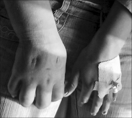
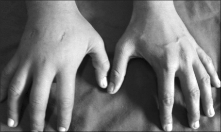
A 16-year-old female patient was referred to our pain clinic from the orthopedic department with persistent pain and swelling of the right forearm. The patient's history revealed her being admitted to a local hospital with urinary tract infection and receiving an infusion of 5% dextrose with 0.9% saline through an IV cannula placed on the right dorsum of the hand five months previous. Later, extravasation of fluid in the surrounding tissue occurred leading to pain and swelling that did not subside but kept increasing. The general practitioner's initial diagnosis was cellulitis. She was treated with analgesics and antibiotics followed by local incision and drainage. Despite symptomatic treatment and surgical intervention, the symptoms further worsened leaving surgical scars (Fig. 1). The primary care physician managed conservatively for almost 5 months before she presented to our hospital. Clinical examination revealed a tender, shinny, swollen limb with allodynia and stiff joints with no evidence of nerve injury suggestive of CRPS type-1. Doppler study ruled out any possibility of vascular disease. A diagnostic stellate ganglion block (SGB) improved the numerical pain rating score (0-10) from 7 to 2 with enhancement of limb mobility. A multimodal approach of treatment including oral medications (gabapentin, amitryptyline, tramadol), a series of SGB with bupivacaine, and limb physiotherapy were initiated. At 6 months follow-up, pain and swelling subsided drastically with marked functional recovery (Fig. 2).
DISCUSSION
Trauma is the most common precipitating event of CRPS type-1, the majority following minor insult to an extremity. The inciting event in our case was inadvertent extravasations of a dextrose solution into tissues surrounding the IV cannula. These extravasations to the interstitial space may have led to an inflammatory response resulting in edema, redness and pain. Rapid swelling and compression of the surrounding tissue may also cause acute limb compartment syndrome [1], reducing tissue perfusion. Swelling, inflammation, tenderness and skin changes have been reported to confound diagnosis of CRPS following snake bite also [2]. These mechanisms' roles cannot be ignored in our case as the current understanding of CRPS pathophysiology involves inflammation and hypoxia along with autonomic and somatic nervous system involvement [3]. However, it is difficult or even impossible to demonstrate a causal relationship between these clinical conditions and CRPS development.
Due to its wide range of precipitating factors and pathophysiologic complexity, CRPS diagnosis is based primarily on established clinical criteria including allodynia, temperature and skin color changes, edema, motor dysfunction and trophic changes [4]. Although special investigations, such as autonomic function tests and bone scans, support the diagnosis of CRPS, these diagnostic modalities add little to the overall accuracy of diagnosing the disease when compared with clinical criteria [5]. Moreover, availability of such modalities is unlikely in every clinical setting. Hence, distinguishing CRPS type-1 clinically from other pain conditions, such as undiagnosed deep vein thrombosis, cellulitis and vasculitis, can be challenging even to specialist pain physicians [6]. The clinical course of CRPS progresses through three stages: acute inflammatory followed by dystrophy, ultimately leading to structural atrophy. However, these stages may overlap with less distinctness [7]. In the acute stage, all classical signs and symptoms of inflammation are present, similar to that of cellulitis. Moreover, in the early course of the disease, all features may not be present making diagnosis difficult [8]. Adolescents are less likely to have overt autonomic signs associated with CRPS, such as hyperhidrosis and trophic changes, compared to adults [7,9]. This may also lead to improper diagnosis.
With a multimodal treatment approach to CRPS, wide ranges of non-pharmacological therapeutic modalities, including intravenous regional blockade, sympathetic blockade and spinal cord stimulation, have been reported with varying results [10,11]. In our case, use of SGB combined with oral medications and limb physiotherapy not only served as therapy, but also contributed to confirming the diagnosis. It is interesting to note that although no special equipment or testing is required for diagnosis of CRPS type-1, an average of 30 months pass and 4.8 different physicians are visited before patients with CRPS are referred to a pain centre even in well-established centres [12,13]. These data show lack of disease awareness among general practitioners who often encounter such patients in the early stage, as in our case.
This case highlights that CRPS often remains under-diagnosed in the hands of non-specialist physicians, particularly if the patient presents with various inflammatory features such as cellulitis, arthritis, etc. Primary care physicians are often the first group of care providers to whom patients go for consultation. Disease awareness among practitioners, its early recognition, appropriate therapy at the beginning and referral to a pain centre shall prevent unnecessary suffering from CRPS and also may prevent the incidence of its development in the community.
 XML Download