

 PDF
PDF Citation
 Print
Print


Neck pain is common in adults and may result from numerous conditions such as neck strain, herniated disc, degenerative disc disease, and whiplash injury. We report a rare case of spontaneous retropharyngeal hematoma causing neck pain diagnosed by magnetic resonance imaging (MRI).
CASE REPORT
A 23-year-old male with persistent neck pain, limited neck motion and mild dysphagia visited our department of pain medicine despite 3 days of treatment with analgesics and muscle relaxants prescribed by local clinics. There was no specific past medical history such as a trauma, upper respiratory infection, administration of anticoagulants or any other medical condition. Upon physical examination, range of neck motion was limited with flexion 50°, extension 20°, both side lateral bending 10° and both side rotation 15°. There was no tenderness, swelling or mass on his neck upon palpation. Examination of the oropharynx was unremarkable. Initial laboratory tests showed white blood cell (WBC) count 13,610/ul (neutrophils, 73.7%; lymphocytes, 15.7%; monocytes, 8.2%), platelet count 251,000/dl, erythrocyte sedimentation rate (ESR) 8 (0-10) mm/hr, and C-reactive protein (CRP) 25.3 (0-8) mg/L. The patient's body temperature was 36.9℃. Thickness of the prevertebral soft tissue was slightly increased on lateral cervical spine radiograph (Fig. 1A). Initial diagnostic impression was retropharyngeal tendinitis or cervical disc disease. Celecoxib 200 mg bid was prescribed and patient symptoms were mildly alleviated after 2 days. Two days later, an MRI was done revealing a prevertebral hematoma with depth 5 mm, length 5 cm spreading from C2 to C4, and an intermediate signal in T1 and high signal in T2 image (Fig. 2A, B). Follow-up laboratory tests showed WBC count returned to normal range, 6,500/uL (neutrophils, 59.9%; lymphocytes, 29.1%; monocytes, 7.2%), platelet count 294,000/dL, ESR 8 mm/hr, and CRP 9.3 mg/L. Based on clinical and radiologic findings, a diagnosis of spontaneous retropharyngeal hematoma was made. The patient was closely followed up in the outpatient department and advised to immediately visit hospital if symptoms such as dyspnea, aggravation of neck pain or dysphagia recurred. Celecoxib 200 mg bid was continued for 3 more days, after which complete remission of symptoms was observed in the outpatient department and follow-up laboratory tests showed a return to normal range: WBC count 6500/uL (neutrophils, 59.9%; lymphocytes, 29.1%; monocytes, 7.2%), platelet count 294,000/dl, ESR 4 mm/hr, and CRP 3.5 mg/L. Normalization of the prevertebral soft tissue in a lateral cervical spine radiograph (Fig. 1B) and nearly complete resolution of a prevertebral hematoma in follow-up T2-weight MR images (Fig. 2C) were also noted. Follow-ups at 1 week and 1 month found the patient remained asymptomatic.
 | Fig. 1(A) Initial lateral cervical spine radiograph shows increased thickness of prevertebral soft tissue: 8 mm at C3 and 10.5 mm at C4. (B) At the 5-day follow-up point, lateral cervical spine radiograph shows a normalization of shadow of prevertebral space: 4.5 mm at C3 and 6 mm at C4.
|
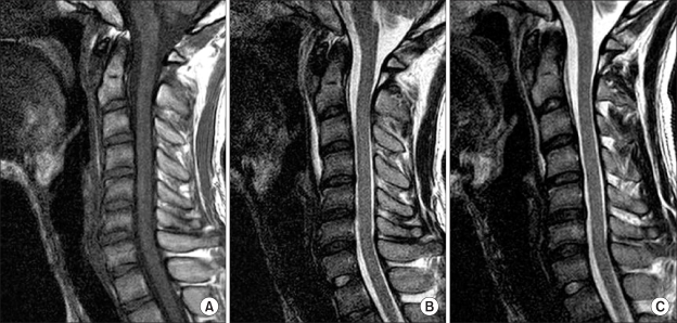 | Fig. 2Magnetic resonance imaging (MRI) shows retropharyngeal hematoma. (A) Sagittal T1 weighted image shows an intermediate signal intensity at the prevertebral space. (B) Sagittal T2 weighted image shows a high signal prevertebral collection with depth 5 mm and length 5 cm, spreading from C2 to C4. (C) Follow-up sagittal T2 weighted image shows a nearly completely absorbed prevertebral collection compared to initial MRI finding.
|
Go to : 
DISCUSSION
Retropharyngeal hematoma is rare, difficult to diagnose early, and may progress to airway obstruction. Etiologies of retropharyngeal hematoma include infection, cervical spine trauma, great vessel trauma, violent head movements, iatrogenic injury associated with cardiac catheterization, cerebral angiography, parathyroid adenoma hemorrhage, and foreign body ingestion [1]. Anticoagulation or hemorrhagic diathesis predisposes an individual to develop retropharyngeal hematoma [2]. Spontaneous retropharyngeal hematoma is defined by the absence of any clear etiology, and there are only a few case reports [1,3,4] in the English literature.
Clinically, spontaneous retropharyngeal hematoma can present as a triad of features including superior mediastinal obstruction, anterior tracheal displacement and bruising on the neck within 48 hours with subsequent spreading to the chest wall [5]. However, in cases with no history of trauma, early diagnosis in an outpatient department may be challenging because of non-specific symptoms, such as neck pain or dysphagia, especially when a hematoma is limited to a prevertebral space. Between prevertebral fascia and posterior wall of the pharynx, the slit-like retropharyngeal space, consisting of loose areolar tissue, is found. Hematomas are assumed to expand within this loose areolar tissue, which may delay symptoms for at least 2-3 hours and possibly cause death. When dyspnea develops due to expansion of a hematoma, procedures such as a tracheostomy or tracheal intubation are required to secure the airway.
Computed tomography (CT) and MRI are used to diagnose a retropharyngeal hematoma, and close observation of prevertebral soft tissue width in lateral cervical spine radiograph is necessary [6,7]. Penning [8] reported that normal prevertebral soft tissue widths were 4.6 mm, 3.2 mm, 3.4 mm, and 5.1 mm at C1, C2, C3, and C4 level in the neutral position, and 14.9 mm, 15.1 mm, 13.9 mm at C5, C6, and C7 level in neutral position. Rojas et al. [9] reported that the upper limits of normal range for thickness of prevertebral soft tissue were 8.5 mm, 6 mm, 7 mm, 18 mm, and 18 mm at C1, C2, C3, C6, and C7, respectively. The upper limit of normal range was not determined for C4 and C5 levels due to variable position of the esophagus and larynx. Furthermore, they reported that mean prevertebral soft tissue thickness were 7 mm at C4 and 12.4 mm at C5 on multi-detector CT images. In our case, thickness of the prevertebral soft tissue was also mildly increased to 8 mm at C3 and 10.5 mm at C4 compared to these reference values. Therefore, we believe these reference values may be useful in early detection of underlying retropharyngeal or cervical spine pathology in clinical situations.
Differential diagnosis of retropharyngeal hematoma includes retropharyngeal tendinitis and retropharyngeal infection. Retropharyngeal tendinitis is due to calcific deposits within the tendons of the longus colli muscles as in calcific deposits in calcific tendinitis of the shoulder. Radiographic findings are characteristically amorphous calcification localized anterior to C1 with associated swelling of the prevertebral (or retropharyngeal) soft tissues from C1 through C4 [10]. Clinical features are sudden onset of severe pain in the neck and throat aggravated by swallowing and movement of the head; this may be associated with mild fever and elevation of ESR. Pain reaches a maximum within 2-5 days then gradually subsides, usually completely within 1-2 weeks. Initial clinical and radiologic findings in this case misled our diagnostic impression toward retropharyngeal tendinitis. Although CT is the method of choice for confirming calcification, MRI was performed to evaluate both calcification and cervical disc disease in this case. Clinical findings of retropharyngeal infection include acute to subacute onset of neck pain, dysphagia, or odynophagia; mildly elevated WBC count; and low-grade fever. ESR may be mildly elevated, and there may be a recent history of upper respiratory infection or minor trauma to the head or neck [11]. Careful history taking, checking for signs of fever, and close follow-up of laboratory tests are important in differential diagnosis of retropharyngeal infection. Additional CT and MRI can also be helpful.
Treatment of retropharygeal hematoma is basically to secure the airway and remove the hematoma. Further securement of the airway may be needed during a distinct airway obstruction sign, and surgical removal and drainage may be necessary in cases highly suggestive of an infection, foreign bodies or continuous hematoma expansion. However, in cases involving small hematomas, close observation may be adequate for patients with no dyspnea [12]. Muñoz et al. [4] reported that in spontaneously developed hematoma, nearly complete absorption was observed after 1 week. In our case, symptoms gradually alleviated and the patient didn't become cyanotic or dyspneic; therefore, we decided to suspend outpatient follow-up and advised the patient to immediately visit hospital if dyspnea or neck pain recurred.
We report a rare case of spontaneous retropharyngeal hematoma managed conservatively. In clinical practice, neck pain is more commonly caused by neck strain, herniated disc, degenerative disc disease and whiplash injury. However, spontaneous retropharyngeal hematoma should also be considered.
Go to :
 XML Download