INTRODUCTION
Video-assisted thoracoscopic surgery (VATS) has many potential advantages over thoracotomy, such as early mobilization, a more cosmetic incision type, less postoperative pain, and a shorter length of hospital stay. Although VATS is a minimally invasive surgery and causes less postoperative pain than thoracotomy, it should be treated carefully in terms of both chronicity and disruption of the patient’s healing process [
1]. Various blocks are performed with the widespread use of ultrasonography (USG) to relieve postoperative pain and reduce the need for opioids in VATS.
The serratus anterior plane block (SAPB) provides analgesia in the chest wall by blocking the lateral branches of the intercostal nerves, usually between the T2-T9 levels [
2]. The paravertebral block (PVB) has been used for many years in the treatment of breast, thorax, and abdominal surgeries; rib fractures; and cancer pain [
3]. The PVB was found to be as effective as a thoracic epidural in postoperative pain control in thoracic surgery [
4]. Both blocks are applied more safely with the increasing use of USG.
The primary aim of our study was to determine whether the USG-guided SAPB is as effective as the thoracic paravertebral block (TPVB) in VATS. Our secondary aim was to evaluate patient and surgeon satisfaction, block application time, first analgesic time, postoperative complications, and length of hospital stay.
Go to :

MATERIALS AND METHODS
1. Study design and patient selection
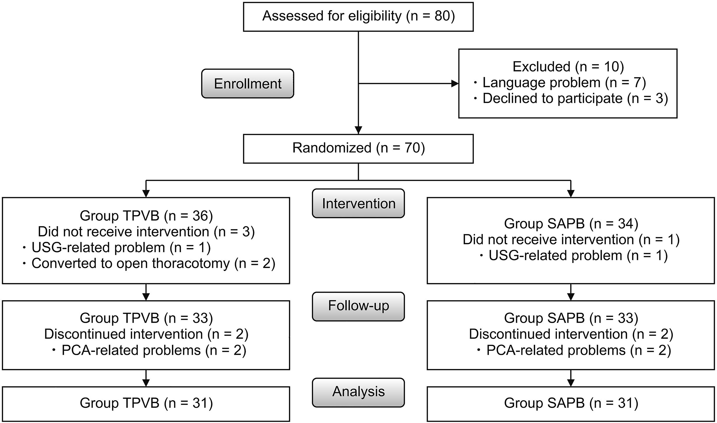
For our study, we obtained approval from the Uludag University Faculty of Medicine Research Ethics Committee (Local Ethics Committee Ethical number: 2018-7/27, Clinical Trials.gov identifier: NCT04235530), and informed patient consent was obtained from all participants. Our study was a single-center, prospective, randomized, double-blind study conducted on patients who underwent VATS for wedge resection. Patients who were aged 18-65 years, were American Society of Anesthesiologists physical status I-II, and were undergoing elective VATS were included in the study. Patients who had a bleeding diathesis, mental or psychiatric disorders, an allergy to the drugs used, any contraindications for SAPB and TPVB application, an inability to speak Turkish, or whose body mass index above 35.0 kg/m2 were not included in the study. Patients who converted to an open thoracotomy, were discharged before 24 hours, had problems with the patient-controlled analgesia (PCA) device, or who experienced block failure (inappropriate local anesthetic distribution and USG image) were also excluded from the study. All patients were informed about the PCA device and visual analog scale (VAS) that were be used in the postoperative period. Patients were randomized into two groups: SAPB (n = 40) and TPVB (n = 40). The randomization list, as well as sealed and opaque envelopes were prepared using a computer program before starting the study by a researcher who was not included in the study.
2. Anesthesia management
We administered 0.01-0.02 mg/kg midazolam to the patients for premedication. Routine monitoring was applied. After monitoring, the patients were intubated after induction with 1-2 mcg/kg of fentanyl, 2-3 mg/kg of propofol, and 0.6 mg/kg of rocuronium. Sevoflurane was used for maintenance in a mixture of 50% air and 50% O2 with a minimum alveolar concentration of 1. At the end of the surgery, the patients were transferred to the postoperative recovery room following extubation. Patients with a Modified Aldrete Score ≥ 9 were transported to the thoracic surgery clinic.
3. Block procedure
The blocks were administered by a single experienced anesthesiologist by USG (MyLab30Gold Cardiovascular; Esaote, Florence, Italy) guidance before the beginning of the surgical procedure, after intubation. After the area where the block was applied was sterilized with an antiseptic solution, the linear probe was wrapped with sterile gloves.
1) Group SAPB (n = 34)
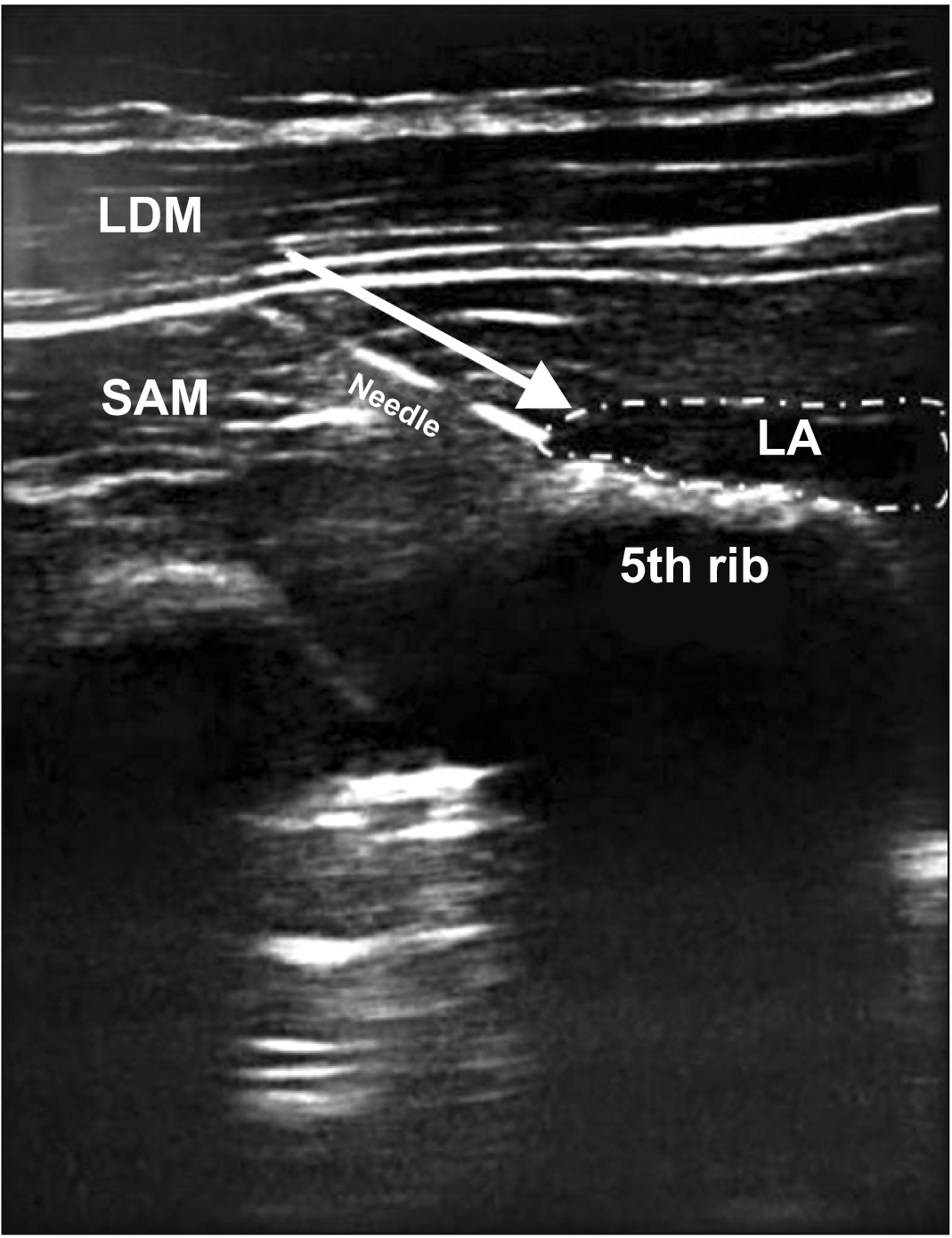
While the patient was in the supine position, a high-frequency linear ultrasound probe was placed horizontally on the mid-axillary line at the level of 4th or 5th ribs on the side of the block. The serratus anterior, latissimus dorsi, and intercostal muscles were identified. The block needle (22-gauge 80 mm, Stimuplex Ultra; B. Braun Melsungen AG, Melsungen, Germany) was advanced below the serratus anterior muscle (SAM) towards the fifth rib (using in-plane technique). The prepared 0.25% bupivacaine was administered at 0.4 mL/kg (max. 20 mL) between the SAM and the rib. It was observed that the solution of local anesthesia was spread between the SAM and the rib (
Fig. 1).
 | Fig. 1Serratus anterior plane block application. LDM: latissimus dorsi muscle, SAM: serratus anterior muscle, LA: local anesthetic. 
|
2) Group TPVB (n = 36)
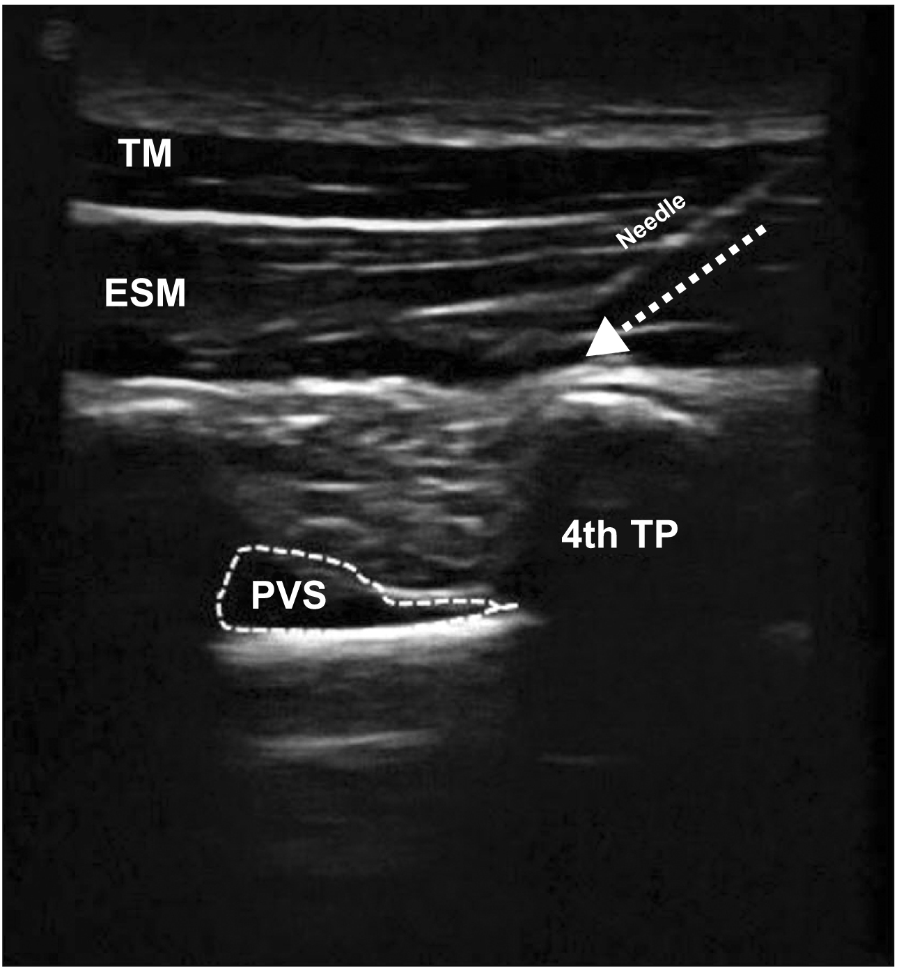
A high-frequency linear ultrasound probe was placed between transverse processes from the T4 level in the paramedian plane while patients were in the lateral decubitus position. The transverse processes, superior costotransverse ligaments, and pleura were visualized. The block needle (22 gauge 80 mm, Stimuplex Ultra; B. Braun Melsungen AG) was advanced until it crossed the superior costotransverse ligament. The prepared 0.25% bupivacaine was administered at 0.4 mL/kg (max. 20 mL) in the thoracic paravertebral space. Depression of the pleura was observed as a result of the spread of local anesthetic (
Fig. 2).
 | Fig. 2Thoracic paravertebral block application. TM: trapezius muscle, ESM: erector spinae muscle, TP: transverse process, PVS: paravertebral space. 
|
4. Analgesia management
After induction, intravenous (IV) 1 g of paracetamol and IV 20 mg of tenoxicam was administered to all patients 10 minutes before the end of the surgery. An IV PCA device (CADD-Legacy® PCA; Smiths Medical, Saint Paul, MN) was used for postoperative pain control. A 54 mL saline + 6 mL tramadol (50 mg/mL) IV solution was prepared. The device was set to a 5 mL bolus dose and a 30 minutes lock time, without basal infusion and loading dose. All patients were administered IV 20 mg tenoxicam every 12 hours in the postoperative period and 1 g paracetamol every 8 hours.
5. Outcomes
The primary outcomes of our study were the total amount of opioid consumption and postoperative VAS and dynamic (during coughing) VAS (DVAS) scores of patients in the recovery room (0 hr) and at postoperative 1, 6, 12, and 24 hours. Secondary outcomes included patient and surgeon satisfaction, block application time, first analgesic time, postoperative complications, and length of hospital stay. The description of the block application time is from needle puncture to the end of local anesthetic injection. The satisfaction of the patients and surgeons was recorded according to the postoperative pain status with a 4-point scale.
6. Statistical analysis
In order to determine the sample size of the study, the minimum sample size was 78 individuals according to the results of the pilot study using a reference power = 0.80 and a confidence interval = 0.95. The SPSS ver. 22.0 (IBM Co., Armonk, NY) was used for statistical analysis. In the descriptive statistics of the data, mean, standard deviation for quantitative data, and percentage values for qualitative data were used. In the distribution of variables, the Kolmogorov–Smirnov normal distribution test was used. Mann–Whitney U, Kruskal–Wallis, and chi-square tests were used in the analysis of data that did not match the normal distribution. P < 0.05 was considered statistically significant as the level of significance in the assessment.
Go to :

DISCUSSION
This prospective, randomized, and double-blinded study was performed on patients who underwent VATS with the intention of wedge resection under general anesthesia. As part of multimodal analgesia, the SAPB or TPVB were applied to patients before the surgery. Although the postoperative opioid consumption of the TPVB group was significantly lower than that of the SAPB group, the consumption of both groups was substantially low at 24 hours (Group SAPB: 31.12 mg, Group TPVB: 18.54 mg). Furthermore, the postoperative median VAS scores of both groups were under 3 and there was no statistically significant difference between the two groups. While the SAPB application time was 182 seconds, the TPVB application time was 300 seconds. The duration of the SAPB application was significantly shorter than that of the TPVB group (P < 0.001).
Piraccini et al. [
5] reported that a PVB was superior to intravenous analgesia in pain control and preservation of postoperative pulmonary function, and it was also equal to thoracic epidural analgesia. We planned our study to investigate whether the SAPB is as effective as the TPVB in pain management after VATS.
SAPB can be applied with two different techniques, deep and superficial. In the superficial technique there is an injection of local anesthetic between the latissimus dorsi muscle and the SAM, while in the deep technique, the injection of local anesthetic is made between the SAM and the external intercostal muscles [
6]. By applying the deep SAPB, the anterior and lateral cutaneous branches of the thoracic intercostal nerves are blocked [
6-
9]. It is known that performing the superficial SAPB also blocks the thoracicus longus nerve and consequently a winged scapula can occur [
10]. Piracha et al. [
11] applied deep SAPB to four patients who had previously undergone the superficial SAPB for post-mastectomy pain syndrome, to compare deep with superficial SAPBs. The patients stated that they benefited more from the second application and were more satisfied. In addition, they concluded that the deep SAPB is more effective. The outcomes of the SAPB may differ depending on the type, volume, concentration, and target point of the local anesthetic. In superficial or deep SAPB applications, 10-30 mL of 0.125%-0.375% ropivacaine or bupivacaine were administered and it was reported to provide adequate analgesia for rib fractures, VATS, thoracotomy, breast surgery [
2,
12,
13]. In order to avoid the development of a winged scapula and local anesthetic systemic toxicity, we administered 20 mL of 0.25% bupivacaine by USG guidance to apply a deep SAPB.
Wang et al. [
14] conducted a retrospective study similar to our study by dividing 123 patients into three groups: SAPB, TPVB, and a control group for postoperative pain treatment in patients who underwent single incision (uniportal) VATS. At postoperative hours 1, 2, 4, and 6, the VAS scores of the SAPB and TPVB groups were found to be significantly lower than those of the control group, but no difference was found at the 24 hr and 48 hr timepoints. There was no significant difference between the SAPB and TPVB groups in terms of VAS scores. In the present study, there were no significant differences in rest and DVAS scores at postoperative hours 0, 1, 6, 12, and 24. The VAS scores were below 3 for both groups.
Wang et al. [
14] stated in their study that the total opioid consumption of the SAPB group was similar to that of the TPVB group, and that the SAPB was as effective as the TPVB. They discussed that the reason for this result was that the operation was performed with a single incision. The TPVB is known to induce deep analgesia depending on the spread of local anesthetics. Local anesthetic injections to the thoracic paravertebral space can block the sympathetic chain by spreading directly to the spinal nerves, laterally to the intercostal nerves, and by spreading through the intervertebral foramina into the epidural space located in the medial region [
15-
17]. In a randomized controlled trial conducted by Aly et al. [
18] for post-thoracotomy analgesia, the SAPB and TPVB were compared in terms of both resting and DVAS scores. In the SAPB group, the DVAS score at the 12th and 18th hours and total morphine consumption were found to be significantly higher. In this study, it was emphasized that the SAPB might not affect the posterior cutaneous branches of the intercostal nerves and does not involve autonomic blockade. In the present study, all operations were performed with a 3-port incision and one of the port incisions was located posteriorly. Therefore, we consider that there is more tramadol consumption in the SAPB group than in the TPVB group. However, the total doses were acceptable for thoracic surgery.
In two meta-analyses investigating the analgesic efficacy of adding the SAPB to general anesthesia, the authors stated that combining the USG-guided SAPB with general anesthesia provides more effective postoperative analgesia in VATS [
2,
19]. They stated that combining the USG-guided SAPB with general anesthesia provides more effective postoperative analgesia. We also believe that the SAPB can be applied effectively as a part of multimodal analgesia in VATS, along with to non-steroidal anti-inflammatory drugs and paracetamol.
Gupta et al. [
20] performed the SAPB and TPVB under USG guidance on patients undergoing modified radical mastectomy under general anesthesia. Morphine consumption, first analgesia time, and VAS scores were recorded in the postoperative period. At the end of the study, while VAS scores were found to be similar, it was determined that the SAPB group consumed more morphine. There was no significant difference between the two groups in terms of the first analgesia time. In the present study, there was similarly no significant difference between the groups in terms of the first analgesia time. The opioid preference in our study was tramadol, a weak opioid with a lower side effect profile. Owing to this preference, we achieved minimal opioid-related adverse effects in our patients.
Aly et al. [
18] compared the SAPB and TPVB for pain after thoracotomy, and no significant difference was found in terms of intraoperative hemodynamic changes, patient satisfaction, and complication incidence. In another study, preoperative the USG-guided TPVB was applied to patients who underwent open cholecystectomy and it was reported that a total spinal block developed after a sudden clouding of consciousness [
21]. In our study, there was no significant difference in terms of intraoperative hemodynamic changes, and the changes were within the clinically acceptable range. There were no complications in either group. In addition, our patient and surgeon satisfaction was high, and there was no significant difference between the two groups.
Aly et al. [
18] compared USG-guided SAPB and TPVB application times and found that the application time was significantly shorter in the SAPB. They mentioned that the SAM is very superficial and easily distinguishable from other structures in the image obtained when the USG probe is placed longitudinally in the mid-axillary line, causing this difference. Similar to this study, the duration of the SAPB application was significantly shorter in the present study. In addition to anatomical placement and distinction, we believe that targeting the rib while advancing the needle makes the performer feel safe and is effective in applying the block without wasting time.
Multimodal analgesia is defined as the administration of two or more analgesic agents, with different modes of action, using one or more routes of administration, and providing superior analgesia with fewer side effects by acting synergistically. Multimodal analgesia strategies are very important for effective postoperative analgesia and rehabilitation [
22]. In our study, we planned to apply a multimodal analgesia regimen with a preoperative truncal block, intraoperative IV paracetamol, as well as non-steroidal anti-inflammatory drugs (NSAIDs), opioids, and postoperative opioids using IV PCA, IV paracetamol, and NSAID administration. We believe that our successful use of multimodal analgesia methods is the reason why opioid consumption in the 24 hours postoperative period was at very low doses.
The limitations of our study include the absence of a control group, the failure to evaluate the effects of the blocks on the onset time, and the affected dermatome levels. Another limitation is that opioid consumption was not measured over time.
In our study, we concluded that the SAPB, applied safely and rapidly as a part of multimodal analgesia in patients who will undergo VATS, is not inferior to the TPVB and can be an alternative to it.
Go to :


 PDF
PDF Citation
 Print
Print


 XML Download