Abstract
Background
Evaluation of the effectiveness of caudal epidural injection on pain, spine mobility, disease activity, and activity of daily living in axial spondyloarthritis (SpA) patients.
Methods
A total sample of 47 patients were registered in this study. They were randomly assigned into 2 groups; Group I received caudal epidural injections, ultrasound-guided, with 1% lidocaine hydrochloride mixed with triamcinolone, whereas Group II did not receive any injections. All participants fulfilled the ASAS criteria for axial SpA. Outcome measures were as follows visual analogue scale, Oswestry disability index (ODI), modified Schober test, lateral lumbar flexion, and Ankylosing Spondylitis Disease Activity Score (ASDAS) with assessment at baseline, 2 weeks, and 8 weeks post-treatment. This clinical trial was registered on clinicaltrials.gov under the number NCT04143165.
Results
There was a significant difference between both groups regarding pain, ODI, spine mobility and ASDAS scores in favor of group I. This effect was at its maximum after 2 weeks. Despite the decline of this effect after 2 months, the difference between the groups remained significant. Higher disease activity, younger age, and shorter disease duration were associated with better outcomes.
Axial spondyloarthritis (SpA) is the principal sort of chronic inflammatory arthritis influencing the axial skeleton. Radiographic axial SpA affects approximately 0.5% of the population worldwide. Inflammatory back pain, radiographic sacroiliitis, and a high frequency of HLA–B27 are its distinctive features. Non-radiographic axial SpA leads to progressive ankylosis of the spine and sacroiliac joint. Disease progression varies widely among patients. Axial SpA represents a great burden on the social, financial, and physical aspects of a patient’s life [1]. Treatment of the spine in axial SpA necessitates a multidisciplinary approach. Patient education, physiotherapy, and exercise, beside medical and surgical treatment, are available options. Non-steroidal anti-inflammatory drugs (NSAIDs) can control the pain and inflammation to some extent. Disease-modifying anti-rheumatic drugs (DMARDs) were ineffective in treating axial SpA. Tumor necrosis factor alpha (TNF-α) inhibitors could slow disease evolution and spinal destruction, and can control extra-articular manifestations.
Caudal or lumbar epidural steroid injections can be used to improve back pain and stiffness, however, there is insufficient supporting evidence for this [2]. TNF-α inhibitors are associated with increased liability of infection, high costs, injection site reaction, development of autoantibodies and drug resistance. Moreover, relapse after withdrawal of the drug is a known complication [3]. Caudal epidural injections have been used for several years in interventional pain medicine, pediatrics, and obstetrics. Its use for pain have been linked to lumbar spinal stenosis, failed back surgery syndrome, and lumbar radiculopathy [4]. Despite the fact that back pain and stiffness are major disabling features of axial SpA, epidural injection was not considered as a treatment modality.
This pilot study is trying to shed light on the role of epidural injections in controlling pain and improving function in axial SpA patients. In addition, we attempted to determine whether it has any effect on disease activity parameters.
All participants registered in this study were informed in detail about the methodology and goals, and then they signed a written consent. The ethics committee of the Minia University Faculty of Medicine, Egypt, approved the study protocol number 688:9/2020. The study was also registered on the clinical trials.gov site under the number NCT04143165. Medical and personal information were kept confidential. The study was performed in accordance with the principles of the Declaration of Helsinki.
This was a prospective randomized controlled trial. Patients who fulfilled ASAS criteria for axial SpA were enrolled in the study through their regular follow-up in the Rheumatology Outpatient Clinic, Minia University Hospital.
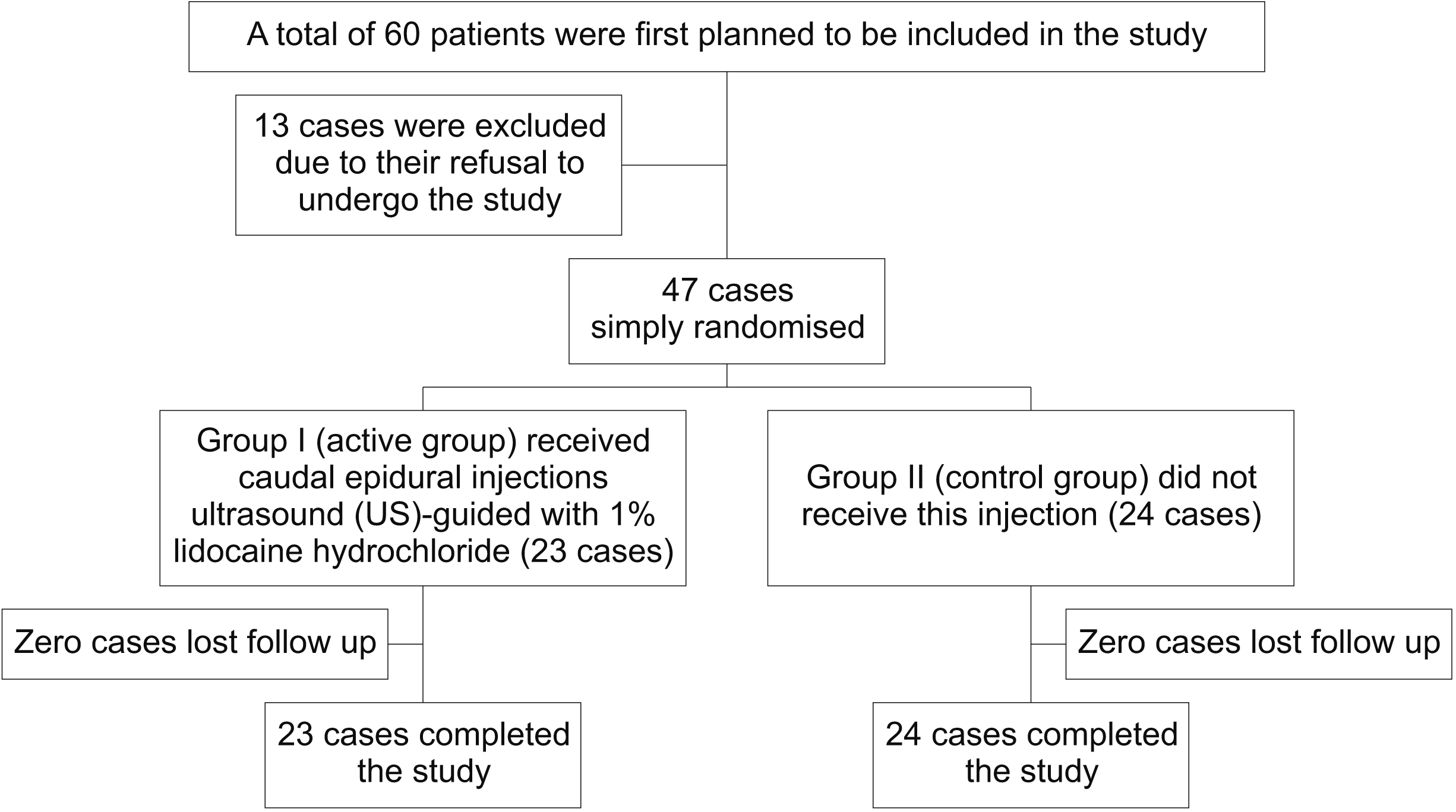
The total number of participants in the present study was 47. They were randomly assigned into 2 groups; Group I (the active group) received ultrasound (US)-guided caudal epidural injections with 1% lidocaine hydrochloride (Xylocaine®; AstraZeneca, Cambridge, UK) 9 mL mixed with 1 mL of triamcinolone acetonide 40 mg (Kenacort®-A 40; Bristol Myers Squibb, New York, NY), whereas Group II (the control group) did not receive this injection [5-7]. Both groups were age- and sex-matched, and both were under treatment with anti-TNF and NSAIDs with or without sDMARDs. Group I was scheduled for epidural injection, and then 2 further appointments were planned for follow-up, after 2 weeks and 8 weeks. Group II was scheduled for follow-up visits at the same intervals. Visual analogue scale (VAS), Oswestry disability index (ODI), modified Schober test, lateral bending test, and the Ankylosing Spondylitis Disease Activity Score (ASDAS) were used on each visit [8].
Epidural injection was performed at baseline by an experienced rheumatologist under US guidance. He carried out clinical evaluation in the 2nd and 3rd visits and was blinded for the initial clinical evaluation. The initial evaluation was done by the other 2 rheumatologists.
Inclusion criteria: Active axial SpA for at least 3 months, with insufficient response or intolerant to ≥ 2 NSAIDs (each taken for ≥ 2 months), and an Ankylosing Spondylitis Disease Activity Score with C-reactive protein (ASDAS-CRP) score ≥ 1.3 [9]. All patients had limitation of spine mobility either in flexion, extension, or lateral binding according to the modified Schober’s and lateral lumbar flexion tests [10,11].
Exclusion criteria: a completely ankylosed spine, infection at the injection site or allergy to lidocaine, present or previous chronic pain conditions (fibromyalgia), and pregnancy.
Randomization was done using 1:1 allocation. For each two participants, the first selected a group number from one box and the next was allocated in the other group. Randomization was guaranteed by the 1st author (Fig. 1).
US-guided injection was performed by a 7.5 to 18 MHz linear probe (Siemens ACUSON P300 US system; Erlangen, Bayren, Germany). Initially, the US scan was performed in the prone position. The transducer was placed axial to the sacrum to examine the sacral hiatus. Then, the probe was rotated to get a sagittal view (Fig. 2). Blood sugar and vital signs were measured before injection, and if the participant had hyperglycemia or abnormal vital signs, injection was suspended.
The injection site was marked and sterilized, and 3 mL of lidocaine 1% was injected to provide local anesthesia before introduction of the needle. Then, the needle was guided by US into the caudal epidural space. The advancement of the needle between the two cornua to the sacral hiatus, and then into the caudal epidural space, was observed through continuous and real-time imaging (Fig. 2). Before injection, aspiration was attempted to avoid intravascular injection. Epidural injection was not performed in participants with a closed sacral hiatus. After the procedure, participants were inspected for complications, vital signs and for blood sugar.
The VAS score was graded from 0 to 10. Grade 0 means no pain and 10 denotes the worst possible pain [12-14]. Significant pain relief was defined as 50% improvement or more [15-18] or a VAS score of 0. Despite the fact that the VAS is a part of the ASDAS, the VAS score was measured to confirm the pain improvement.
In this score, 5 sections are assessed: back pain (score from 0 to 10), morning stiffness duration (score from 0 to 10), the patient global assessment (score from 0 to 10), peripheral pain/swelling (score from 0 to 10), and C-reactive protein (calculated in mg/L). The score was determined using the ASDAS calculator. ASDAS improvement was considered to be when the score reduction was ≥ 1.1 [9].
This score consists of 10 domains, which measure pain strength, the activities of daily living, sleep, and other features. Each domain has 6 possible answers. A score of 0 denotes the best level and 5 denotes the worst. The total ODI score ranges from 0 to 100. Minimal disability lies between 0% and 20%, mild disability between 20% and 40%, severe disability between 40% and 60%, crippling disability between 60% and 80%, and > 80% means bedridden. The use of this score was validated in axial SpA, and it had a strong relation to Bath Ankylosing Spondylitis Functional Index (BASFI) and Bath Ankylosing Spondylitis Disease Activity Index (BASDAI) scores [19].
The modified Schober’s test and lateral lumbar flexion tests were used to assess spinal mobility [10,11].
Blood sugar was measured before injection, and if the participant had hyperglycemia, injection was suspended. After the procedure, participants were inspected for complications, vital signs and blood sugar.
We were planning a study of independent cases and controls with 1 control(s) per case. Prior data indicate that the failure rate among controls is > 0.8 [20]. If the true failure rate for experimental subjects is to be < 0.5, we would need to study at least 14 experimental subjects and at least 14 control subjects to be able to reject the null hypothesis that the failure rates for experimental and control subjects are equal with a probability (power) of 0.95. The Type I error probability associated with the test of this null hypothesis is 0.9. We used an uncorrected chi-squared statistic to evaluate this null hypothesis. We included 47 cases, which is larger than the lower limit (28 total; 14 cases and 14 controls) which we calculated before the study.
Data were analyzed using IBM SPSS version 25 (IBM Co., Armonk, NY). Data were expressed as mean ± standard deviation, number, and percentage. Mean and standard deviation were used as descriptive values for quantitative data. Pearson’s Chi square test was used to compare percentages of qualitative variables, and Fisher’s exact test was used instead for non-parametric data. The student t-test was used to compare the means between two groups, and the paired t-test was used to compare means of the same variable at different periods of time. The Pearson correlation test was used to compare two quantitative variables. A P value of less than 0.05 was considered significant.
The mean age of the participants was around 40 years. Around 60% of them were males. However, in the non-radiographic SpA, the male to female ratio was nearly 1:1, while in radiographic SpA, the male to female ratio was around 2:1. The mean disease duration was around 4 years among non-radiographic axial SpA compared to 8 years among ankylosing spondylitis (AS) patients. No significant differences were detected between the two study groups regarding demographic and clinical data (Table 1).
Baseline comparison between the two groups (active and control) showed non-significant differences regarding the results of the VAS, ASDAS, ODI, modified Schober’s test and lateral bending test. All of these measures showed significant improvement after 2 weeks in the active group (compared to baseline values), and also significant differences compared to the control group. This improvement was more obvious as early as 2 weeks after injection regarding the VAS, ASDAS, and ODI. The effect was more sustained, for as long as 8 weeks, for the modified Schober’s test and lateral bending test, proved by the non-significant difference between their values at 2 and 8 weeks (Table 2).
The comparison of the degree of improvement in VAS scores and ASDAS (the difference between baseline and 2 weeks measures), with clinical and laboratory data, revealed that VAS improvement was significantly associated with younger age, shorter disease duration, and higher CRP. On the other hand, ASDAS improvement was significantly associated with younger age, female sex, and the presence of bone marrow edema (Table 3).
We used univariate binary logistic analysis for possible predisposing factors for improvement using the mean VAS and ASDAS changes. The outcome of the regression analysis was improvement, defined as a reduction of > 50% of the mean VAS or ASDAS improvement of > 1.1. We found that younger age and higher CRP may significantly predict improvement of VAS scores, while younger age, higher CRP, and positive bone marrow edema may significantly predict improvement of ASDAS. These factors were then included in multivariate binary logistic regression analysis, which showed that there were no independent predisposing factors for improvement in VAS scores, while younger age was the only independent predisposing factor for ASDAS improvement (Table 4).
Complications of injection in the active group did not exceed 1%. These included injection site pain, radicular pain, headache, and flushing.
Caudal epidural injection is an approved treatment for chronic low back pain. Pain and limited spine mobility are major morbidities influencing quality of life and disease progression in axial SpA. To the best of our knowledge, this is the first study to shed light on the value of caudal epidural injection for spinal pain, mobility, and activity of daily living in axial SpA patients.
In our study, the active group outweighed the control group with respect to pain, function, and disease activity outcomes. In addition, amelioration was more in those having earlier disease, younger age, shorter disease duration, and a more active disease. Negative effects were minor and limited to less than one-fifth of the active group. Pain showed the best improvement, followed by spinal mobility measures, ODI score, and then ASDAS. With respect to carry-on effect, spine mobility was followed by ASDAS, ODI and finally pain.
BASFI was not used in this study because cervical mobility was not expected to improve with caudal epidural injection, and this would negatively impact the endpoints of the study. It was replaced by the Schober’s and lateral bending tests to yield more realistic results, reflecting the changes in lumbar spine mobility only.
There is evidence supporting the anti-inflammatory effect of local anesthetics. In addition, local anesthetics can inhibit leukocyte adhesion, phagocytosis, degranulation and migration [21]. Likewise, TNF, leukotrienes, and IL1 release are inhibited by local anesthetics. Moreover, the release of lysosomal enzymes from activated polymorphonuclear leukocytes is minimized by local anesthetics. This effect is dose-dependent and reversible [22]. It binds to the prostaglandin E2 receptors and inhibits prostaglandins production, thus, alleviating inflammation. It can also suppress adrenergic neurotransmission, bradykinin, and substance p, which are pro-inflammatory neurotransmitter [23,24]. Although adding steroids to the anesthetic in managing low back pain did not show a significant difference, yet its use in axial SpA could be beneficial due to the inflammatory nature of the disease [25,26]. Steroids were also effective in controlling inflammation of the sacroiliac joint in axial SpA [27]. Local infiltration anesthesia can improve wound healing and minimize inflammation and infection [28]. Rectal lidocaine gel can improve ulcerative proctitis on clinical and histological levels [29]. Moreover, in vivo studies have confirmed that local anesthetics induce significant inhibition of burn edema and improve blood supply to the burn injury [30].
As a matter of fact, there is no parallel study to compare our results with. Stav et al. [27] have performed a case series study. They registered 9 patients with Bechterew’s disease and limited spine mobility and treated them with cervical and lumbar epidural injections of methyl prednisolone and local anesthetic. Pain and range of motion improved dramatically. This improvement continued for nearly one year. Multiple limitations of this study encompass the restricted number of participants, the classification criteria used are not revealed, magnetic resonance imaging (MRI) was not used for diagnosis but only computed tomography and x-ray, the disease duration was relatively long with a mean of 18 years, all participants were only under treatment with NSAIDs, and none of them received biologics [27]. In addition, another case series considered cervical epidural injection of steroids in patients who presented with pain and stiffness due to spondyloarthropathy. They were refractory to conservative treatment. They registered only 3 patients, 1 with AS and 2 with psoriatic arthritis. Disease duration ranged from 6 to 35 years. ASAS criteria were not used for diagnosis. Besides, disease activity scores were also not considered. They considered symptomatic improvement as outcome measures without sharp definition of the improvement criteria. Improvement was between 50% and 90%. This improvement persisted for at least 5 months [31]. In an additional study done by Manchikanti et al. [32], 140 participants were allocated into 2 groups: group 1 received only lidocaine 0.5%, while group 2 received lidocaine 0.5% and dexamethasone. Injection was fluoroscopy-guided, and all participants had post lumbar surgery syndrome. They were followed up for 2 years and showed significant improvement in pain and function. Although no significant difference was observed between the groups, improvement was better in the steroid group. The maximum improvement was sustained until 3 months, with gradual decline in the next follow-up visits, but it was still significant. They evaluated pain by the VAS and function with the ODI [32]. A systematic review of caudal epidural injections with or without steroids in managing chronic pain secondary to variable etiologies was conducted by another research group. All the enrolled studies were done using fluoroscopy. They deemed pain relief and functional improvement as the main outcome measures. Local anesthetic and steroids showed good evidence in controlling pain due to lumbar disc herniation, whereas the evidence was fair for local anesthetic only. The evidence for its use in ameliorating chronic axial pain and non-discogenic back pain was also fair. However, scarcity in the literature was an impediment [33].
Monti and her colleagues [34] tried to estimate the prevalence of inactive disease in the cohort of AS patients treated with anti-TNF. About half of those with radiographic axial SpA achieved that goal according to ASDAS-CRP. They enrolled 218 patients, and 165 of them had radiographic axial SpA. Treatment duration was at least 6 months. Median disease duration for their patients was 9.3 years (with radiographic axial SpA). They indicated that the ASDAS was related more to clinical and functional improvement than other disease activity scores [34]. Another study by Lubrano and his work group [35] aimed at assessing physical function improvement in axial SpA patients with BASFI and BASMI scores. They enlisted 183 patients with axial SpA, and 156 of them had radiographic axial SpA. The minimum follow-up period was 6 months. BASMI and BASFI scores showed significant improvement. This was noticed more with shorter disease duration, especially in the case of the BASMI [35]. In a survey conducted in Germany regarding the efficacy of NSAIDs in axial SpA, nearly 20% of the included patients achieved complete pain control with NSAIDs, and about 60% reported pain reduction [36]. In another trial, ASAS partial remission criteria were fulfilled in 14.7%, 17.6%, and 9.1% of radiographic axial SpA patients, depending on the NSAID dose [37]. In INFAST (infliximab as first line therapy in patient with early active axial spondyloarthritis trial), early (less than 3 years) axial SpA patients (either radiographic or non-radiographic) were randomly selected to take either infliximab and naproxen or a placebo plus naproxen. In the placebo group, 16% of the patients obtained ASAS partial remission at week 6, and 35% patients achieved the same at week 28 [38]. In comparison, in another study with more advanced AS and a mean disease duration of 10 years, anti-TNF could attain ASAS partial remission in 17%-23% patients at 24 weeks. The placebo group in those studies achieved partial remission in only 1 to 6% of cases [39-41]. The predictors of good responders to anti-TNF include younger age, shorter disease duration, less functional disability, TNF naivety, high CRP, and active axial inflammation on MRI [42].
The exact mechanism that illuminates how epidural injection can control disease progress is not well known. Usually, spinal stiffness begins in the lumbosacral spine and advances in a cephalic direction [43]. Caudal epidural injection or sacroiliac injection could theoretically impede this crawl, especially if given in the early stage of the disease. Steroids may provide relief in nociceptive spinal pain [27]. Stiffness stimulates more pain, which further provokes more stiffness in a vicious circle. Breaking this circle could hinder disease progress. Further, pain amelioration improves spine mobility, and this subsequently can turn off or delay spine calcification [32,44]. This could explain why caudal epidural injection is more effective in earlier disease or with shorter disease duration.
In a recent study by Renson and his colleagues [45], 77% of female patients had sacroiliac bone marrow edema 10 days after labor and 46% of them still had bone marrow edema after 6 months. This percentage went down to 12% after 12 months. They pointed out that epidural anesthesia has a significant effect on reduction of this bone marrow edema. This finding strongly supports our results and signifies the role of epidural anesthesia in controlling sacroiliac joint inflammation [45].
There are limitations in this study. First, the number of study participants was limited. This may be because some participants considered injections an invasive maneuver and did not accept or adhere to the follow-up time schedule. The addition of sacroiliac injection to this technique could be of great benefit. Yet, the application of triple injection in one setting is time consuming and riskier. Further, the application of this injection in the following visits could lengthen the study duration, and several participants might drop out of the study in follow-up visits. Second, a particulate steroid (triamcinolone) was used in our study. There is a potential risk of injecting particulate steroids in the epidural space. However, particulate steroids have been used in performing epidural blocks by many researchers for treating chronic low back pain. They have also been used for treating pain and stiffness in Bechterew’s syndrome [26,46-48]. No serious complications were registered in the present study. Finally, fluoroscopy-guided injection is more accurate than US-guided injection; nevertheless, it is time consuming. Radiation hazards, expensive equipment and trained staff are barriers that limit the use of fluoroscopy in this study.
In conclusion, caudal epidural injection is effective in controlling pain, stiffness, and disease activity in axial SpA patients. Pain showed the best improvement, and spine mobility persisted the longest. This effect is more prominent in those with early disease, shorter duration, younger age, and higher ASDAS. This effect is comparable to the therapeutic effect of biological treatment. More research is mandatory to assess the long-term effect of caudal epidural injection in axial SpA.
Notes
REFERENCES
1. Ward MM, Deodhar A, Gensler LS, Dubreuil M, Yu D, Khan MA, et al. 2019; 2019 update of the American College of Rheumatology/Spondylitis Association of America/Spondyloarthritis Research and Treatment Network recommendations for the treatment of ankylosing spondylitis and nonradiographic axial spondyloarthritis. Arthritis Rheumatol. 71:1599–613. DOI: 10.1002/art.41042. PMID: 31436036. PMCID: PMC6764882.
2. Shahinyan A, Margaryan L, Samuel S. Abd-Elsayed A, editor. 2019. Ankylosing spondylitis. Pain: a review guide. Springer International Publishing;Cham: p. 1239–41. DOI: 10.1007/978-3-319-99124-5_265.
3. Antoni C, Braun J. 2002; Side effects of anti-TNF therapy: current knowledge. Clin Exp Rheumatol. 20(6 Suppl 28):S152–7. PMID: 12463468.
4. Walia AA, Tran H, DuBose D. Deer TR, Pope JE, Lamer TJ, Provenzano D, editors. 2019. Caudal epidural injection. Deer's treatment of pain: an illustrated guide for practitioners. Springer International Publishing;Cham: p. 455–60. DOI: 10.1007/978-3-030-12281-2_55.
5. Lee JJ, Nguyen ET, Harrison JR, Gribbin CK, Hurwitz NR, Cheng J, et al. 2019; Fluoroscopically guided caudal epidural steroid injections for axial low back pain associated with central disc protrusions: a prospective outcome study. Int Orthop. 43:1883–9. DOI: 10.1007/s00264-019-04350-w. PMID: 31168645.
6. Chen CP, Lew HL, Tang SF. 2015; Ultrasound-guided caudal epidural injection technique. Am J Phys Med Rehabil. 94:82–4. DOI: 10.1097/PHM.0000000000000047. PMID: 24722013.
7. Doo AR, Kim JW, Lee JH, Han YJ, Son JS. 2015; A comparison of two techniques for ultrasound-guided caudal injection: the influence of the depth of the inserted needle on caudal block. Korean J Pain. 28:122–8. DOI: 10.3344/kjp.2015.28.2.122. PMID: 25852834. PMCID: PMC4387457.
8. Quack C, Schenk P, Laeubli T, Spillmann S, Hodler J, Michel BA, et al. 2007; Do MRI findings correlate with mobility tests? An explorative analysis of the test validity with regard to structure. Eur Spine J. 16:803–12. DOI: 10.1007/s00586-006-0264-z. PMID: 17143634. PMCID: PMC2200719.
9. Machado PM, Landewé R, Heijde DV. Assessment of SpondyloArthritis international Society (ASAS). 2018; Ankylosing Spondylitis Disease Activity Score (ASDAS): 2018 update of the nomenclature for disease activity states. Ann Rheum Dis. 77:1539–40. DOI: 10.1136/annrheumdis-2018-213184. PMID: 29453216.
10. Assassi S, Weisman MH, Lee M, Savage L, Diekman L, Graham TA, et al. 2014; New population-based reference values for spinal mobility measures based on the 2009-2010 National Health and Nutrition Examination Survey. Arthritis Rheumatol. 66:2628–37. DOI: 10.1002/art.38692. PMID: 24782356. PMCID: PMC4146670.
11. Haywood KL, Garratt AM, Jordan K, Dziedzic K, Dawes PT. 2004; Spinal mobility in ankylosing spondylitis: reliability, validity and responsiveness. Rheumatology (Oxford). 43:750–7. DOI: 10.1093/rheumatology/keh169. PMID: 15163832.
12. Elsaman AM, Radwan AR, Mohammed WI, Ohrndorf S. 2016; Low-dose spironolactone: treatment for osteoarthritis-related knee effusion. A prospective clinical and sonographic-based study. J Rheumatol. 43:1114–20. DOI: 10.3899/jrheum.151200. PMID: 27036390.
13. Carlsson AM. 1983; Assessment of chronic pain. I. Aspects of the reliability and validity of the visual analogue scale. Pain. 16:87–101. DOI: 10.1016/0304-3959(83)90088-X. PMID: 6602967.
14. Crichton N. 2001; Visual analogue scale (VAS). J Clin Nurs. 10:706. DOI: 10.1046/j.1365-2702.2003.00777.x. PMID: 12919219.
15. Khan MA, van der Linden S. 2019; Axial spondyloarthritis: a better name for an old disease: a step toward uniform reporting. ACR Open Rheumatol. 1:336–9. DOI: 10.1002/acr2.11044. PMID: 31777810. PMCID: PMC6857996.
16. Zochling J. 2011; Measures of symptoms and disease status in ankylosing spondylitis: Ankylosing Spondylitis Disease Activity Score (ASDAS), Ankylosing Spondylitis Quality of Life Scale (ASQoL), Bath Ankylosing Spondylitis Disease Activity Index (BASDAI), Bath Ankylosing Spondylitis Functional Index (BASFI), Bath Ankylosing Spondylitis Global Score (BAS-G), Bath Ankylosing Spondylitis Metrology Index (BASMI), Dougados Functional Index (DFI), and Health Assessment Questionnaire for the Spondylarthropathies (HAQ-S). Arthritis Care Res (Hoboken). 63 Suppl 11:S47–58. DOI: 10.1002/acr.20575. PMID: 22588768.
17. van der Heijde D, Landewé R, Feldtkeller E. 2008; Proposal of a linear definition of the Bath Ankylosing Spondylitis Metrology Index (BASMI) and comparison with the 2-step and 10-step definitions. Ann Rheum Dis. 67:489–93. DOI: 10.1136/ard.2007.074724. PMID: 17728332.
18. Hawker GA, Mian S, Kendzerska T, French M. 2011; Measures of adult pain: Visual Analog Scale for Pain (VAS Pain), Numeric Rating Scale for Pain (NRS Pain), McGill Pain Questionnaire (MPQ), Short-Form McGill Pain Questionnaire (SF-MPQ), Chronic Pain Grade Scale (CPGS), Short Form-36 Bodily Pain Scale (SF-36 BPS), and Measure of Intermittent and Constant Osteoarthritis Pain (ICOAP). Arthritis Care Res (Hoboken). 63 Suppl 11:S240–52. DOI: 10.1002/acr.20543. PMID: 22588748.
19. O'Shea FD, Riarh R, Anton A, Inman RD. 2010; Assessing back pain: does the Oswestry Disability Questionnaire accurately measure function in ankylosing spondylitis? J Rheumatol. 37:1211–3. DOI: 10.3899/jrheum.091240. PMID: 20395642.
20. Apáthy A, Penczner G, Licker E, Eiben A, Bálint G, Genti G, et al. 1999; [Caudal epidural injection in the management of lumbosacral nerve pain syndromes]. Orv Hetil. 140:1055–8. Hungarian. PMID: 10339997.
21. Stewart GJ, Knight LC, Arbogast BW, Stern HS. 1980; Inhibition of leukocyte locomotion by tocainide, a primary amine analog of lidocaine: at study with 111indium-labeled leukocytes and scanning electron microscopy. Lab Invest. 42:302–9. PMID: 6767137.
22. Cassuto J, Sinclair R, Bonderovic M. 2006; Anti-inflammatory properties of local anesthetics and their present and potential clinical implications. Acta Anaesthesiol Scand. 50:265–82. DOI: 10.1111/j.1399-6576.2006.00936.x. PMID: 16480459.
23. Hogan QH, Stadnicka A, Bosnjak ZJ, Kampine JP. 1998; Effects of lidocaine and bupivacaine on isolated rabbit mesenteric capacitance veins. Reg Anesth Pain Med. 23:409–17. DOI: 10.1097/00115550-199823040-00017. PMID: 9690595.
24. Reyes-García MG, García-Tamayo F. 2009; A neurotransmitter system that regulates macrophage pro-inflammatory functions. J Neuroimmunol. 216:20–31. DOI: 10.1016/j.jneuroim.2009.06.024. PMID: 19732963.
25. Manchikanti L, Cash KA, McManus CD, Pampati V, Fellows B. 2012; Fluoroscopic caudal epidural injections with or without steroids in managing pain of lumbar spinal stenosis: one-year results of randomized, double-blind, active-controlled trial. J Spinal Disord Tech. 25:226–34. DOI: 10.1097/BSD.0b013e3182160068. PMID: 22652990.
26. Stav A, Ovadia L, Sternberg A, Alfici R, Landau M. 1994; Cervical and lumbar epidural steroid injections for pain relief in patients with Bechterew's syndrome. Preliminary results. Pain Clin. 7:283–9.
27. Lee SW, Song Y, Lee S, Joo KB, Sung IH, Kim TH. 2015; AB0755 An intraarticular sacroiliac corticosteroid injections in ankylosing spondylitis. Ann Rheum Dis. 74 Suppl 2:1151. DOI: 10.1136/annrheumdis-2015-eular.3778.
28. Eriksson AS, Sinclair R, Cassuto J, Thomsen P. 1992; Influence of lidocaine on leukocyte function in the surgical wound. Anesthesiology. 77:74–8. DOI: 10.1097/00000542-199207000-00011. PMID: 1610012.
29. Björck S, Dahlström A, Ahlman H. 1989; Topical treatment of ulcerative proctitis with lidocaine. Scand J Gastroenterol. 24:1061–72. DOI: 10.3109/00365528909089256. PMID: 2595267.
30. Jönsson A, Brofeldt BT, Nellgård P, Tarnow P, Cassuto J. 1998; Local anesthetics improve dermal perfusion after burn injury. J Burn Care Rehabil. 19(1 Pt 1):50–6. DOI: 10.1097/00004630-199801000-00011. PMID: 9502024.
31. Gungor S, Aiyer R, Erkan D. 2018; Cervical epidural injection in the management of refractory pain and stiffness in spondyloarthropathy: a case report series. Pain Pract. 18:532–8. DOI: 10.1111/papr.12616. PMID: 28742241.
32. Manchikanti L, Singh V, Cash KA, Pampati V, Datta S. 2010; Management of pain of post lumbar surgery syndrome: one-year results of a randomized, double-blind, active controlled trial of fluoroscopic caudal epidural injections. Pain Physician. 13:509–21. PMID: 21102963.
33. Parr AT, Manchikanti L, Hameed H, Conn A, Manchikanti KN, Benyamin RM, et al. 2012; Caudal epidural injections in the management of chronic low back pain: a systematic appraisal of the literature. Pain Physician. 15:E159–98. PMID: 22622911.
34. Monti S, Todoerti M, Codullo V, Favalli EG, Biggioggero M, Becciolini A, et al. 2018; Prevalence of Ankylosing Spondylitis Disease Activity Score (ASDAS) inactive disease in a cohort of patients treated with TNF-alpha inhibitors. Mod Rheumatol. 28:542–9. DOI: 10.1080/14397595.2017.1367076. PMID: 28880727.
35. Lubrano E, Perrotta FM, Manara M, D'Angelo S, Ramonda R, Punzi L, et al. 2020; Improvement of function and its determinants in a group of axial spondyloarthritis patients treated with TNF inhibitors: a real-life study. Rheumatol Ther. 7:301–10. DOI: 10.1007/s40744-020-00197-5. PMID: 32062827. PMCID: PMC7211226.
36. Amor B, Dougados M, Listrat V, Menkes CJ, Roux H, Benhamou C, et al. 1995; Are classification criteria for spondylarthropathy useful as diagnostic criteria? Rev Rhum Engl Ed. 62:10–5. PMID: 7788318.
37. van der Heijde D, Baraf HS, Ramos-Remus C, Calin A, Weaver AL, Schiff M, et al. 2005; Evaluation of the efficacy of etoricoxib in ankylosing spondylitis: results of a fifty-two-week, randomized, controlled study. Arthritis Rheum. 52:1205–15. DOI: 10.1002/art.20985. PMID: 15818702.
38. Sieper J, Lenaerts J, Wollenhaupt J, Rudwaleit M, Mazurov VI, Myasoutova L, et al. 2014; Efficacy and safety of infliximab plus naproxen versus naproxen alone in patients with early, active axial spondyloarthritis: results from the double-blind, placebo-controlled INFAST study, Part 1. Ann Rheum Dis. 73:101–7. DOI: 10.1136/annrheumdis-2012-203201. PMID: 23696633. PMCID: PMC3888606.
39. van der Heijde D, Dijkmans B, Geusens P, Sieper J, DeWoody K, Williamson P, et al. 2005; Efficacy and safety of infliximab in patients with ankylosing spondylitis: results of a randomized, placebo-controlled trial (ASSERT). Arthritis Rheum. 52:582–91. DOI: 10.1002/art.20852. PMID: 15692973.
40. van der Heijde D, Kivitz A, Schiff MH, Sieper J, Dijkmans BA, Braun J, et al. 2006; Efficacy and safety of adalimumab in patients with ankylosing spondylitis: results of a multicenter, randomized, double-blind, placebo-controlled trial. Arthritis Rheum. 54:2136–46. DOI: 10.1002/art.21913. PMID: 16802350.
41. Inman RD, Davis JC Jr, Heijde Dv, Diekman L, Sieper J, Kim SI, et al. 2008; Efficacy and safety of golimumab in patients with ankylosing spondylitis: results of a randomized, double-blind, placebo-controlled, phase III trial. Arthritis Rheum. 58:3402–12. DOI: 10.1002/art.23969. PMID: 18975305.
42. Poddubnyy D, Gensler LS. 2014; Spontaneous, drug-induced, and drug-free remission in peripheral and axial spondyloarthritis. Best Pract Res Clin Rheumatol. 28:807–18. DOI: 10.1016/j.berh.2014.10.005. PMID: 25488786. PMCID: PMC4289210.
43. Braun J, Yu DTY. Stone JH, editor. 2009. Ankylosing spondylitis. A clinician's pearls and myths in rheumatology. Springer;London: p. 57–65. DOI: 10.1007/978-1-84800-934-9_7.
44. Badsha H, Tak PP. 2008; Can Islamic prayers benefit spondyloarthritides? Case report of a patient with ankylosing spondylitis and increased spinal mobility after an intensive regimen of Islamic prayer. Rheumatol Int. 28:1057–9. DOI: 10.1007/s00296-008-0557-0. PMID: 18324379.
45. Renson T, Depicker A, De Craemer AS, Deroo L, Varkas G, de Hooge M, et al. 2020; High prevalence of spondyloarthritis-like MRI lesions in postpartum women: a prospective analysis in relation to maternal, child and birth characteristics. Ann Rheum Dis. 79:929–34. DOI: 10.1136/annrheumdis-2020-217095. PMID: 32299794.
46. Murakibhavi VG, Khemka AG. 2011; Caudal epidural steroid injection: a randomized controlled trial. Evid Based Spine Care J. 2:19–26. DOI: 10.1055/s-0031-1274753. PMID: 23230402. PMCID: PMC3506149.
47. Prost S, Farah K, Mazas S, Pesenti S, Fuentes S, Tropiano P, et al. 2020; Sacral hiatus corticosteroid injection in the management of radicular pain in adults. Orthop Traumatol Surg Res. 106:1191–3. DOI: 10.1016/j.otsr.2020.05.011. PMID: 32917581.
48. Iqbal MR, Ali L, Shah SHA, Iqbal M, Waseem SM, Hamid S. 2020; Comparison of analgesic efficacy of transforaminal vs caudal epidural approaches for unilateral lumbar radiculopathy. Pak Armed Forces Med J. 70 Suppl 1:S135–39.
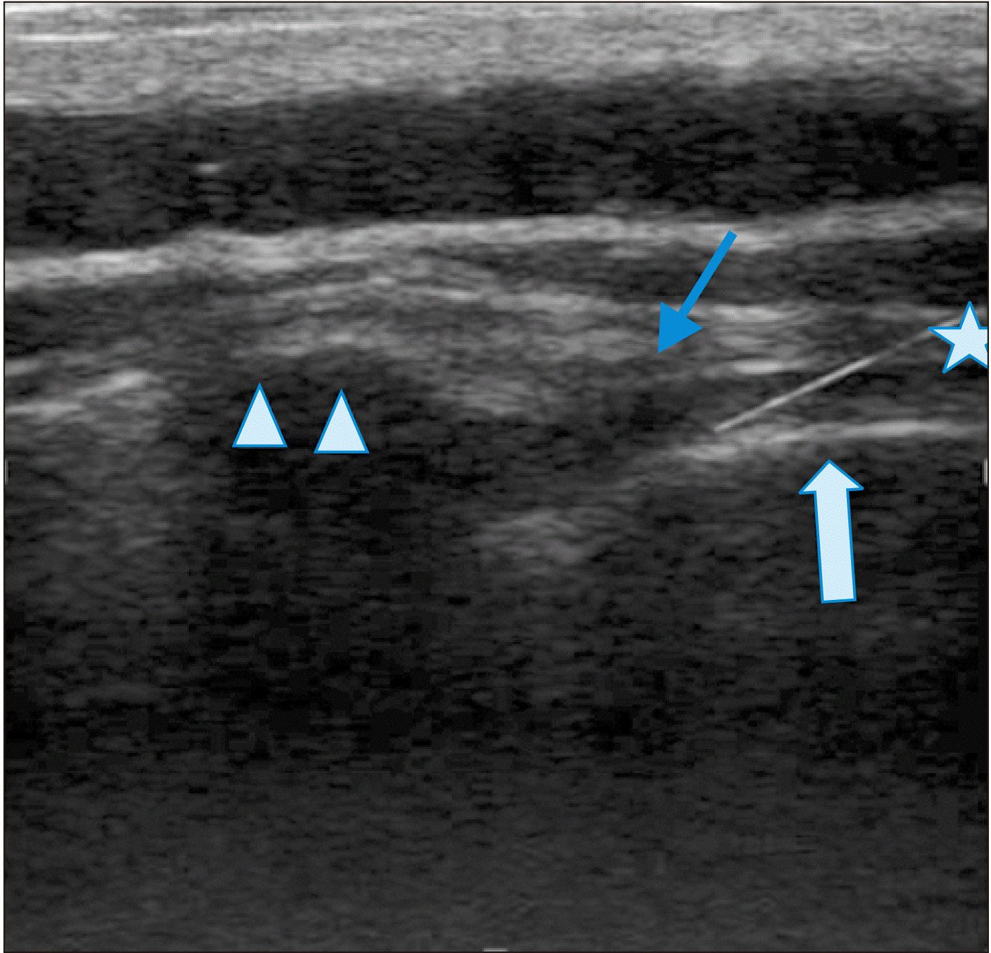
Table 1
Table 2
Table 3
Table 4
Variable | Univariable analysis, OR (95% CI) | P value | Multivariable analysis, OR (95% CI) | P value | |
---|---|---|---|---|---|
VAS item | Age | 0.844 (0.722-0.986) | 0.033* | 0.879 (0.744-1.038) | 0.128 |
Male sex | 1.333 (0.336-5.287) | 0.682 | |||
Disease duration | 0.768 (0.569-1.037) | 0.085 | |||
CRP | 1.047 (1.005-1.092) | 0.028* | 1.034 (0.989-1.081) | 0.141 | |
BM edema | 1.333 (0.358-4.965) | 0.668 | |||
ASDAS item | Age | 0.692 (0.542-0.884) | 0.003* | 0.685 (0.519-0.904) | 0.008* |
Male sex | 0.327 (0.085-1.265) | 0.105 | |||
Disease duration | 0.894 (0.676-1.183) | 0.432 | |||
CRP | 1.062 (1.016-1.111) | 0.007* | 1.022 (0.964-1.083) | 0.462 | |
BM edema | 5.750 (1.307-25.294) | 0.021* | 6.052 (0.806-45.431) | 0.080 |