

 PDF
PDF Citation
 Print
Print


INTRODUCTION
Over the past 10 years, the number of interventional pain procedures using a fluoroscope has increased exponentially [1] because fluoroscopy is essential for accurate procedures with minimal complications. As a result, there has been a growing concern about occupational radiation exposure [2–4]. When an epidural block is performed without fluoroscopy (i.e., blind technique; loss-of-resistance [LOR] method), there is a 13%–30% chance for the needle to be misplaced outside of the epidural space [5,6].
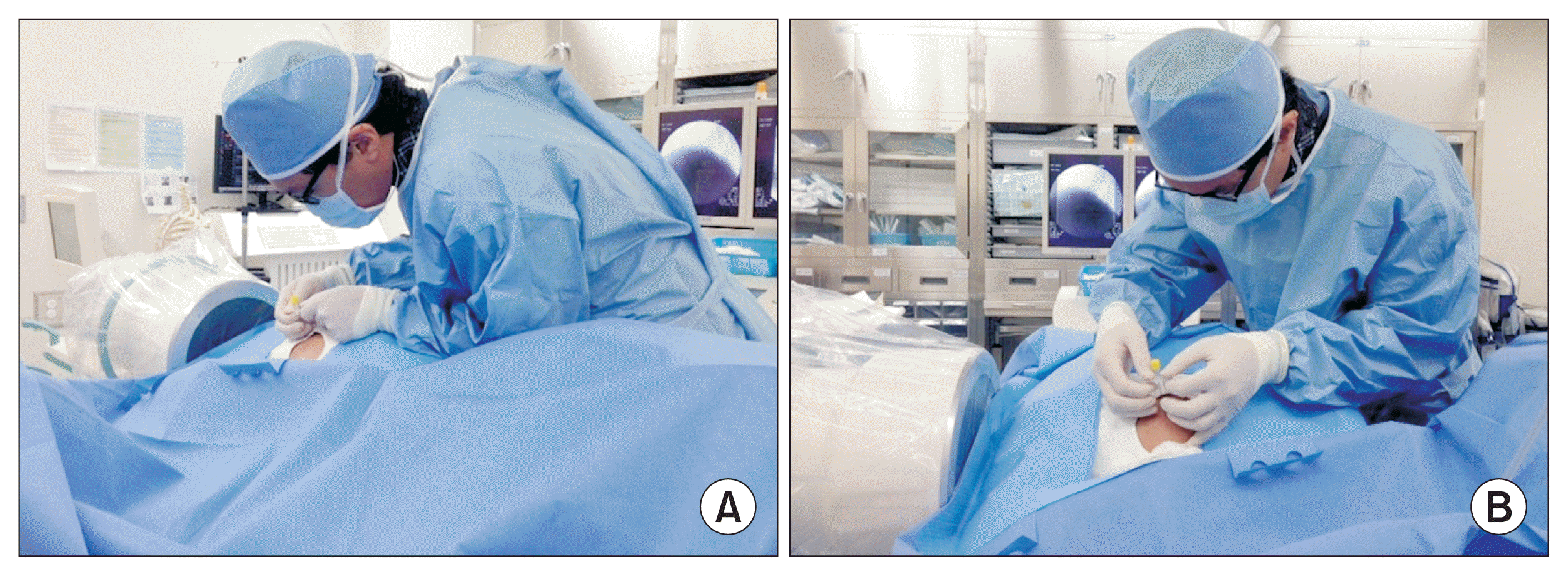
The cervical epidural block (CEB) is often used to treat patients with cervical herniated intervertebral discs and spinal stenosis [7,8]. When CEB is performed, it is essential to practice caution because the epidural space is narrower than the space in other locations, and severe complications, including spinal cord injury and paraplegia, can occur [9–11]. Therefore, CEB needle insertions must be performed under fluoroscopic guidance to practice utmost caution for ensuring an accurate procedure. Unfortunately, this places the pain physician’s head near the fluoroscope, placing the physician’s eyes and thyroid at a higher risk of radiation exposure compared with other techniques (Fig. 1).
Hiroshima, Nagasaki, and Chernobyl nuclear disaster survivors [12–14] and physicians of interventional cardiology [15,16] and radiology [17,18] have shown a higher risk of cataract development as a direct result of crystalline lens radiation exposure. Subsequently, the International Commission on Radiological Protection (ICRP) lowered the yearly maximum radiation dose to the eye from 150 to 20 mSv [19]. The yearly maximum radiation dose to the thyroid remains at 500 mSv [20].
Even though the dangers of radiation exposure are being continuously re-evaluated, no study has reported the risk of radiation exposure for pain physicians performing CEB. Therefore, the primary objectives of this study were to measure actual radiation exposure to a pain physician’s eyes and thyroid during CEB to reveal the risks of radiation exposure and to determine the need for radiation protection. Secondary study objectives were to examine the efficacy of lead-based protection in minimizing radiation exposure and to compare radiation exposure between two pain physicians with different experience levels.
Go to : 
MATERIALS AND METHODS
This prospective study was approved by the Institutional Review Board of Seoul National University Bundang Hospital (No. B-1407/258-002) and was registered in the Clinical Research Information Service (No. KCT0001292). All study conduct adhered to the tenets of the Declaration of Helsinki, and all study subjects provided written informed consent to participate in the study.
1. Subjects
This study examined head and neck radiation exposure over 2 months (October and November 2014) in pain physicians performing CEB. Study subjects included a highly experienced professor (> 2,000 CEB cases over 4 years, height 171.5 cm, and weight 68 kg) and a relatively inexperienced fellow (approximately 100 cases over 3 months of training, height 175.3 cm, and weight 72 kg). Before their CEBs, patients were randomized into two groups using a computer-generated random list by independent physicians (nurses from the operating room). One group received CEBs carried out by the fellow and the other group by the professor.
2. Lead-based radiation protection
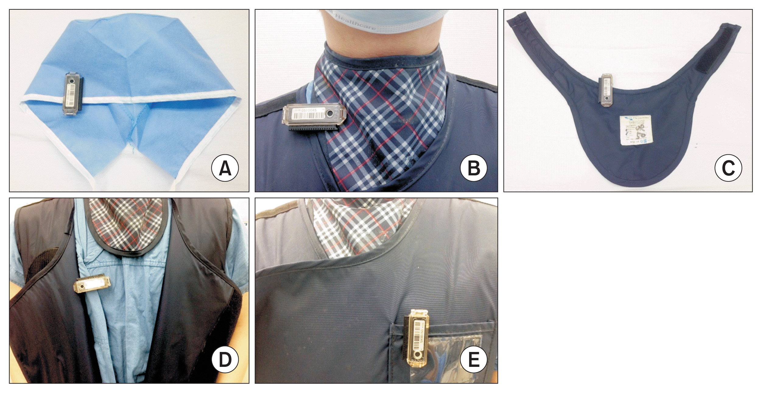
The physicians wore a lead thyroid protector and a lead apron during all procedures. The thyroid protector was a conventional collar (0.5 mm lead) that sufficiently covered the region between the sternal notch and the chin when properly worn. The lead apron was the coat type and was made of 0.3 mm lead in the front and 0.25 mm lead in the back. The apron provided 360 degrees of shielding over the upper thighs and torso.
3. Radiation exposure measurements
Seven thermoluminescent dosimeters (TLD; UD-802; Panasonic, Osaka, Japan) were used to measure radiation doses. Five of the TLDs were placed on the physician in the following locations during the CEBs: inside the thyroid protector, outside the thyroid protector, inside the lead gown chest area, outside the lead gown chest area, and on the forehead (Fig. 2). Our pain center does not have glass type TLDs, so the forehead dosimeter was used to estimate ocular radiation exposure. The forehead TLD was placed as close as possible to the physician’s eye without obstructing the field of vision. The two remaining TLDs served as control badges and were placed on a wall shelf at a distance of 250 cm horizontally and 200 cm vertically from the fluoroscopic table. The others were placed more than 100 m outside the operating room. Control badges remained in their positions for the full 2-month study period.
Data on the CEB procedural time, radiation exposure time, and radiation absorption dose (RAD) were collected as standard fluoroscopy data (Ziehm Vision; Ziehm Imaging GMBH, Nuremberg, Germany). The distance between the X-ray beam and the physician (Fig. 3) was measured during imaging after needle manipulation. Once the physician positioned himself right next to the patient and got onto the foot stool, he was told to minimize body movement and to not step down from the stool. The pain physician did not wear leaded eyewear and the lead apron, thyroid protector, and 5 physician-placed TLDs were stored outside of the operating room when not in use. Patient age, sex, height, and body weight were also recorded.

4. CEB
Before the procedure, an intravenous catheter was placed. All CEBs were fluoroscopy-guided and performed under sterile conditions while vital signs (i.e., blood pressure, pulse oximeter, and electrocardiogram) were monitored. The patient was placed in the prone position on a radiological table, and the pain physician placed himself right next to the patient using a foot stool (the operating bed was positioned in the high optimal fluoroscopy placement). Once placed next to the patient, the pain physician remained on the foot stool. Bending and/or extending of the back and neck and moving of the hands were allowed. The physician was allowed to stay away from the patient while checking the fluoroscopic image after manipulating the needle. Therefore, the physician only minimally moved his body during the procedure and kept a fairly constant posture. Other than the thyroid protector and lead apron, no other protective devices (e.g., leaded curtain or a leaded glass shield) were worn.
After positioning the patient for fluoroscopy, the site of the skin puncture was locally anesthetized with 1% lidocaine. A 20-gauge Tuohy Needle (Tae-Chang Industrial Co., Gongju, Korea) was inserted between the C6 and C7 vertebrae using a midline approach determined with anteroposterior fluoroscopy images. When the needle was firmly engaged, positioning was confirmed by examining the lateral view on fluoroscopy. The needle was advanced using a LOR technique in order to identify the epidural space. After obtaining LOR, contrast media (Iohexol, 300 mg iodine/mL; GE Healthcare, Piscataway, NJ) was injected through the needle for confirmation of position in the cervical epidural space. After all the scout films were checked, radiation exposure time and RAD were measured. It should be noted that real-time fluoroscopy was used in continuous mode during contrast media injection.
5. Statistical analyses
In the pilot study of our pain clinic, the radiation dose to which the professor was exposed was 32% of that to which the fellow was exposed. The pilot study was conducted on C-arm fluoroscopy-guided procedures during a period of 3 months. Our study included fluoroscopy-guided cervical spine procedures, and thus, we expected a higher radiation dose than that in lumbar spine procedures. We expected the radiation dose to which the professor was exposed to be 60% of that to which the fellow was exposed, approximately. A sample size of 45 patients per group was calculated to be needed, with a significance level of 0.05 (α = 0.05) and a power of 80% (β = 0.20), allowing for a 10% drop out rate.
Patient demographic data (age, sex, height, and weight) as well as data on the procedure time, radiation exposure time, absorbed dose, and distance from the X-ray field were collected. Measured radiation doses recorded by the TLDs over 2 months were converted to annual equivalent doses for all analyses. Data are expressed as mean ± standard deviation, and all statistical analyses were performed using IBM SPSS Statistical software ver. 20.0 (IBM Corp., Armonk, NY). The t-tests were used to determine the statistical significance of differences in means. Statistical significance was defined as P < 0.05.
Go to :
RESULTS
In total, 100 CEB procedures were performed on 100 patients during the 2-month study period. Fifty cases were performed by the fellow and 50 cases were performed by the professor. Patient demographic data is summarized in Table 1. The CEB procedural time, radiation exposure time, and radiation absorption time were not significantly different between the two physicians. However, the distance from the X-ray field to the physician was significantly greater for the professor than for the fellow (P = 0.03).
Table 1
Patient Demographics and Radiation-related Procedural Data
![]()
The forehead TLD indicated a radiation exposure of 0.74 and 0.62 mSv for the fellow and professor, respectively. The radiation dose recorded on the outside of the lead apron (fellow: 0.71 mSv, professor: 0.64 mSv) was similar to that recorded at the forehead for both physicians (Table 2). The TLDs placed on the inside of the apron showed radiation exposure values similar to those of the control TLD, but the TLDs placed inside the thyroid protector had slightly higher readings than did the control TLD. Therefore, lead-based protection provided a reduction in radiation exposure by 22%–35%, reducing exposure to levels near those of the control TLDs (Table 2). All exposure values from the 2-month period were scaled up to determine the equivalent radiation exposure for 600 CEB procedures, our center’s approximate annual CEB number per physician. All exposure values were well below the annual acceptable radiation exposure dose established by the ICRP (Fig. 4).
 | Fig. 4Annual equivalent dose (approximately 600 cases) was calculated from 2-month (100 cases) exposure data, as measured using thermoluminescent dosimeters. Annual maximum permissible radiation doses set by the International Commission on Radiological Protection are 500 and 20 mSv for the thyroid and crystalline lens, respectively. In: inside, Out: outside.
|
Table 2
Radiation Dose Measured with Thermoluminescent Dosimeters (TLDs)
![]()
Go to :
DISCUSSION
Fluoroscopy is widely used in interventional pain management procedures. As a result, studies concerning the radiation exposure of pain physicians are now being published [21,22]. Fortunately, most studies regarding radiation exposure during fluoroscopy-guided interventions have concluded that exposure levels are well below the yearly limit established by the ICRP [23–25]. However, exposure to low levels of ionizing radiation over the long-term cannot be accurately predicted. These long-term, low-level ionizing radiation exposures may not acutely destroy cells, but may lead to cell damage and genetic mutations that can lead to sequelae years later [3]. Epidemiologic studies from the Hiroshima, Nagasaki, and Chernobyl nuclear disasters revealed that low-dose radiation exposure may lead to early cataract development [12–14]. Therefore, pain physicians should understand the risks associated with radiation exposure during fluoroscopy-guided interventions and take proper precautions to minimize occupational radiation exposure.
We chose to examine radiation exposure during the CEB for several reasons. The CEB would be associated with more radiation exposure compared with other procedures because, in our hospital, the CEB is carried out in close proximity to the radiation beam, although we acknowledge that the procedures followed by pain physicians differ. In our study, the distance between the physician and the X-ray field was only 37.5 ± 2.1 cm (Table 1) for the fellow. Such proximity may increase the physician’s radiation exposure because radiation that scatters off the patient’s body is more likely to reach the pain physician’s head [26]. Indeed, we found that the forehead TLD recorded the highest radiation dose, reaching exposure levels as high as 4.44 mSv/yr (converted from 100 cases over 2 months to approximately 600 cases over 1 yr; Fig. 4).
Both examined physicians had similar radiation exposures on the forehead and outside of the apron. In contrast, the TLD placed outside of the thyroid protector recorded values almost as low as those recorded inside the apron. This unusual result may be explained by the habit of pain physicians flexing their heads to better visualize an injection site. This results in tucking of the chin, possibly blocking the thyroid from radiation exposure. Although higher compared to the control and lead-protected TLDs, other TLDs located outside of lead-based protective devices, even the forehead TLD, had radiation exposures well below ICRP limits. The physicians participating in this study did not wear leaded eyewear protection because many pain physicians refrain from wearing protective eye gear during procedures, and we wanted our results to be representative of the usual clinical setting.
Our secondary objective was to examine the efficacy of lead-based protection gear and whether or not it affected radiation exposure in the clinical setting. In our study, a lower radiation dose was measured in the TLDs placed inside of the lead-based protection gear compared with the outer TLDs (Table 2). Therefore, we conclude that lead-based protective gear is effective and stress the importance of shielding in keeping pain physician radiation exposure to a minimum.
The ICRP has three main principles for reducing unnecessary occupational radiation exposure, which are justification, optimization, and dose limitation [20]. Justification ensures that “more good is done than harm” when using harmful radiation. Optimization means that radiation doses should be kept “as low as possible,” and should consider social, medical, and economic implications. Dose limitation involves the three factors of time, distance, and shielding [27].
Reducing exposure time proportionally decreases radiation. Skilled physicians generally perform procedures more quickly and have the advantage of reduced exposure time. In our study, the radiation exposure time of the professor was shorter than that of the fellow, but this difference was not statistically significant. However, the distance between the X-ray field and the professor was significantly greater than the distance between the fellow and the X-ray field. Given that radiation exposure is inversely proportional to the square of the distance, inexperienced pain physicians should be educated on the importance of radiation safety, which includes keeping as large a distance from the X-ray field as possible.
The term “shielding” literally means to shield one’s body from the radiation rays, usually by wearing leaded products (e.g., aprons, gloves, and goggles). Leaded aprons that are 0.5 mm thick are known to reduce radiation exposure by up to 99% [28], and leaded eyewear is known to reduce radiation exposure by up to 70% [29]. However, we observed much lower protection rates in the current study (lead apron: 34%–35%, thyroid protector: 22%–32%). This may have resulted from the thinner apron (0.3 mm lead in the front) used by our hospital and from possible defects in our protective lead-based gear. In support of the product defect theory, Oyar and Kışlalıoğlu [30] found that, in his hospital, only 15.3% of the aprons were providing normal protection levels and that 68.2% of aprons were not stored and cared for properly. In fact, even normally-functioning aprons had folds and creases, causing leaks where radiation could enter. Unfortunately, our study physicians did not use new aprons or thyroid protectors. Therefore, it is possible that the thyroid protectors and lead aprons used in the current study could have had defects, causing a skewing of our results.
Many physicians are not properly educated on the importance of protective equipment and the harm that occupational radiation exposure can cause [31–33]. A pilot study on fellows with at least one year of clinical practice experience showed that a mere 33% of physicians had received proper radiation safety training. The importance of radiation safety training was evident because the educated group was more likely to use protective gear [34]. The use of gear in an educational environment is particularly important because radiation exposure time has been estimated to be 2–14 times higher than that in environments where only experienced physicians perform procedures [35].
Our study showed that eyes are more susceptible than the thyroid to radiation. This concurs with the ICRP’s recent decision to lower the yearly maximum radiation dose to the eye from 150 to 20 mSv. In contrast to apron and thyroid protectors, which are widely used among pain physicians, the actual use of leaded eyewear is unknown. During fluoroscopy-guided procedures, pain physicians almost always place themselves near the patient, increasing the risk of ocular radiation exposure from scattered radiation from the patient. Thus, the use of leaded eyewear must be enforced. Furthermore, the ideal position of the physician’s head to minimize ocular radiation exposure would be 90 degrees from the scattered radiation. Without optimal head positioning, leaded eyewear may be useless. Therefore, appropriate tilting of the head should also be considered [36]. In conclusion, the use of lead-based protection must be enforced, and clinical experience affects physician radiation, specifically because inexperienced physicians position themselves closer to the radiation beam.
The main limitation of our study was that radiation exposure was only examined during the CEB procedure. Radiation exposure during only one type of procedure is likely not representative of the annual radiation dose for pain physicians performing multiple fluoroscopy-guided techniques at multiple hospitals. Further research including a variety of techniques would give a more realistic annual radiation dose. In addition, our study calculated annual radiation doses using only two months of data. Further studies that collect radiation exposure data over a full year are needed.
There was a difference in the distance to the X-ray field during the procedure but no significant difference in the radiation exposure between the professor and the fellow during the CEBs. Even though the distance between the X-ray beam and the pain physician is small, radiation exposure can be minimized if proper protectors (thyroid protector, leaded apron, and eyewear) are worn. Further, we should consider that exposure to radiation varies depending on the physical condition of the pain physician and the method of the procedure. However, it remains uncertain whether or not long-term health effects occur from frequent low-dose radiation exposure, and safety precautions must always be followed.
Go to :
 XML Download