

 PDF
PDF Citation
 Print
Print


INTRODUCTION
Carpal tunnel syndrome (CTS) is a very common peripheral neuropathy of the upper extremity and is characterized by the entrapment of the median nerve [1]. CTS typically leads to paresthesia and pain in the radial-half of the ring finger, first two fingers, and the thumb. Sensory deficits and paresthesia frequently involve the entire palm area [2]. Diagnosis of CTS is based upon physical examination (Phalen’s sign, Tinel’s sign, and modified Phalen’s sign), electrodiagnostic studies, clinical symptoms, and ultrasonography (USG) [3]. Recently, carpal tunnel magnetic resonance imagings (CTMRI) have been demonstrated as a good diagnostic tool for the diagnosis of CTS [4–6]. CTMRI are noninvasive and can clearly depict the anatomical detail of peripheral nerves along with the pathological changes related to compression. The bowing of the flexor retinaculum, flattening of the medial nerve within the carpal tunnel, and the swelling of the nerve in the proximal and distal carpal tunnel are morphologically important anatomical features of the carpal tunnel area [7]. The morphological changes in the median nerve resulting from prolonged increased mechanical pressure within the carpal tunnel have already been demonstrated [6]. Apparently, the median nerve cross-sectional area (MNCSA) is considered a useful morphological parameter for the evaluation of CTS [4,8–12]. However, there have only been limited studies involving investigation of the anatomical basis of median nerve flattening. Thus, to evaluate the connection between the median nerve diameter and CTS, we measured the median nerve thickness (MNT). In the past, MNT has not been examined for any possible associations with CTS. Moreover, none of the reported studies have evaluated the clinical optimal cut-off value of the MNCSA and MNT. In the present work, we have compared the accuracy of using MNCSA and MNT for the diagnosis of CTS, using CTMRI to identify the most sensitive parameter.
Go to : 
MATERIALS AND METHODS
The present original research was registered at the Catholic Kwandong University College of Medicine, Republic of Korea (IS18RISI0013) and the Institutional Review Boards reviewed and approved the current research protocol. We reviewed patients who visited our Hand and Wrist Orthopedic Center from March 2014 to June 2018, and who were diagnosed with CTS. All the enrolled patients for the diagnosis of CTS were confirmed by two experienced, board-certified musculoskeletal radiologists. The CTS group included 20 patients (6 males and 14 females) with a mean age of 49.8 ± 13.4 years (range, 24–59 yr).
The inclusion criteria for the CTS group were: 1) positive CTS sign (Tinel’s sign, Phalen’s test or modified Phalen’s test) and 2) CTMRI taken within 12 months of the first diagnose and available for review. Exclusion criteria were: 1) history of wrist fracture; 2) double crush syndrome; 3) history of any wrist surgery; 4) wrist infection; 5) tumors in the carpal tunnel; and 6) chronic renal failure. To compare the MNCSA and MNT between patients with and without CTS, we enrolled control subjects who underwent CTMRI without having had CTS. The control group consisted of 20 patients (9 male and 11 female) with a mean age of 40.6 ± 10.7 years (range, 28–54 yr) (Table 1). The MNCSA and MNT in the control group were similarly examined at the hook of the hamate level (Table 1).
Table 1
Comparison of the Characteristics of Control and CTS Groups
![]()
1. MRI scanning protocol
All the CTMRI scans were performed at the Department of Hand and Wrist Orthopedic Center, Catholic Kwandong International St. Mary’s Hospital, Incheon, Republic of Korea. In both groups, the CTMRIs were obtained by following the same technique. CTMRI examinations were performed on 3T Avanto (Siemens Medical Solutions, Erlangen, Germany) with Achieva 3T scanners (Philips, Amsterdam, Netherlands). The CTMRI examination was conducted using transverse turbo spin echo T1-weighted images obtained with a slice thickness = 3.0 mm, 893-msec/13-msec repetition time/echo time, 0.9 mm intersection gap, 120 × 120 field of view, and 512 × 333 matrix. All CTMRI data were transferred from the central MRI center to our INFINITT PACS (INFINITT Healthcare System, Seoul, Korea).
2. Image analysis
The MNCSA was measured on the transverse angled sections through the whole cross-sectional area at the hook of the hamate level (Fig. 1A).
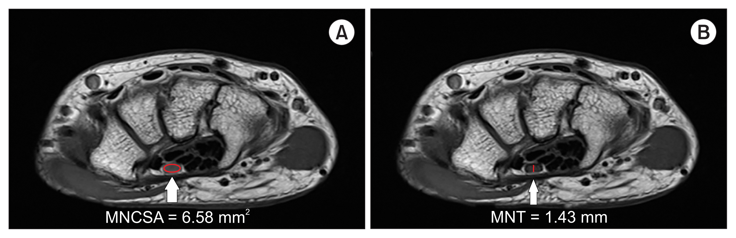
The MNT was measured based on the most compressed median nerve (Fig. 1B).
3. Statistical analyses
Data are expressed as mean ± standard deviation. Differences in demographic characteristics between the control and CTS groups were analyzed using unpaired Student’s t-tests. The validity of the MNCSA and MNT for the diagnosis of disease was estimated based on the cut-off values, the area under the curve (AUC), Receiver operating characteristics (ROC) curves, sensitivity, and specificity. The AUC was calculated independently in the final results to demonstrate the additional value gained from the addition of each parameter. All P-values of < 0.05 were interpreted as being statistically significant. IMB SPSS ver. 22.0 (IBM Corp., Armonk, NY) was used for statistical analysis.
Go to :
RESULTS
There were no statistically significant differences in the basic demographic data between the two groups (Table 1). The mean MNCSA was 9.01 ± 1.94 mm2 in the control group and 6.58 ± 1.75 mm2 in the CTS group. The mean MNT was 2.18 ± 0.39 mm in the control group and 1.43 ± 0.28 mm in the CTS group. CTS patients had significantly lower MNCSA (P < 0.001) and MNT (P < 0.001) (Table 1). Regarding the validity of both MNCSA and MNT as predictors of CTS, the ROC curve showed that the most suitable cut-off value for the MNT was 1.76 mm, with 85.0% sensitivity, 85.0% specificity, and an AUC of 0.94 (95% confidence interval [CI], 0.87–1.00) (Table 2, Fig. 2). The best cut-off point of the MNCSA was 7.72 mm2, with 75.0% sensitivity, 75.0% specificity, and an AUC of 0.82 (95% CI, 0.69–0.95) (Table 3, Fig. 2).
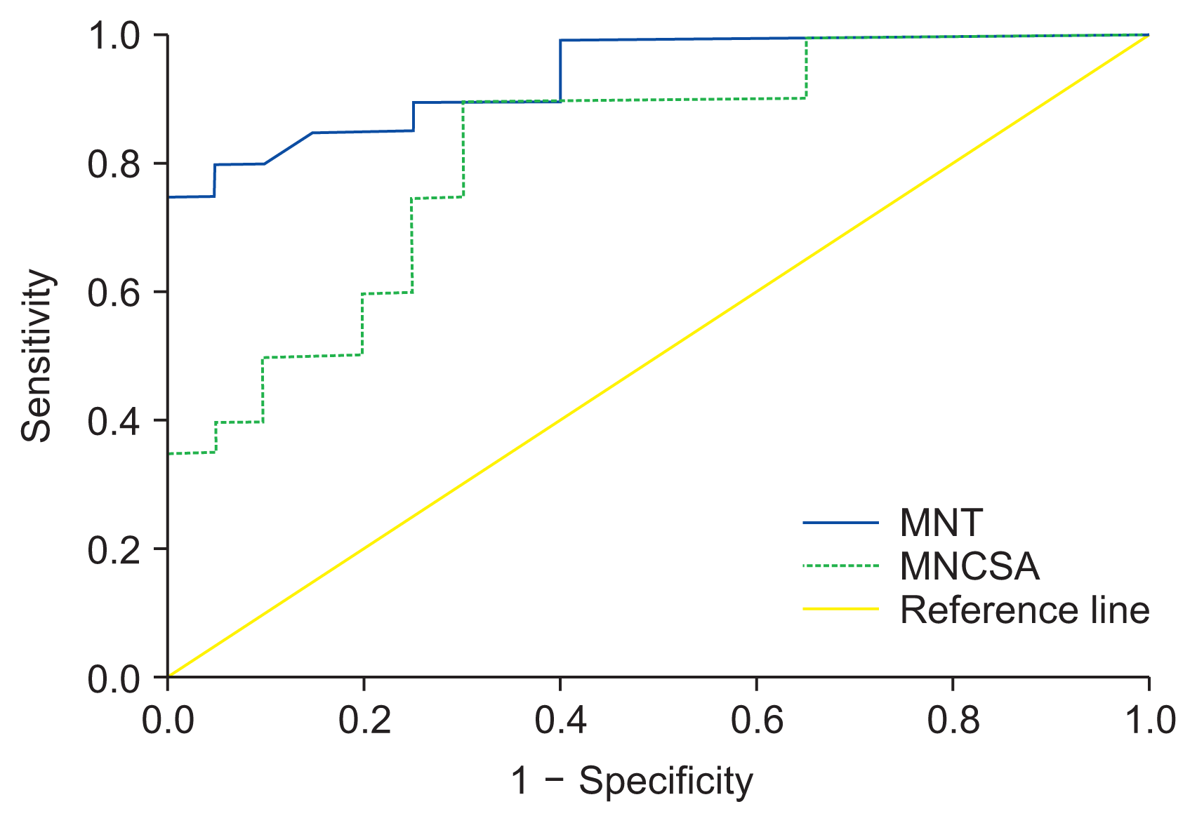 | Fig. 2Receiver operating characteristic curve of the median nerve thickness (MNT) and median nerve cross-sectional area (MNCSA) for prediction of carpal tunnel syndrome. The most suitable cut-off point of MNT was 1.76 mm vs. 7.49 mm2 of MNCSA, with sensitivity 85% vs. 80%, specificity 85% vs. 80% and an area under the curve 0.94 vs. 0.82, respectively.
|
Table 2
Sensitivity and Specificity of Each Cut-off Point of the MNT
| MNT (mm) | Sensitivity (%) | Specificity (%) |
|---|---|---|
| 1.33 | 25.0 | 100.0 |
| 1.44 | 60.0 | 100.0 |
| 1.55 | 75.0 | 95.0 |
| 1.76a | 85.0 | 85.0 |
| 1.95 | 95.0 | 60.0 |
| 2.14 | 100.0 | 55.0 |
![]()
Table 3
Sensitivity and Specificity of Each Cut-off Point of the MNCSA
| MNCSA (mm2) | Sensitivity (%) | Specificity (%) |
|---|---|---|
| 4.75 | 20.0 | 100.0 |
| 6.36 | 40.0 | 90.0 |
| 7.29 | 55.0 | 80.0 |
| 7.72a | 75.0 | 75.0 |
| 7.78 | 80.0 | 70.0 |
| 8.44 | 90.0 | 55.0 |
![]()
Go to :
DISCUSSION
CTS is a debilitating neuropathy that is frequently encountered in the primary care unit. It is the most common entrapment disorder of the upper extremity, affecting approximately 3.0% of the general adult population [6,13–16]. Females are three times more likely to have CTS than males, and the severity and prevalence have been reported to increase with age [17,18]. The hallmarks of CTS are paresthesia and pain in the distribution of the median nerve, which includes the radial half of the ring finger, the palmar aspect of the thumb and index, and middle finger [2]. CTS is diagnosed conventionally based on the past history of disease along with neurophysiological and physical examination findings. Electrophysiological testing is considered a standard for the diagnosis of CTS [5,19,20]. Recently, CTMRI has been demonstrated to be more advantageous than electrophysiological testing [5,6,12]. CT-MRI can visualize the median nerve at high resolution and represents an effective imaging method for visualization of tendons, ligaments, carpal bones, muscles, and other wrist morphological structures [4,6,12]. Several types of research have used CTMRI for the evaluation of CTS and have described characteristic CTMRI findings in CTS patients [4,6,12,19,21]. The reported results include lesions with increased signal intensity on T2-weighted images, swelling of the median nerve in the distal and proximal carpal tunnel, bowing of the flexor retinaculum, and flattening of the medial nerve within the carpal tunnel. High signal intensity is considered a major indicator of CTS and is a unique feature of CTMRI [22,23]. Musluoğlu et al. [24] suggested that CTMRI can be useful for the diagnosis of difficult cases of CTS. CTMRI provides detailed anatomical morphology that correlates well with electrophysiological results with regards to the severity of CTS. Kleindienst et al. [6] reported that CTMRI demonstrates a staging of median nerve compression and the results may positively contribute to making therapeutic decisions. The signal change in the median nerve was measured from the end of the carpal tunnel to the distal radius.
Both MRI and USG are useful tools for diagnosing and grading CTS. Of these, USG is used widely because it is more convenient and economical. However, MRI still has some advantages over USG. USG is an operator dependent modality, and therefore its result can be more susceptible to bias. On the other hand, MRI helps pinpoint the relevant factors causing CTS. Furthermore, in severe to extreme CTS, the demarcation of the median nerve is poor and the signal intensity of the nerve shows fasciculation, and therefore MRI is more useful in advanced CTS [7,16,25]. The MNCSA is also a useful morphological parameter for the evaluation of CTS. Many previous researches have used image modalities and focused on a quantitative analysis of the MNCSA in the carpal tunnel, and reported that MNCSA measurements could be useful in assessing CTS [4,9,11,12,26,27]. Ikeda et al. [4] demonstrated that CTMRI is a valuable examination for imaging the cross-sectional area of the median nerve and has potential as a diagnostic modality for CTS. However, there are limited studies investigating the anatomical basis of median nerve flattening. Furthermore, its clinical applications are limited due to this lack of studies. Accordingly, to evaluate the connection between the median nerve flattening and CTS, we measured the MNT. The MNT has not been examined for its associations with CTS. We hypothesized that MNT is a valuable diagnostic morphologic parameter in CTS.
Eventually, our research data demonstrated the association between MNT and CTS. CTS patients had significantly lower MNT than the control groups. In our study, the best cut-off point for MNT was 1.76 mm, with 85.0% sensitivity, 85.0% specificity, and an AUC of 0.94 (95% CI, 0.87–1.00). Moreover, the best cut-off value for the MNCSA was 7.72 mm2, with 75.0% sensitivity, 75.0% specificity, and an AUC of 0.82 (95% CI, 0.69–0.95). We consider this value as the standard for both the MNT and MNCSA, as there exists no study about optimal cut-off values for both MNT and MNCSA. In the present work, we have demonstrated a significant association between both MNCSA and MNT and CTS; and MNT was identified as a more sensitive measurement parameter. We thought that MNT could be a precise, clear, and objective measurement parameter to evaluate median nerve flattening. In our present research, MNT was measured from transverse T1-weighted turbo spin echo CTMRIs. To the best of our knowledge, none of the published reports have demonstrated an association between MNT and CTS as a morphologic parameter on CTMRI. We choose the hook of the hamate level to measure both the MNCSA and MNT. Previous studies revealed the increase in the cross-sectional area of the median nerve at the pisiform level and flattening of the median nerve at the hook of the hamate level [28]. Consequently, the hook of the hamate level of the carpal tunnel narrows the carpal tunnel space leading to compression of the median nerve. Chow et al. [29] investigated the relationship between CTS and anatomic variations of the hook of the hamate. The researchers indicated a significant increase in the incidence of a variant hook of hamate in the CTS group compared with the non-CTS group.
The current study has several limitations. First, diagnosis of CTS was made by only physical tests, but not by electromyography and nerve conduction studies. The CTS diagnosis would have been more accurate if symptoms, physical tests, and neurotransmission tests were considered together [2]. Second, there might be some errors associated with the measurement of the MNT and MNCSA on MRI. Although we tried to maintain a high quality of morphologic measurement in the transverse T1-weighted wrist MRIs that best showed the median nerve at the hook of hamate level, the measurement of MNT and MNCSA in the single MRI slice could be inhomogeneous because of the differences in the cutting angle in MRIs resulting from technical limits and individual anatomic variation. Also, the MRI evaluator’s blindness to the diagnosis is another weakness of this study. Third, there are several levels which could be used to measure the median nerve using MRI, such as the distal radioulnar joint, the body of the scaphoid, the tubercle of the scaphoid, and the hook of the hamate. Although analysis at the hook of the hamate level is reliable and accurate for diagnosis, the results might still be biased. If data that includes different levels can be obtained from CTS patients and compared with a control group, much better information can be obtained. Fourth, the cause of CTS has multiple sources, including the transcarpal ligament, soft tissues, and flexor retinaculum [30,31]; however, we only focused on the median nerve. Fifth, because this study is not randomized and is a case-control group, an adjustment of baseline characteristics should be considered to compare the two groups.
Despite these limitations, this is the first research to document the association between MNT and CTS. MNT is proposed as a reliable and simple measurement tool with high sensitivity for the evaluation of CTS.
In conclusion, MNT is proposed as a new sensitive morphological parameter for the evaluation of CTS. It is anticipated that this new adjuvant measurement technique will be helpful for the assessment of patients with CTS.
Go to :
 XML Download