Journal List > Korean J Pain > v.32(4) > 1159222

Elberry, Sharkawi, and Wahba: Antinociceptive and anti-inflammatory effects of N-acetylcysteine and verapamil in Wistar rats

Abstract

Background

Antinociceptive anti-inflammatory drugs have many adverse effects. The goal of this investigation is to study the probable anti-inflammatory and analgesic effects of verapamil and N-acetylcysteine (NAC) in experimental rats.

Methods

Adult male Wistar rats were randomly divided into 4 groups in the antinociceptive study, each containing 6 rats; the normal control group, which received saline (1 mL/kg); the diclofenac group, which received diclofenac sodium (5 mg/kg); the NAC group, which received NAC (125 mg/kg); and the verapamil group, which received verapamil (8 mg/kg). In the anti-inflammatory study, 5 groups were used, the 4 previous groups with the addition of an edema control group, received saline and were subjected to formalin test. Hot plate latency time was recorded for antinociceptive evaluation. Paw edema thickness and biochemical parameters were recorded for anti-inflammatory evaluation.

Results

Administration of NAC showed significant prolongation of hot plate latency time at 1 hour when compared to the control group while verapamil showed a significant prolongation of hot plate latency time at 1 and 2 hours when compared to the control group and NAC group values. Administration of NAC and verapamil significantly decreased paw edema thickness at 2, 4, and 8 hours when compared to edema control values. Regarding biochemical markers, NAC and verapamil significantly decreased serum nitric oxide synthase, C-reactive protein, and cyclooxygenase-2 levels compared to the edema control value. In accordance, a marked improvement of histopathological findings was observed with both drugs.

Conclusions

NAC and verapamil have antinociceptive and anti-inflammatory effects comparable to diclofenac sodium.

INTRODUCTION

Pain and inflammation are common manifestations of many diseases. Pain is an unpleasant, sensory and emotional experience associated with actual or potential tissue damage, and originates from inflammation and the inflammatory responses [1]. Inflammation is a nonspecific response that has a beneficial effect on the host. Several experimental studies have shown that reactive oxygen species (ROS) contribute to pain [2]. Proinflammatory cytokines including tumor necrosis factor (TNF)-α play significant roles in neuronal reaction and inflammation [3]. Nonsteroidal anti-inflammatory drugs, such as diclofenac, are the most commonly used antinociceptive and antiinflammatory drugs, since they are effective in the management of pain, fever, redness, and edema resulting from inflammatory mediator release [4]. Although diclofenac is usually well tolerated by patients, its use is associated with some adverse drug reactions, such as gastrointestinal mucosal damage, renal toxicity, bleeding, and cardiovascular side effects [5].
Finding different types of drugs to induce the same antiinflammatory and antinociceptive effects with fewer side effects became a priority. N-acetylcysteine (NAC) acts both as a glutathione (GSH) precursor and shows anti-inflammatory and antioxidant effects [6]. By regulation of the redox status in the cell, it can interfere with several signaling pathways and decrease the cytokine production in the early inflammatory response [7]. Verapamil belongs to the dihydropyridine family of calcium channel blockers that block voltage-dependent L-type calcium channels, and is used clinically to treat hypertension, angina, and cardiac arrhythmias [8]. Previous studies have also found that verapamil inhibits activation of superoxide production in neutrophils and macrophages [9]. Increased calcium influx through L-type calcium channels causes activation of signaling cascades, such as ROS and activation of proinflammatory cytokines [10]. Previous studies showed that verapamil elicited neuroprotection action and has a significant anti-inflammatory and anti-angiogenesis effects [11]. The aim of the present work was to study the probable anti-inflammatory and antinociceptive effects of verapamil and NAC in rats.

MATERIALS AND METHODS

1. Drugs and chemicals

Diclofenac sodium (Diclac 75 mg; Minapharm, Giza, Egypt), NAC (Acetylcysteine 600 mg; SEDICO, Cairo, Egypt), verapamil (Isoptin 80 mg; Kahira Pharmaceuticals & Chemical Industries Company, Cairo, Egypt), isotonic saline (Otsuka Pharmaceutical Company, Cairo, Egypt), and formalin (Formaldehyde sol. 38%–40%; Al-Nasr Pharma Chemicals, Cairo, Egypt) were used in the current study.

2. Animals

Experiments were performed on adult male Wistar rats weighing 220 to 280 g in accordance with the guidelines of the Ethics Committee at Beni-Suef University (# 018-63), which complies with Guidelines on Ethical Standards for Investigation of Experimental Pain in Animals [12]. The animals were housed and maintained in a well ventilated, temperature-controlled room at 22°C ± 2°C with a relative humidity of 50% to 60% and a 12-hour light-dark cycle with free access to food and water. They were acclimatized for 1 week and randomly allocated into groups. Care was taken to avoid stressful conditions and to minimize animal suffering, and all experimental procedures were performed between 9 and 11 am. All drugs were administered orally using a metallic tube, and the rat was forced to swallow them one hour before the hot plate or formalin test.

3. Antinociceptive evaluation

The rats were divided randomly into 4 groups, 6 rats in each group; the normal control group received saline (1 mL/kg), the diclofenac group received diclofenac sodium (2 mg/kg) [13], the NAC group received NAC (150 mg/kg) [14], and the verapamil group received verapamil (8 mg/kg) [15].
The hot plate method was used to investigate the nociception in rats as described by Langerman et al. [16]. Each rat was placed in a hot plate analgesia meter (TSE Systems, Bad Homburg vor der Höhe, Germany) and the metal surface was maintained at 50°C ± 1°C. The latency times in seconds taken to nocifensive behavior including hind paw withdrawal or licking, and jumping was recorded prior to the treatment, 1, and 2 hours after the treatment [17]. Rats showing a pretreatment reaction time greater than 30 seconds in the hot plate test were excluded from the experiment (cut-off latency) [18].

4. Anti-inflammatory evaluation

1) Formalin test

The rats were divided randomly into 5 groups as in 4 groups in the hot plate test in addition to a paw edema group. The normal control group received saline (1 mL/kg), the paw edema control received saline (1 mL/kg), the diclofenac group received diclofenac sodium (2 mg/kg) [13], the NAC group received NAC (150 mg/kg) [14], and verapamil group received verapamil (8 mg/kg) [15]. Compounds were administered via the oral route just prior to induction of inflammation. The inflammation was induced in rats of all groups except the normal control group by subcutaneous injection of 0.05 mL, 10% formalin into the plantar surface of the right hind paws using a 25-gauge needle [19]. The anti-inflammatory activity was then calculated based on paw-volume changes at 1, 2, 4, and 8 hours after formalin injection using vernier caliper. Blood samples were collected 6 hours after the formalin injection.

2) Biochemical examination

Blood samples (up to 2 mL) were collected by orbital puncture under inhalation anesthesia using alcohol, chloroform, and ether in a ratio of 1:2:3. In serum separating tubes, the blood samples were centrifuged at 3,000 rpm for 15 minutes to separate the sera and were stored at 80°C for biochemical analyses [20]. Reduced GSH and myeloperoxidase (MPO) were determined according to standard methods using diagnostic kits from BioSystems S.A. (Barcelona, Spain). Assessment of serum TNF-α, inducible nitric oxide synthetase (iNOS), cyclooxygenase enzyme (COX-2), and C-reactive protein (CRP) were carried out by enzyme-linked immunosorbent assay (ELISA) using kit purchased from DRG International Inc. (Mountainside, NJ).

5. Histopathological examination of the paw tissue

After collection of blood samples, all animals were euthanized. The right and left paws were dissected from the rats and kept intact for histological examination and the soft tissues of paw were isolated carefully. Paw tissues were fixed immediately and decalcified in ethylenediaminetetraacetic acid [21]. Then, the tissue specimens were dehydrated, embedded in paraffin, and sectioned into 5 μm portions, and stained using hematoxylin and eosin (H&E) for histopathological examination using a research microscope provided with an HD camera (Leica DM2500 M; Leica Microsystems GmbH, Wetzlar, Germany). A histopathologic score is used for inflammation, congestion, and edema on a 7-grade scale from 0 to 6 (0 = normal and 6 = the most severe inflammation).

6. Statistical analysis

Data were expressed for each parameter investigated as a mean ± standard error of the mean. One-way analysis of variance (ANOVA) was used to compare the different groups. All statistics were carried out using SPSS ver. 15 (SPSS Inc., Chicago, IL). Differences were considered statistically significant when the P value was < 0.05.

RESULTS

1. Antinociceptive effect and hot plate latency time

The present study showed that administration of a single dose of NAC produced a significant prolongation of the hot plate latency time at 1 hour after treatment when compared to the normal control group values. Moreover, administration of a single dose of verapamil showed a significant prolongation of the hot plate latency time at 1 and 2 hours after treatment, which is comparable to diclofenac group values (Table 1).

2. Anti-inflammatory effect

1) Paw edema

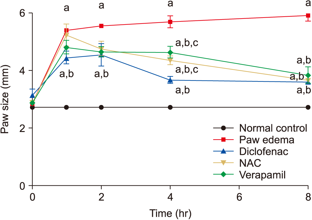
The present study revealed that administration of NAC and verapamil significantly decreased paw edema thickness at 2, 4, and 8 hours when compared to edema control values. Both NAC and verapamil decreased paw edema thickness to the same values of diclofenac at 2 and 8 hours (Fig. 1).

2) Serum biochemical markers

The current investigation showed that formalin-induced paw edema significantly decreased the serum levels of GSH, while TNF-α, MPO, NOS, CRP, and COX-2 increased significantly when compared to normal control values. The alterations in NOS, CRP, COX-2, and TNF-α were alleviated significantly when compared to paw edema values (Table 2, Fig. 2).
On the other hand, verapamil decreased significantly the elevated MPO level when compared with the paw edema value, while NAC decreased this elevation, but non-significantly. Moreover, diclofenac, NAC, and verapamil failed to alleviate the serum GSH level (Table 2).

3. Histopathological results

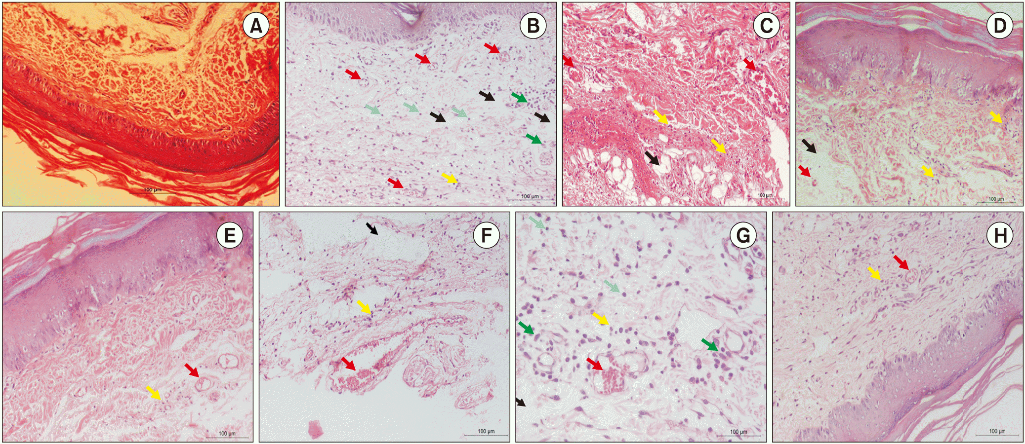
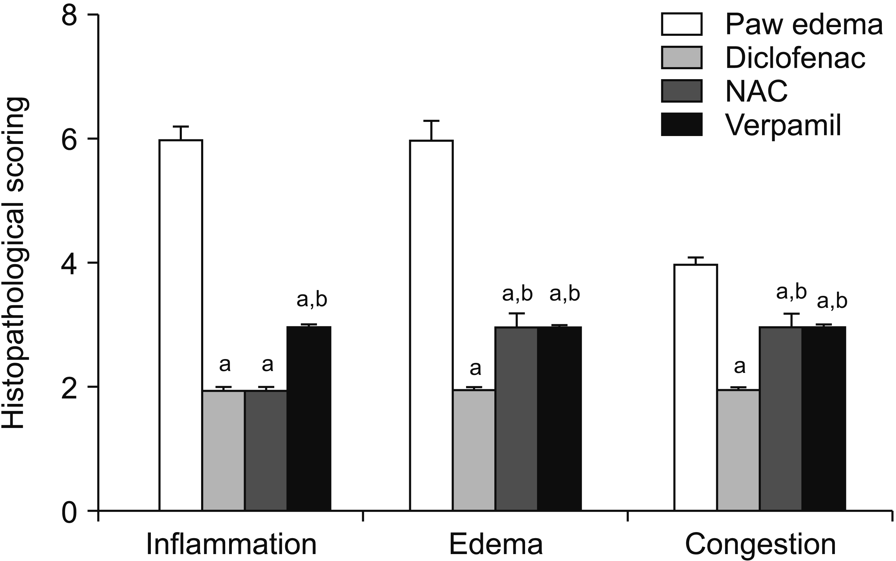
Histopathological examination of the hind paw of the normal control rats showed normal dermal and epidermal architecture with no signs of inflammation (Fig. 3A). On injection of formalin, the histopathological study revealed severe inflammation evidenced by mononuclear infiltration of inflammatory cells, severe edema, mild congestion, and the highest score (Fig. 3B). On the other hand, injection of diclofenac sodium ameliorated the edema, inflammation, and congestion and ameliorated the histopathological scoring (Figs. 3A, 3D, 4). Rats which received NAC (Fig. 3E, F) and verapamil (Fig. 3G, H) showed low histopathological scoring and mild inflammation evidenced by subsiding edema and fewer inflammatory cells with less congestion and presence of macrophages that engulf debris with necrosed tissue (Fig. 3G, H).

DISCUSSION

The hot plate test and other tests that apply heat stimuli to the hind paws are considered to integrate supraspinal pathways, as rats with spinal transection do not withdraw the hind limbs in the hot plate test [22]. The results of the present study revealed that NAC has an antinociceptive effect evidenced by a significant prolongation of hot plate latency time when compared to normal control values. Despite that, NAC significantly prolonged the hot plate latency time when compared to normal control values but this prolongation was less than that of diclofenac sodium values. These results are in agreement with Horst et al. [23] who reported that NAC has an anti-hyperalgesic effect in rats with neuropathic pain mostly attributed to modulation of the antioxidant redox system, reduction in several neuroinflammatory molecules, and a decrease in nitric oxide (NO). Similarly, the current study showed that verapamil produced a significant prolongation of hot plate latency time when compared to normal control values and the NAC group but still inferior to diclofenac sodium group values. These results are in agreement with Abdollahi et al. [24] who reported that verapamil induced a significant anti-nociception in both phases of a formalin test model. Verapamil, a calcium channels blocker, resulted in impaired calcium influx, a necessary process for activation of N-methyl-D-aspartate receptors of glutamate which induces the local release of both prostaglandin E2 (PGE2) and NO, thereby relieving pain [24].
On the other hand, formalin significantly increased paw edema thickness in the present study after 1, 2, 4, and 8 hours, which is in agreement with the results of Lee and Jeong [25]. This edema is mainly caused by vascular changes which are mediated by release of substance P, prostanoids, 5-hydroxytryptamine, and histamine [26]. The present study showed that NAC significantly alleviated paw edema thickness after 2, 4, and 8 hours, which is in agreement with the results of Lasram et al. [27] and Farshid et al. [28]. Moreover, the present study showed that verapamil produced a statistically significant alleviation in paw edema thickness after 2, 4, and 8 hours. This result is in agreement with the results of Martinez et al. [29].
The alleviation by NAC was be explained in previous studies by reduction of neutrophil infiltration and inhibition of COX products such as prostaglandins, and also via inhibiting nuclear factor-κB (NF-κB) and modulating cytokine synthesis [30]. In addition, the mitigation by verapamil can be explained as verapamil inhibits activation of superoxide production in neutrophils and macrophages, and it can suppress the release of pro-inflammatory mediators, such as TNF-α and NO [9]. This is in accordance with the results of the current study which revealed histopathological improvement of the inflammatory reactions and neutrophil recruitment produced by formalin injection, as well as alleviation of the serum level changes of TNF-α, NOS, CRP, and COX.
Despite both NAC and verapamil showing a significant anti-inflammatory effect compared to the edema control group, neither NAC nor verapamil was superior to diclofenac in ameliorating paw edema after 2 and 4 hours. However, after 8 hours the ameliorative effect of both NAC and verapamil on paw edema thickness values was nearly the same as that of diclofenac values. Unfortunately, the results of the present study showed that diclofenac, NAC, and verapamil failed to improve the decreased level of GSH in the edema control group. This can be explained by the short duration of the experiment and nonspecific method that is used in the current study, as GSH was measured in plasma and not tissues.
In the current study, treatment with either diclofenac or NAC failed to produce any significant ameliorative effect on the increased level of MPO in the edema control group. The diclofenac effect in the present study is in agreement with the previous results of Pireddu et al. [31]. On the other hand, verapamil significantly ameliorated the increased serum MPO level when compared to the edema control group and decreased it to nearly the normal control values. These results are in agreement with the results of Messiha and Abo-Youssef [32]. This could be attributed to its calcium channel blocking activity in addition to attenuation of chemo-attractant release by Kupffer cells [33].
Huang et al. [34], showed similar results regarding the effect of NAC on COX expression. On the other hand, Wang et al. [35] reported that verapamil significantly inhibited COX-2 level in inflammatory arthritis in mice, which could be explained, since Ca2+ influx leads to the up-regulation of COX-2 expression and PGE2 release via the activation of the protein kinase A, so, a Ca2+ influx inhibitor decreases COX-2 expression [36]. Previous studies showed that inflammation stimulates the release of TNF-α, which, in turn, stimulates the production of COX, thus stimulating the local production of sympathetic amines [37]. In addition, the present work showed that NAC significantly decreased the elevated serum TNF-α when compared to the edema control group values, and this decrease was nearly equal to that in the diclofenac group. This is in agreement with the results of Pathak et al. [38]. However, Talasaz et al. [39] reported that NAC did not make any significant effect on the serum TNF-α level in patients following an ST-segment elevation myocardial infarction. The effect of NAC could be due to inhibition of NF-κB which regulate cytokine transcription as TNF-α [40]. According to the results of the present study, verapamil treatment significantly decreased the high TNF-α values observed in the edema control group. Even verapamil was superior to diclofenac in ameliorating the high level of TNF-α, and it decreased the level nearly to the normal control values. These results are in agreement with the work of Wang et al. [35] who showed that verapamil significantly inhibited TNF-α both in vitro and in mice models of arthritis. The mechanism of action of verapamil’s effect is possibly via inhibition of NF-κB [41].
Regarding to the findings of the present work, NAC, verapamil, and diclofenac significantly reduced the elevated serum NOS level observed in the edema control group. There was no significant difference between the three groups. Guibas et al. [42] showed that NAC significantly inhibited the accumulation of inflammatory cells and down regulated iNOS expression in a rat model of allergic rhinitis. The effect of NAC on NOS can be attributed mostly to the inhibitory effect of NAC on one of the events leading to iNOS protein expression [43]. The effect of verapamil on NOS in the present results may be due to inhibition of inflammatory cytokines [10].
Regarding the findings of the present work, NAC, verapamil, and diclofenac sodium significantly reduced the elevated serum CRP level observed in the edema control group. There was no significant difference between the 3 groups. The results of the present study regarding the NAC group are in agreement with Saddadi et al. [44].
The NAC was shown to inhibit the production of interleukin (IL)-6 from macrophages which is related to the anti-oxidative stress effects of NAC [45]. Since CRP increases following IL-6 secretion by macrophages and T cells, NAC results regarding CRP can be explained by its effect on IL-6 [46]. Bakris [47] reported that verapamil significantly reduced CRP and lowered albuminuria in kidney disease progression. The effect of verapamil on CRP may be because it can modulate T-helper cell type 2 associated cytokine secretion, such as IL-5 and IL-6 [48] which stimulate hepatic secretion of CRP following IL-6 secretion by macrophages and T cells [46].
In conclusion, NAC and Verapamil were found to have antinociceptive and anti-inflammatory effects. This antinociceptive effect can be attributed to COX inhibition and an antioxidant effect. Further clinical and long-term studies are needed to confirm their clinical effectiveness and safety.

Notes

Author contributions: Ahmed Abdullah Elberry: Study conception; Souty Mouner Zaky Sharkawi: Methodology; Mariam Rofaiel Wahba: Investigation.

CONFLICT OF INTEREST

No potential conflict of interest relevant to this article was reported.

REFERENCES

1. Swarup A, Agarwal R, Malhotra S, Dube AS. A comparative study of efficacy of gabapentin in inflammation induced neuropathic animal pain models with conventional analgesic diclofenac. Int J Res Med Sci. 2016; 4:1429–32. DOI: 10.18203/2320-6012.ijrms20161204.
crossref
2. Trevisan G, Benemei S, Materazzi S, De Logu F, De Siena G, Fusi C, et al. TRPA1 mediates trigeminal neuropathic pain in mice downstream of monocytes/macrophages and oxidative stress. Brain. 2016; 139:1361–77. DOI: 10.1093/brain/aww038. PMID: 26984186.
crossref
3. Hasanvand A, Ahmadizar F, Abbaszadeh A, Amini-Khoei H, Goudarzi M, Abbasnezhad A, et al. The antinociceptive effects of rosuvastatin in chronic constriction injury model of male rats. Basic Clin Neurosci. 2018; 9:251–60. DOI: 10.32598/bcn.9.4.251. PMID: 30519383. PMCID: PMC6276538.
crossref
4. Howard L. Fields. Pain free: modern drugs and neuropathic pain. J Korean Med Sci. 2002; 22:360–7.
5. McGettigan P, Henry D. Use of non-steroidal anti-inflammatory drugs that elevate cardiovascular risk: an examination of sales and essential medicines lists in low-, middle-, and high-income countries. PLoS Med. 2013; 10:e1001388. DOI: 10.1371/journal.pmed.1001388. PMID: 23424288. PMCID: PMC3570554.
crossref
6. Tardiolo G, Bramanti P, Mazzon E. Overview on the effects of N-acetylcysteine in neurodegenerative diseases. Molecules. 2018; 23:E3305. DOI: 10.3390/molecules23123305. PMID: 30551603. PMCID: PMC6320789.
crossref
7. Triggle DJ. Calcium-channel drugs: structure-function relationships and selectivity of action. J Cardiovasc Pharmacol. 1991; 18(Suppl 10):S1–6. DOI: 10.1097/00005344-199118101-00002. PMID: 1724996.
8. Hotchkiss RS, Bowling WM, Karl IE, Osborne DF, Flye MW. Calcium antagonists inhibit oxidative burst and nitrite formation in lipopolysaccharide-stimulated rat peritoneal macrophages. Shock. 1997; 8:170–8. DOI: 10.1097/00024382-199709000-00004. PMID: 9377163.
crossref
9. Liu Y, Lo YC, Qian L, Crews FT, Wilson B, Chen HL, et al. Verapamil protects dopaminergic neuron damage through a novel anti-inflammatory mechanism by inhibition of microglial activation. Neuropharmacology. 2011; 60:373–80. DOI: 10.1016/j.neuropharm.2010.10.002. PMID: 20950631. PMCID: PMC3014428.
crossref
10. Kalonia H, Kumar P, Kumar A. Attenuation of proinflammatory cytokines and apoptotic process by verapamil and diltiazem against quinolinic acid induced Huntington like alterations in rats. Brain Res. 2011; 1372:115–26. DOI: 10.1016/j.brainres.2010.11.060. PMID: 21112316.
crossref
11. Eteraf-Oskouei T, Mikaily MS, Najafi M. Anti-inflammatory and anti-angiogenesis effects of verapamil on rat air pouch inflammation model. Adv Pharm Bull. 2017; 7:585–91. DOI: 10.15171/apb.2017.070. PMID: 29399548. PMCID: PMC5788213.
crossref
12. Zimmermann M. Ethical guidelines for investigations of experimental pain in conscious animals. Pain. 1983; 16:109–10. DOI: 10.1016/0304-3959(83)90201-4. PMID: 6877845.
crossref
13. Yuan J, Ma H, Cen N, Zhou A, Tao H. A pharmacokinetic study of diclofenac sodium in rats. Biomed Rep. 2017; 7:179–82. PMID: 28781777. PMCID: PMC5526189.
crossref
14. Horst A, de Souza JA, Santos MCQ, Riffel APK, Kolberg C, Partata WA. Effects of N-acetylcysteine on spinal cord oxidative stress biomarkers in rats with neuropathic pain. Braz J Med Biol Res. 2017; 50:e6533. DOI: 10.1590/1414-431x20176533. PMID: 29069230. PMCID: PMC5649872.
crossref
15. Tamaddonfard E, Erfanparast A, Taati M, Dabbaghi M. Role of opioid system in verapamil-induced antinociception in a rat model of orofacial pain. Vet Res Forum. 2014; 5:49–54. PMID: 25568692. PMCID: PMC4279656.
16. Langerman L, Zakowski MI, Piskoun B, Grant GJ. Hot plate versus tail flick: evaluation of acute tolerance to continuous morphine infusion in the rat model. J Pharmacol Toxicol Methods. 1995; 34:23–7. DOI: 10.1016/1056-8719(94)00077-H. PMID: 7496043.
crossref
17. Espejo EF, Mir D. Structure of the rat’s behaviour in the hot plate test. Behav Brain Res. 1993; 56:171–6. DOI: 10.1016/0166-4328(93)90035-O. PMID: 8240711.
crossref
18. Vittalrao AM, Shanbhag T, Kumari M, Bairy KL, Shenoy S. Evaluation of antiinflammatory and analgesic activities of alcoholic extract of Kaempferia galanga in rats. Indian J Physiol Pharmacol. 2011; 55:13–24. PMID: 22315806.
19. Yoon MH, Choi JI. Pharmacologic interaction between cannabinoid and either clonidine or neostigmine in the rat formalin test. Anesthesiology. 2003; 99:701–7. DOI: 10.1097/00000542-200309000-00027. PMID: 12960556.
crossref
20. Van Herck H, Baumans V, Brandt CJ, Hesp AP, Sturkenboom JH, van Lith HA, et al. Orbital sinus blood sampling in rats as performed by different animal technicians: the influence of technique and expertise. Lab Anim. 1998; 32:377–86. DOI: 10.1258/002367798780599794. PMID: 9807751.
crossref
21. Yamamoto-Fukud T, Shibata Y, Hishikawa Y, Shin M, Yamaguchi A, Kobayashi T, et al. Effects of various decalcification protocols on detection of DNA strand breaks by terminal dUTP nick end labelling. Histochem J. 2000; 32:697–702. DOI: 10.1023/A:1004171517639. PMID: 11272810.
22. Giglio CA, Defino HL, da-Silva CA, de-Souza AS, Del Bel EA. Behavioral and physiological methods for early quantitative assessment of spinal cord injury and prognosis in rats. Braz J Med Biol Res. 2006; 39:1613–23. DOI: 10.1590/S0100-879X2006001200013. PMID: 17160271.
crossref
23. Horst A, Kolberg C, Moraes MS, Riffel AP, Finamor IA, Belló-Klein A, et al. Effect of N-acetylcysteine on the spinal-cord glutathione system and nitric-oxide metabolites in rats with neuropathic pain. Neurosci Lett. 2014; 569:163–8. DOI: 10.1016/j.neulet.2014.03.063. PMID: 24704379.
crossref
24. Abdollahi M, Nikfar S, Abdoli N. Potentiation by nitric oxide synthase inhibitor and calcium channel blocker of aspartame-induced antinociception in the mouse formalin test. Fundam Clin Pharmacol. 2001; 15:117–23. DOI: 10.1046/j.1472-8206.2001.00013.x. PMID: 11468021.
crossref
25. Lee IO, Jeong YS. Effects of different concentrations of formalin on paw edema and pain behaviors in rats. J Korean Med Sci. 2002; 17:81–5. DOI: 10.3346/jkms.2002.17.1.81. PMID: 11850594. PMCID: PMC3054816.
crossref
26. Damas J, Liégeois JF. The inflammatory reaction induced by formalin in the rat paw. Naunyn Schmiedebergs Arch Pharmacol. 1999; 359:220–7. DOI: 10.1007/PL00005345. PMID: 10208309.
crossref
27. Lasram MM, Lamine AJ, Dhouib IB, Bouzid K, Annabi A, Belhadjhmida N, et al. Antioxidant and anti-inflammatory effects of N-acetylcysteine against malathion-induced liver damages and immunotoxicity in rats. Life Sci. 2014; 107:50–8. DOI: 10.1016/j.lfs.2014.04.033. PMID: 24810974.
crossref
28. Farshid AA, Tamaddonfard E, Yahyaee F. Effects of histidine and N-acetylcysteine on diclofenac-induced anti-inflammatory response in acute inflammation in rats. Indian J Exp Biol. 2010; 48:1136–42. PMID: 21117455.
29. Martinez LL, Aparecida De Oliveira M, Fortes ZB. Influence of verapamil and diclofenac on leukocyte migration in rats. Hypertension. 1999; 34:997–1001. DOI: 10.1161/01.HYP.34.4.997. PMID: 10523397.
crossref
30. Berk M, Malhi GS, Gray LJ, Dean OM. The promise of N-acetylcysteine in neuropsychiatry. Trends Pharmacol Sci. 2013; 34:167–77. DOI: 10.1016/j.tips.2013.01.001. PMID: 23369637.
crossref
31. Pireddu R, Caddeo C, Valenti D, Marongiu F, Scano A, Ennas G, et al. Diclofenac acid nanocrystals as an effective strategy to reduce in vivo skin inflammation by improving dermal drug bioavailability. Colloids Surf B Biointerfaces. 2016; 143:64–70. DOI: 10.1016/j.colsurfb.2016.03.026. PMID: 26998867.
crossref
32. Messiha BA, Abo-Youssef AM. Protective effects of fish oil, allopurinol, and verapamil on hepatic ischemia-reperfusion injury in rats. J Nat Sci Biol Med. 2015; 6:351–5. DOI: 10.4103/0976-9668.160003. PMID: 26283828. PMCID: PMC4518408.
crossref
33. Ray SD, Kamendulis LM, Gurule MW, Yorkin RD, Corcoran GB. Ca2+ antagonists inhibit DNA fragmentation and toxic cell death induced by acetaminophen. FASEB J. 1993; 7:453–63. DOI: 10.1096/fasebj.7.5.8462787. PMID: 8462787.
34. Huang FM, Chou LS, Chou MY, Chang YC. Protective effect of NAC on formaldehyde-containing-ZOE-based root-canalsealers-induced cyclooxygenase-2 expression and cytotoxicity in human osteoblastic cells. J Biomed Mater Res B Appl Biomater. 2005; 74:768–73. DOI: 10.1002/jbm.b.30283. PMID: 15934011.
crossref
35. Wang W, Li Z, Meng Q, Zhang P, Yan P, Zhang Z, et al. Chronic calcium channel inhibitor verapamil antagonizes TNF-α-mediated inflammatory reaction and protects against inflammatory arthritis in mice. Inflammation. 2016; 39:1624–34. DOI: 10.1007/s10753-016-0396-1. PMID: 27438468.
crossref
36. Kusama K, Yoshie M, Tamura K, Imakawa K, Isaka K, Tachikawa E. Regulatory action of calcium ion on cyclic AMP-enhanced expression of implantation-related factors in human endometrial cells. PLoS One. 2015; 10:e0132017. DOI: 10.1371/journal.pone.0132017. PMID: 26161798. PMCID: PMC4498924.
crossref
37. Ferreira SH, Lorenzetti BB, Poole S. Bradykinin initiates cytokine-mediated inflammatory hyperalgesia. Br J Pharmacol. 1993; 110:1227–31. DOI: 10.1111/j.1476-5381.1993.tb13946.x. PMID: 8298813. PMCID: PMC2175796.
crossref
38. Pathak S, Stern C, Vambutas A. N-Acetylcysteine attenuates tumor necrosis factor alpha levels in autoimmune inner ear disease patients. Immunol Res. 2015; 63:236–45. DOI: 10.1007/s12026-015-8696-3. PMID: 26392121. PMCID: PMC4651738.
crossref
39. Talasaz AH, Khalili H, Jenab Y, Salarifar M, Broumand MA, Darabi F. N-Acetylcysteine effects on transforming growth factor-β and tumor necrosis factor-α serum levels as profibrotic and inflammatory biomarkers in patients following ST-segment elevation myocardial infarction. Drugs R D. 2013; 13:199–205. DOI: 10.1007/s40268-013-0025-5. PMID: 24048773. PMCID: PMC3784054.
crossref
40. Park JH, Kang SS, Kim JY, Tchah H. The antioxidant N-acetylcysteine inhibits inflammatory and apoptotic processes in human conjunctival epithelial cells in a high-glucose environment. Invest Ophthalmol Vis Sci. 2015; 56:5614–21. DOI: 10.1167/iovs.15-16909. PMID: 26305534.
crossref
41. Li G, Qi XP, Wu XY, Liu FK, Xu Z, Chen C, et al. Verapamil modulates LPS-induced cytokine production via inhibition of NF-kappa B activation in the liver. Inflamm Res. 2006; 55:108–13. DOI: 10.1007/s00011-005-0060-y. PMID: 16673153.
crossref
42. Guibas GV, Spandou E, Meditskou S, Vyzantiadis TA, Priftis KN, Anogianakis G. N-acetylcysteine exerts therapeutic action in a rat model of allergic rhinitis. Int Forum Allergy Rhinol. 2013; 3:543–9. DOI: 10.1002/alr.21145. PMID: 23307410.
crossref
43. Bergamini S, Rota C, Canali R, Staffieri M, Daneri F, Bini A, et al. N-acetylcysteine inhibits in vivo nitric oxide production by inducible nitric oxide synthase. Nitric Oxide. 2001; 5:349–60. DOI: 10.1006/niox.2001.0356. PMID: 11485373.
crossref
44. Saddadi F, Alatab S, Pasha F, Ganji MR, Soleimanian T. The effect of treatment with N-acetylcysteine on the serum levels of C-reactive protein and interleukin-6 in patients on hemodialysis. Saudi J Kidney Dis Transpl. 2014; 25:66–72. DOI: 10.4103/1319-2442.124489. PMID: 24434384.
crossref
45. Nascimento MM, Suliman ME, Silva M, Chinaglia T, Marchioro J, Hayashi SY, et al. Effect of oral N-acetylcysteine treatment on plasma inf lammatory and oxidative stress markers in peritoneal dialysis patients: a placebo-controlled study. Perit Dial Int. 2010; 30:336–42. DOI: 10.3747/pdi.2009.00073. PMID: 20190028.
crossref
46. Pepys MB, Hirschfield GM. C-reactive protein: a critical update. J Clin Invest. 2003; 111:1805–12. DOI: 10.1172/JCI200318921. PMID: 12813013. PMCID: PMC161431.
crossref
47. Bakris GL. Implications of albuminuria on kidney disease progression. J Clin Hypertens (Greenwich). 2004; 6(11 Suppl 3):18–22. DOI: 10.1111/j.1524-6175.2004.04065.x. PMID: 15538107.
crossref
48. Bleier BS, Kocharyan A, Singleton A, Han X. Verapamil modulates interleukin-5 and interleukin-6 secretion in organotypic human sinonasal polyp explants. Int Forum Allergy Rhinol. 2015; 5:10–3. DOI: 10.1002/alr.21436. PMID: 25330767.
crossref

Fig. 1
The effect of N-acetylcysteine (NAC) and verapamil on formalin-induced paw edema after 1, 2, 4, and 8 hours of formalin injection. Data are expressed as mean ± standard error of the mean. aSignificantly different from normal control value. bSignificantly different from Paw edema value. cSignificantly different from diclofenac value (P < 0.05 is considered significant).
KJP-32-256-g001.gif
Fig. 2
The effect of 2 doses of diclofenac sodium, N-acetylcysteine (NAC), and verapamil on the formalin-induced paw edema serum alterations of C-reactive protein (CRP) (A), and cyclooxygenase-2 (COX-2) (B). Data are expressed as mean ± standard error of the mean (P < 0.05 is considered significant). aSignificantly different from normal control value. bSignificantly different from Paw edema value.
KJP-32-256-g002.gif
Fig. 3
(A) Section in rat paw of normal control group showing normal dermal and epidermal architecture with no signs of edema or inflammatory cells. (B) Section in paw of rat injected with formalin (group II): showing deep dermal layer dispersed by edema (black arrows) and infiltrated by abundant mononuclear inflammatory cells (green arrows = macrophages, blue arrows = lymphocytes, and yellow arrow = neutrophils) and congestion (red arrows). (C, D) Sections in paw of rats received diclofenac sodium showing edema (black arrows), few inflammatory cells (yellow arrows) and congestion (red arrows) but somewhat less than paw edema group. (E, F) Sections in paw of rats received N-acetylcysteine showing subsiding edema (black arrow) and very few inflammatory cells (yellow arrows) with congestion (red arrows) but less than group paw edema group. (G, H) Section in paw of rats which treated by verapamil showing subsiding congestion (red arrows) and edema (black arrow) with congestion (red arrows), and few inflammatory (blue arrows = lymphocytes, and yellow arrows = neutrophils) with macrophages (green arrows) that engulf debris and necrosed tissue (H&E, × 100).
KJP-32-256-g003.gif
Fig. 4
The effect of diclofenac, N-acetylcysteine (NAC), and verapamil on formalin-induced paw edema histopathological scoring. Data are expressed as mean ± standard error of the mean. aSignificantly different from Paw edema value; bSignificantly different from diclofenac value (P < 0.05 is considered significant).
KJP-32-256-g004.gif
Table 1
The Effect of N-acetylcysteine and Verapamil on Hot Plate Latency Time in Rats
Variable Latency time (sec)

Pre-treatment Post-treatment

1 hr 2 hr
Normal control 15.00 ± 0.41 16.00 ± 0.25 16.00 ± 0.65
Diclofenac 16.00 ± 0.82 29.00 ± 1.50a 27.00 ± 1.10a

Values are presented as mean ± standard error of the mean (6 rats in each group).

a Significantly different from normal control value.

Table 2
The Effect of 2 Doses of Diclofenac Sodium, N-acetylcysteine and Verapamil on the Formalin-Induced Paw Edema Serum Alterations of Reduced GSH, TNF-α, MPO, NOS
Variable GSH (mg/dL) TNF-α (mg/dL) MPO (mg/dL) NOS (mg/dL)
Normal control 67.48 ± 1.16 20.98 ± 1.88 2.22 ± 0.24 10.30 ± 0.76
Paw edema 33.28 ± 5.17a 97.48 ± 6.02a 7.46 ± 1.84a 33.04 ± 5.39a

Values are presented as mean ± standard error of the mean (6 rats in each group).

GSH: glutathione, TNF-α: tumor necrosis factor-alpha, MPO: myeloperoxidase, NOS: nitric oxide synthase.

a Significantly different from normal control value.

TOOLS
Similar articles