

 PDF
PDF Citation
 Print
Print


LETTER TO THE EDITOR
Vertebral compression fractures (VCFs) can be associated with severe pain. The cornerstone of pain management in such patients is medical, including bed rest, optimal bone metabolism therapy with anti-resortives and calcitonin, and pharmacological pain relief based on narcotic analgesics [1]. Unfortunately, when using opioids, the presence of side effects like nausea, constipation, and cognitive impairment is common [2,3].
Vertebroplasty and kyphoplasty [4] are more invasive options for patients with severe pain and refractory to pharmacological and conservative management. However, a recent Cochrane review does not support the routine use of vertebral augmentation techniques (VAT) in patients with VCFs compared with comprehensive medical management [5]. But, what can be offered to patients who do not respond to non-surgical therapies and are not candidates for VAT for medical reasons? The pain associated with VCFs can arise from posterior elements (the lamina and facet joints), particularly the joints above the fracture for an anterior wedge compression, the joints below the fracture for vertical compression, and potentially both joints above and below for lateral wedge compression fractures, and therefore procedures at the medial branch of dorsal ramus can alleviate pain [6].
To perform this procedure, the use of fluoroscopy is mandatory, especially in the thoracic levels, to confirm the proper placement of the needle and the injectate. The erector spinae plane (ESP) block is a new procedure originally described for neuropathic thoracic pain [7]. The retrolaminar block shares some elements with the ESP block and at some articles argue a very similar mechanism. Recent anatomical studies identified differences among the 2 techniques [8]. The ESP or the retrolaminar block have been used for other clinical scenarios [7] but not for acute pain relief in vertebral fractures. We would like to report the successful use of the ESP block and retrolaminar block in a patient with difficulty in controlling pain secondary to multiple vertebral fractures.
A 74-years-old female patient with a previous diagnosis of severe osteoporosis (T score < 2.5) experienced a trauma from a fall from her own height. Besides her osteoporosis history, her medical background included a previous esophageal perforation, severe gastro-esophageal reflux disease and a chronic obstructive pulmonary disease. During the first day after the trauma she experienced severe dynamic pain in her thoracic spine without motor or sensory involvement. An urgent magnetic resonance image showed multiple collapses of old vertebrae, including T5 and T11 (no edema was reported), but in T6 (Fig. 1) an acute/subacute fracture with retropulsion of the posterior wall was observed, without changes in the signal of the spinal cord. She could not tolerate intravenous hydromorphone, intravenous oxycodone, or oral pregabalin. The only medication acceptable to her was intravenous paracetamol (1 g, tid), intravenous magnesium (2 g, bid), and nasal calcitonin (200 μg nasal/day), but her visual analogue scale (VAS) score was up to 9/10 during movement.
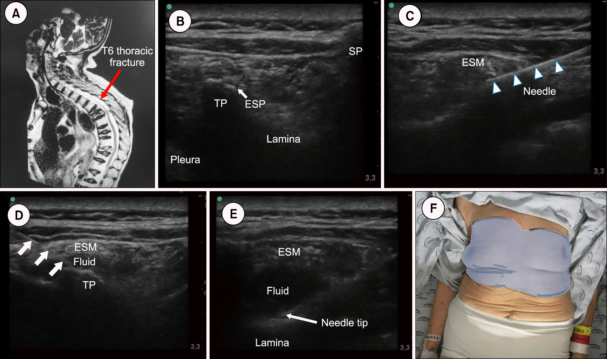 | Fig. 1(A) Thoracic spine magnetic resonance imaging showing acute/subacute T6 vertebral fracture. (B) Sonographic image for short axis erector spinae plane (ESP) block. (C) Needle pathway to target on the junction of the transverse process (TP) and the erector spinae muscle. (D) Fluid below the erector spinae muscle (ESM). (E) Needle tip at the retrolaminar level, showing fluid pushing the ESM. (F) Cold and pinprick response after the block. SP: spinous process.
|
A vertebral augmentation procedure was not offered due to patient preferences and her severe pulmonary condition. She was referred to our interventional pain management service for evaluation. Initially a medial branch block was proposed, but the patient could not assume a prone position, therefore a fluoroscopic guidance procedure was not possible. Taking in to account this problem, a bilateral ESP block and a retrolaminar block were scheduled. Using a highfrequency-linear transducer (Turbo M®; Sonosite, Bothell, WA) the thoracic spine around the lower-border of the scapula was scouted (Fig. 1) identifying the spinous process, lamina, and the transverse process. This was used as an anatomical landmark for the ESP block, because the muscle is above it. The patient was only able to be in a tilt-position, making the previously described ESP block in the long axis difficult to perform. Therefore a short-axis approach was chosen. Using a conventional a septic preparation with 2%-clorhexidine-70% alcohol, the skin was disinfected and local anesthesia was applied to decrease pain during the puncture. Using an in-plane technique with an echogenic needle (Sonoplex®; Pajunk, Geisingen, Germany) the tip of the transverse process was reached, while the ESP was properly identified using hydrolocation with saline. Using 15 mL of 0.375% levo-bupivacaine plus 10 mg of triamcinolone were administered after judicious aspiration (Fig. 1). Later 5 mL of the same mixture was delivered at the retrolaminar level with a good displacement of the erector spinae muscle (Fig. 1). The procedure was performed bilaterally. The patient experienced a T3 to T8 block, confirmed by a decreased response to cold and pinprick. Her VAS score decreased to 0/10 during the first 24 hours and 2/10, 2/10, 4/10, and 5/10 for the second, third, fourth, and fifth day, respectively. She was discharged from Clinica Las Americas with a 4.3 μg/hr buprenorphine patch and acetaminophen 500 mg tid. She was followed for 6 weeks with an 80% positive global perceived effect.
This case illustrate the potential role of the ESP block and the retrolaminar block in patients with severe pain associated with vertebral fractures who are not candidates for medial branch blocks or VAT. We combined both procedures to impact the posterior elements, the intercostal elements, and the foraminal structures.
To our knowledge there are no reports of the ESP block or retrolaminar blocks being used for pain relief in vertebral fractures patients. Further studies/case series are needed to address each block separately in VCF patients.
Go to : 
 XML Download