

 PDF
PDF Citation
 Print
Print


INTRODUCTION
Discogenic pain is a common cause of disability and is assumed to be a major cause of non-specific low back pain. Among patients with low back pain, 39% had an internal disc disruption, with concordant pain provocation at discography indicating the discogenic origin of their pain [1].
Discogenic pain arises from granulation tissue with nerve growth deep into the annulus fibrosus and nucleus pulposus, combined with neovascularization in the posterior part of the painful disc [2–4]. Growth factors, including basic fibroblast growth factor and transforming growth factor-beta 1, macrophages, and mast cells may also play a key role in the repair of the injured annulus fibrosus and subsequent disc degeneration [5].
Discogenic pain can usually be successfully treated by non-surgical interventions such as pain medication, physical therapy, and exercise. However, severe, chronic discogenic pain that causes functional limitations may require invasive treatments including intradiscal steroid injection, intradiscal electrothermal annuloplasty, percutaneous radiofrequency ablation of the ramus communicans, nucleoplasty, or surgery [6–11]. Several authors have reported the efficacy of endoscopic lumbar annuloplasty and nucleoplasty for treatment of patients with discogenic pain [12–14].
Painful granulation tissue can be manually removed by using small forceps, or eliminated by laser or radiofrequency ablation. Intradiscal radiofrequency annuloplasty (IDRA) combines percutaneous manual discectomy with nuclear ablation and annular modification using radiofrequency equipment. Two previous articles have reported the efficacy of IDRA for contained lumbar disc herniation [15,16]. Tansforaminal laser annuloplasty (TFLA) is another recent development in annuloplasty that can be used both intradiscally and extradiscally.
To our knowledge, there are no published comparisons of IDRA and TFLA in the treatment of discogenic low back pain. This study was conducted to compare the therapeutic success of IDRA and TFLA.
Go to : 
MATERIALS AND METHODS
Our Institutional Review Board approved the study protocol (IRB No.: WSH 18-02-11) and all patients provided written informed consent. The patients were assigned to the TFLA or IDRA group depending on the type of procedure they had received. The procedure was decided by the surgeon’s preference. One surgeon performed TFLA and another surgeon performed IDRA.
Inclusion criteria were 1) axial low back pain, 2) single level, 3) intolerance to sitting, 4) clear evidence of an annular tear appearing as a high intensity zone on spinal magnetic resonance imaging (MRI), 5) positive pain provocation on discography, and 6) back pain that had not responded to conservative treatments (pharmacotherapy and physical therapy) within the previous 4 to 6 weeks. Exclusion criteria were 1) low back pain elicited by pressure on paraspinal muscles, 2) herniated intervertebral disc, 3) spinal stenosis, 4) spondylolisthesis, 5) pain due to infection, 6) bleeding tendency, and 7) previous back surgery.
Data collected included patient age, gender, and duration of pain. The main outcome measures included pain score, as determined by the numeric rating scale (NRS), and Oswestry disability index (ODI) at baseline and at 1- and 6-month follow-up visits. Success was defined as reduction in the NRS scores of ≥ 50% or an ODI reduction of ≥ 40%.
1. TFLA procedure
TFLA was performed using the transforaminal epiduroscopic laser annuloplasty system (TELA®, Lutronics Corp., Goyang, Korea) with patients under local anesthesia. The point of entry was determined prior to the procedure by measuring from the midline (usually 12 to 14 cm) on MRI.
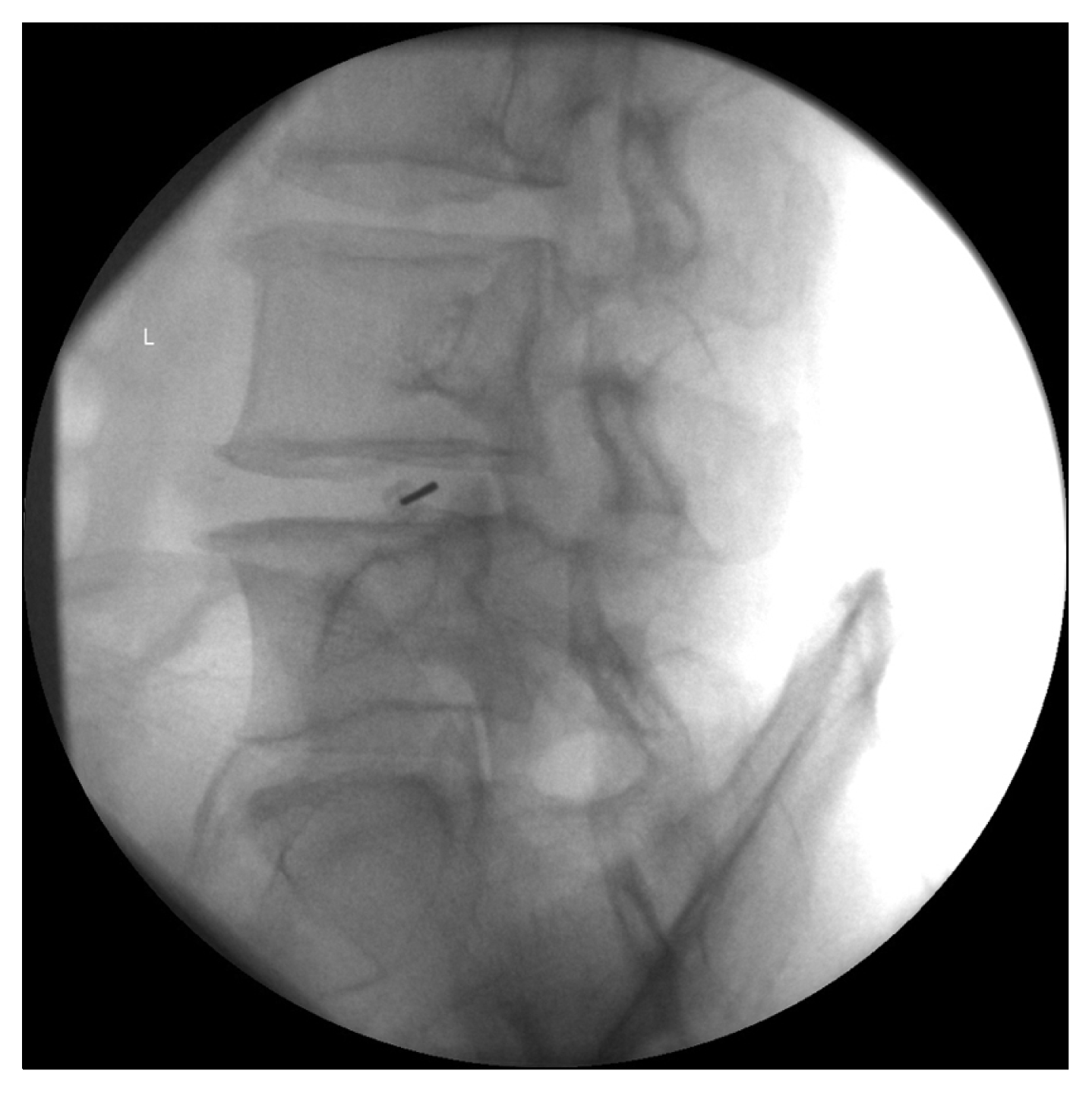
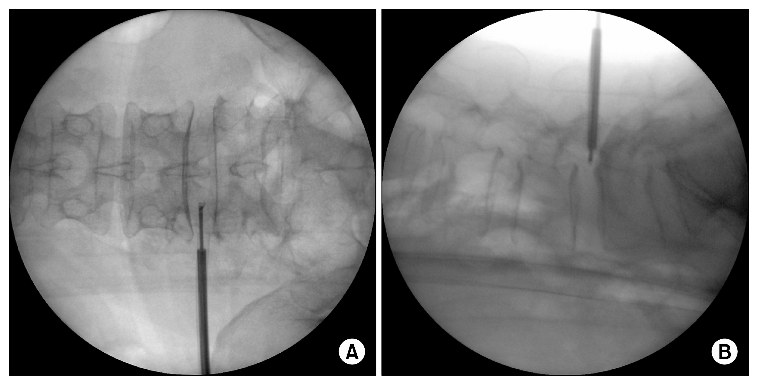
A conventional posterolateral approach on the side of the pathology was used. The patient was placed in a prone position and the presumptive skin entry and needle track were infiltrated with 1% lidocaine. A 15-gauge spinal needle was inserted just lateral to the superior articular pillar (SAP) toward the target level (Fig. 1). The needle was advanced under fluoroscopic guidance until it contacted the SAP and was then carefully advanced along the SAP toward the ventral epidural space. After needle placement in the anterior epidural space was confirmed on the anteroposterior and lateral radiographic views (Fig. 2), the guide wire was inserted and a small incision (0.7 cm) was made in the overlying skin. A cannula and a soft tissue dilator were then advanced over the guide wire into the ventral epidural space under fluoroscopic monitoring.

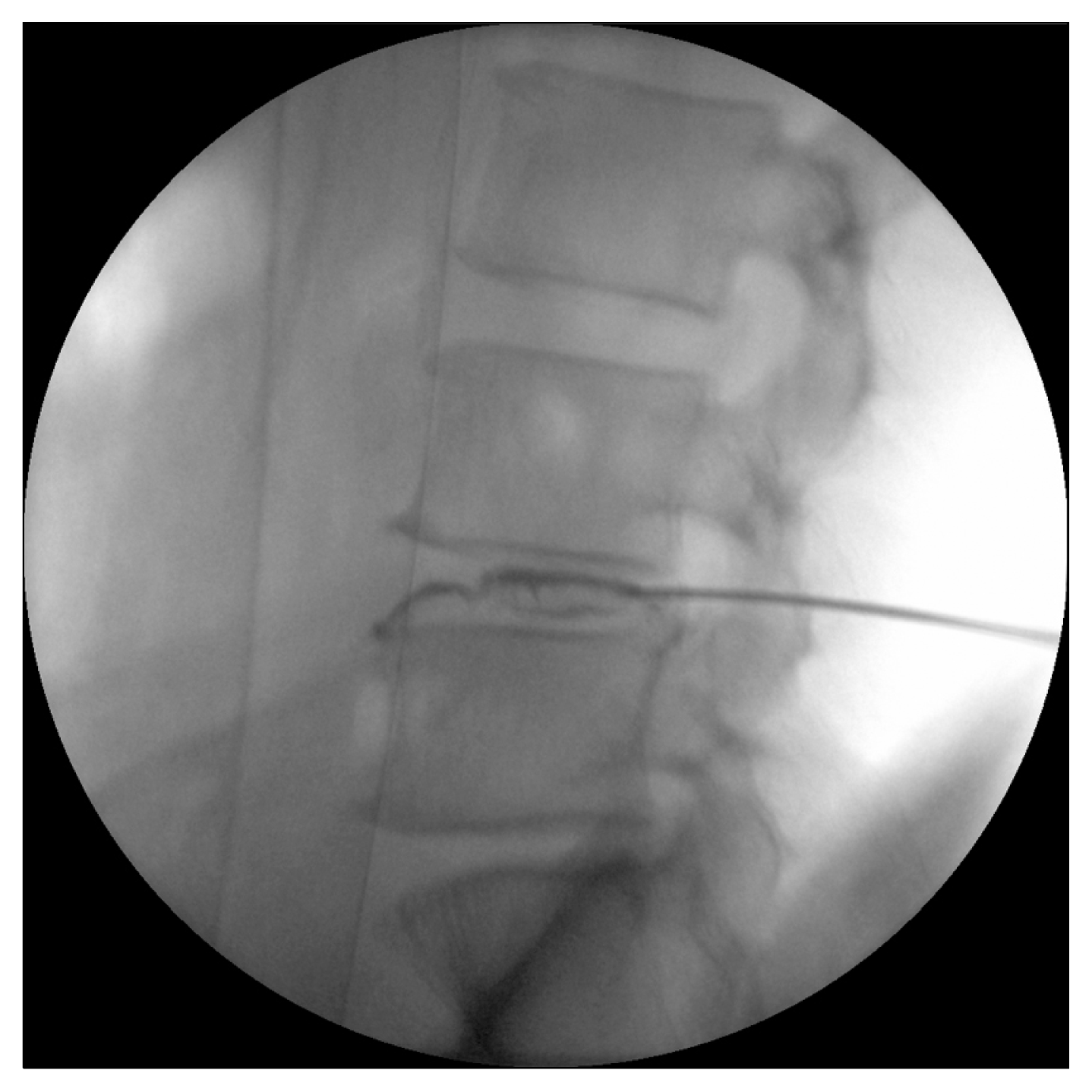
After cannula placement, the epiduroscope (NeedleView CH scope®, Lutronics; outer diameter 3.4 mm) was introduced, and placement in the ventral epidural space near the targeted disc was confirmed by lateral C-arm fluoroscopic view. Epidural fat and transforaminal ligaments were removed via forceps to ensure anatomic visualization, adhesiolysis was performed using the laser, and the annulus was then punctured at the annular foraminal zone with a sharpened wire. Forceps removal and laser ablation of the interposed granulation and nucleus pulposus was performed through the annular puncture (Nd:YAG laser with side-firing fiber side, set at 2.5 to 10 W) (Fig. 3). The epiduroscope was angled and the epidural space was dissected beyond the midline to the contralateral annulus followed by extradiscal laser annuloplasty (Nd:YAG laser with side-firing fiber side, set at 2.5 to 10 W). The operative site was irrigated via the cannula with antibiotic solution and normal saline during all procedures. Patients were observed postoperatively for neurologic deficits or other procedure-related problems, and were typically discharged on the same day or within 24 hours.
2. IDRA procedure
All IDRA procedures were performed in the operating room using the Disc-FX® system (Elliquence, Baldwin, NY) with the patients under local anesthesia in a prone position. Before the procedure, the needle tip was positioned at a point on the medial-to-lateral pedicular line on the anteroposterior fluoroscopic projection, and at the posterior vertebral line on the lateral projection.
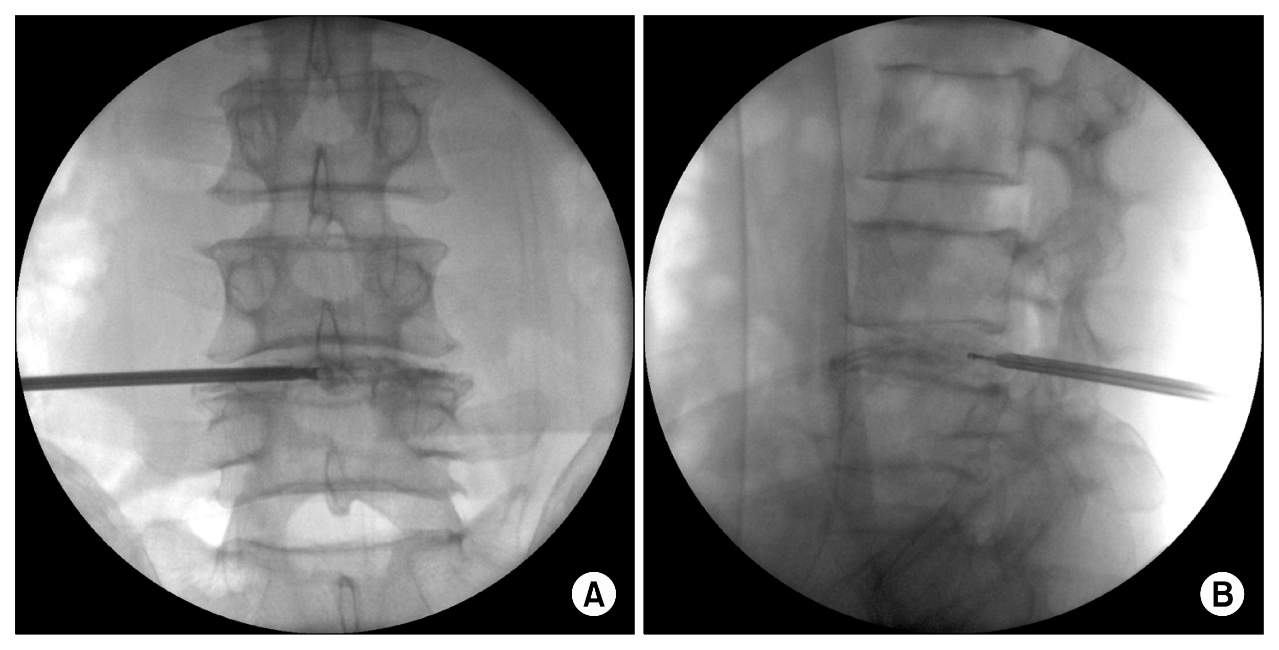
The skin entry point was measured from the midline using MRI. The skin entry point and needle track were infiltrated with 1% lidocaine and a 15-guage spinal needle was inserted at the previously identified entry point and directed lateral to the SAP toward the target. The needle was advanced into the nucleus pulposus and placement was confirmed on anteroposterior and lateral radiographic views, after which a discogram was obtained (Fig. 4).
After removal of the stylet, a guide wire was inserted through the spinal needle and a 0.7 cm longitudinal skin incision was made over the guidewire to allow insertion of the cannula and soft tissue dilator, which were advanced over the guidewire and into the annulus under continuous fluoroscopic monitoring.
3. Statistical analysis
Independent t-test, chi-square, analysis of variance, and likelihood ratio test were used to analyze the data, equating statistical significance with type I error rates of < 0.05. All computations were performed with standard software (IBM SPSS ver. 23, IBM Corp., Armonk, NY).
Go to :
RESULTS
The retrospective study included 80 patients who were grouped according to procedure (TFLA = 37 vs. IDRA = 43 patients). A summary of patient characteristics is provided in Table 1.
In all procedures, the NRS and ODI were significantly decreased over time (Tables 2, 3). Mean post-treatment pain scores at months 1 and 6 were significantly lower (P < 0.01) in both groups, and between-group differences were not significant (Table 2). ODI scores were also significantly decreased compared with baseline, but between-group differences were not significant (Table 3). At 6 months post-procedure, 26 patients (70.3%) in the TFLA group and 25 patients (58.1%) in the IDRA group reported > 50% reduction in pain score (P = 0.19), and there was no significant difference between groups in the number of patients reporting > 40% reduction in ODI (P = 0.28) (Table 4). In the trend test, there was no difference between both treatments (Table 5).
Table 2
Changes of Numeric Rating Score Scale According to Procedures
| Procedure | Pre-treatment | Post-treatment (mo) | P value* | |
|---|---|---|---|---|
|
|
||||
| 1 | 6 | |||
| TFLA (n = 37) | 7.6 ± 0.8 | 4.0 ± 1.6 | 3.5 ± 1.3 | < 0.01 |
| IDRA (n = 43) | 7.3 ± 0.8 | 3.7 ± 1.8 | 3.6 ± 1.8 | < 0.01 |
| P value | 0.104 | 0.473 | 0.654 | |
![]()
Table 3
Changes of Oswestry Disability Index According to Procedures
| Procedure | Pre-treatment | Post-treatment (mo) | P value* | |
|---|---|---|---|---|
|
|
||||
| 1 | 6 | |||
| TFLA (n = 37) | 57.1 ± 5.6 | 24.5 ± 10.0 | 23.8 ± 9.9 | < 0.01 |
| IDRA (n = 43) | 57.2 ± 10.0 | 25.2 ± 9.6 | 22.1 ± 8.4 | < 0.01 |
| P value | 0.964 | 0.706 | 0.432 | |
![]()
Table 4
The Number of Patients Who Obtained The Percentage Improvement of Pain at Post-treatment 6 Months
| Reduction | TFLA (n = 37) | IDRA (n = 43) | P value* |
|---|---|---|---|
| Numeric rating scale | 0.186 | ||
| < 50 | 11 (29.7) | 18 (41.9) | |
| ≥ 50 | 26 (70.3) | 25 (58.1) | |
| Oswestry disability index | 0.284 | ||
| < 40 | 5 (13.5) | 9 (20.9) | |
| ≥ 40 | 32 (86.5) | 34 (79.1) |
![]()
No serious complications, including epidural bleeding, dural or neural injuries, or infection, were recorded in either group.
Go to :
DISCUSSION
Outcomes of this study indicate that significant pain relief was achievable by either IDRA or TFLA. These comparable results corroborate previous data [15,17,18]. However, even if there was no statistical difference between the two groups, patients who improved more than 50% were more likely to have TFLA.
Nucleoplasty has proven effective in patients presenting with discogenic back pain [11], and results of a 1-year pilot study support the efficacy of intradiscal electrothermal annuloplasty as a minimally invasive treatment for chronic discogenic low back pain [19].
The focus of discogenic pain is the damaged posterior annulus fibrosus and the free nerve endings inside the annulus [3,20,21]. Nucleo-annuloplasty using the Disc-FX® system allows removal of granulation tissue with forceps and ablation or cauterization of annular tears using the steerable Trigger-Flex® probe. Radiothermal ablation techniques have proven effective for a variety of endoscopic spinal and neurosurgical procedures [22,23]. In this study, the IDRA procedures were performed under C-arm fluoroscopy, not endoscopically, and consequently there may have been less thermo-coagulation and indirect decompression.
In our study, in TFLA, more than 50% of patients improved. TFLA can be used to directly shrink and coagulate interposed granulation tissue associated with tears in the posterior annulus [24]. The laser can also be used to block the sensory nerves surrounding the annulus fibrosus both inside and outside the disc, and we hypothesized that TFLA would be more effective than IDRA for treatment of discogenic pain, because TFLA allows direct removal of granulation tissue within the disc and coagulation of the meningeal branches of the spinal nerve (sinuvertebral nerves) outside the disc. Although we did not detect a difference between the efficacy of the two procedures in this study, previous studies have reported that laser annuloplasty is effective for discogenic pain [13,14].
We used an Nd:YAG laser. The advantages of the Nd:YAG laser include easier control, a simpler procedure, and proven safety and efficacy under spinal epiduroscopic guidance [25]. TFLA involves a precisely targeted decompression in the annulus, without touching the central and anterior regions of the disc, and while preserving the healthy nucleus as much as possible. The surgeon can apply the laser energy directly to the lesion under the endoscopic view via intraoperative fluoroscopy.
One limitation of our study is that the significant improvements in pain were not corroborated by any secondary outcomes. A second is that the follow-up period was only 6 months, so we do not have mid- or long-term follow-up results. A third limitation is that there was no post-procedure MRI. In addition, our study was retrospective.
In conclusion, annuloplasty is a reasonable treatment option for carefully selected patients with lower back and radicular pain of discogenic origin, and that TFLA (both intradiscal and extradiscal) might be superior to intradiscal procedures in patients with discogenic low back pain.
Go to :
 XML Download