Journal List > Korean J Pain > v.23(2) > 1159176

Lee, Kim, Lee, Shin, Lee, Park, Choi, Kim, and Park: The Neurological Safety of Epidural Pamidronate in Rats

Abstract

Background

Pamidronate is a potent inhibitor of osteoclast-mediated bone resorption. Recently, the drug has been known to relieve bone pain. We hypothesized that direct epidural administration of pamidronate could have various advantages over oral administration with respect to dosage, side effects, and efficacy. Therefore, we evaluated the neuronal safety of epidurally-administered pamidronate.

Methods

Twenty-seven rats weighing 250-350 g were equally divided into 3 groups. Each group received an epidural administration with either 0.3 ml (3.75 mg) of pamidronate (group P), 0.3 ml of 40% alcohol (group A), or 0.3 ml of normal saline (group N). A Pinch-toe test, motor function evaluation, and histopathologic examination of the spinal cord to detect conditions such as chromatolysis, meningeal inflammation, and neuritis, were performed on the 2nd, 7th, and 21st day following administration of each drug.

Results

All rats in group A showed an abnormal response to the pinch-toe test and decreased motor function during the entire evaluation period. Abnormal histopathologic findings, including neuritis and meningeal inflammation were observed only in group A rats. Rats in group P, with the exception of 1, and group N showed no significant sensory/motor dysfunction over a 3-week observation period. No histopathologic changes were observed in groups P and N.

Conclusions

Direct epidural injection of pamidronate (about 12.5 mg/kg) showed no neurotoxic evidence in terms of sensory/motor function evaluation and histopathologic examination.

INTRODUCTION

Bisphosphonates are pyrophosphate derivatives that bind to the inorganic matrix of bone, and thereby strongly inhibit bone resorption [1,2]. This class of drugs was previously considered for treatment of osteoporosis for conditions in which hormone replacement therapy is restricted. In recent years, however, it has been reported that bisphosphonates have analgesic properties for non-bone-related pains in various preclinical animal models [3-8]. Bonabello et al. [3] showed both a peripherally and centrally mediated analgesic action of 4 bisphosphonates. They used intravenous and intracerebroventricular injection for observing the peripheral and central effect of bisphosphonates and both showed a dose-dependent antinociception. In addition, some biological effects of bisphosphonates are mediated through CNS dendritic cells and microglia [9]. Based on these, the epidural administration of bisphosphonates can be an alternative for the treatment of non-bone-related pain. Currently, bisphosphonates are used clinically by oral administration or intravenous injection. The major disadvantage of orally administrated bisphosphonates is their poor absorption in the gastrointestinal tract [9]. Thus, a relatively high dose of bisphosphonates must be given by intravenous infusion for a rapid and considerable effect. However, intravenous administration causes gastrointestinal disturbances, including nausea, vomiting, and diarrhea, as well as susceptibility to electrolyte imbalance, renal dysfunction, anemia, and hypertension. Moreover, This class of drugs is expensive, and requires hospitalization for long-term intravenous injection [1,2]. Neuroaxial block, such as epidural administration of the drug is expected to reduce dosage, as compared with an oral or intravenous infusion, thereby minimizing systemic adverse effects and maximizing treatment efficacy [10]. This type of administration method has been of great interest in the field of pain medicine. Because it is a non-invasive method, it can be administered long-term and the risk of neurotoxicity is lower.
Although the drug is used safely for systemic administration, it has been well established that animal experiments must be performed to assess efficacy and safety when the drug is administered to the subarachnoid or epidural space [10]. Especially, when the safety is confirmed, we can conduct the preclinical experiment for the efficacy of a drug. Thus, we selected pamidronate, which is one of the most commonly used bisphosphonates for intravenous infusion, for administration into the epidural space in rats. We then performed behavioral observations and histopathologic analysis to assess neurotoxicity.

MATERIALS AND METHODS

1. Study subjects

Following approval of the Institutional Review Board of Animal Experiments, male Sprague-Dawley (SD) rats weighing approximately 250-350 g were divided into group A (alcohol injection group), group P (pamidronate injection group) and group N (normal saline injection group). Each group was comprised of 9 rats, and a total of 27 rats were examined in the current study. All rats were bred following a 1-week adaptation period, with a 12-hr photo cycle. All rats showing abnormalities during behavioral observation were excluded from the current analysis.

2. Placement of a catheter in the epidural space

SD rats were anesthetized with inhaled oxygen (3 L/min) and 4% sevoflurane. Approximately 3 minutes later, 2-3% sevoflurane was administered with a mask, and thereby anesthesia was maintained. An epidural catheter was prepared by making a knot at 2.5 cm from the tip of a 17 cm micro-plastic catheter (PE-10; Natsume Co, Japan). An epidural puncture was then performed. That is, skin was incised at a length of 2-3 cm and the area adjacent to the supraspinous ligament was dissected. Between the 13th thoracic spine and the first lumbar spine, the ligamentum flava was exposed with no damage. Using microsurgical scissors, a small hole was made in the ligamentum flava. A catheter was then inserted and progressed toward the tail at a distance of approximately 2.5 cm, toward the tip of a catheter placed between the 4/5 lumbar spine. Animals were excluded from the current experiment if blood or CSF was aspirated through the catheter. The remaining part of the catheter passed through the dermal layer and was then extracted in the junction between the cervical and thoracic spine. All scars were rinsed with saline. A catheter was passed through the center of a knot using a 4-0 silk suture, and tightly fixed to the adjacent tissues. Using 1-2 drops of surgical glue (alpha cyanoacrylate; Aron-Alpha, Toagosei, Japan), the puncture site was sealed. Through a catheter, 2% lidocaine at 0.15 ml was gradually injected. Anterior legs showed normal movement, but posterior legs did not move temporarily. If rats died of respiratory distress when given lidocaine, it was considered subarachnoid or intravascular injection. Following recovery from anesthesia, ambulatory posture and vertebral anomaly were examined. Rats showing signs of motor nerve damage, were excluded from the current experiment. Each rat was then isolated and raised in stabilized conditions [11,12].

3. Drug infusion to the epidural space

Three days following insertion of the catheter into the epidural space, ambulatory posture, vertebral deformity, and abnormal behavior were examined. Further laboratory procedures progressed in rats with no abnormal findings. Under general anesthesia, animals in group P were injected with pamidronate (Panorin®, pamidronate disodium 15 mg/1 ml Ampule, Hanlim, Seoul, Korea) 3.75 mg, dissolved in saline. In group A, 40% alcohol was injected, and in group N, saline was gradually injected at a dose of 0.3 ml for approximately 1 minute, except for the volume of a catheter. All drugs were newly prepared prior to injection and managed using an aseptic method. Upon recovery from anesthesia, rats were isolated to be safely bred at a 12-hr interval during day and night.

4. Assessment of sensory and motor nerve disturbance through behavioral observation

Acute and chronic toxicity were assessed 2, 7, and 21 days after injection. Rats were evaluated for sensory and motor nerve disturbance and abnormal behavior at the same time each day by 1 investigator blinded to the experimental procedure.

1) Pinch-toe test:

A hind paw was pinched using a forceps (01-1155, Solco, Seoul, Korea). Then, using a pinch toe test, motor and sensory nerve damage was assessed [13-15]. When the hind paw was pinched by a forceps to such an extent that resistance of bone could be perceived (deep pinch), observed normal avoidance responses were as follows:
  1. Avoidance of lower extremities

  2. Crying

  3. Attempt by the animal to bite the forceps

In the lower limbs on both sides, if all 3 of the above categories were observed at a minimum 5-minute interval up to 3 times during a maximum of 6 seconds (cut off time), the case was determined to be normal. Otherwise, corresponding cases were determined to have an abnormal response that indicated the possibility of motor and sensory nerve damage [13].

2) An assessment based on ambulatory pattern and lower limb deformity:

To examine motor nerve disorders, with the application of motor function based on ambulatory pattern and the deformity of lower extremities [16], all rats were divided into the following grades. Grade 1 = normal gait with no evidence of motor paresis; Grade 2 = normal gait with slight hindpaw deformity, plantar flexion of toes; Grade 3 = slight gait disturbance with motor weakness and/or an inverted hindpaw; and Grade 4 = prominent limping gait with dropped hindpaw. Rats corresponding to > Grade 2 were all considered to have motor nerve injury.

5. Histopathologic examination

Tissue samples were collected from the spinal cord on day 2 (3 rats/group) for assessment of acute toxicity, and on day 7 (3 rats/group) and 21 (3 rats/group) for chronic toxicity. Tissue preparation was performed using the same method, for which 4% paraformaldehyde in 0.1 M phosphate buffer was injected into the heart. Euthanasia was thus induced and the cadaver was fixated. In the center of the area where the tip of a catheter was placed, approximately 3 segments of the spinal cord, both superiorly and inferiorly, including the nerve root, were removed. Tissue samples were fixated in a 10% formalin solution for 3 days and then decalcified using 10% formic acid for 2 weeks. Tissue specimens were rinsed with a buffer solution (pH 7.4) and then fixated using 2% osmium tetroxide, which was dissolved in a buffer solution for 30 minutes. Dehydration was performed using a graded ethanol, followed by an embedding of the tissue sample using epoxy resin (agar 100) between teflon-sprayed slides and thick acetate foil with a thickness of 100 µm. Tissue samples containing the ventral and dorsal horns of the spinal cord were dissected and then re-embedded for fine sectioning. Tissue samples were then stained using hematoxylin-eosin and Luxol fast blue dyes for light microscopy.
Histopathological findings for assessment of neurotoxicity include chromatolysis in the motor neurons of the ventral horn of the spinal cord, neuritis, meningeal inflammation, adhesion in periosteum, dura mater, and medulla, enlargement of dura mater, peripheral neuropathy, myelin loss, and local infarction. To rule out bias, histopathologic change was analyzed by 1 pathologist blinded to the study.

6. Statistical analysis

Assessment of motor and sensory nerve disorders and histopathologic examination of the spinal cord were performed and their results were tested using the Chi-square test between the 3 groups. Any cases with a significant difference were analyzed using Fisher's exact test between the 2 groups, which was followed by Bonferroni correction (SigmaStat ver. 2.0, Jandel corporation). A value of P < 0.05 was considered statistically significant.

RESULTS

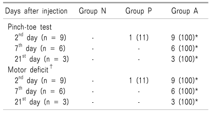
In the P group, with the exception of 1 rat, and in the N group, no rats showed motor or sensory change, or any behavioral change such as aggressive behavior or crying. Normal reactions to the pinch-toe test included avoidance, crying, and attempts to bite the forceps. All rats in group N and all rats, except 1, in group P had a normal gait at the time point for all examinations (Table 1). During the overall period, however, most rats in Group A showed decreased activity and appetite. In group A, all rats responded with some of the 3 aforementioned reactions, or had no reaction to the pinch-toe test, and had a gait disturbance of Grade 3 or more (P < 0.05, Table 1).
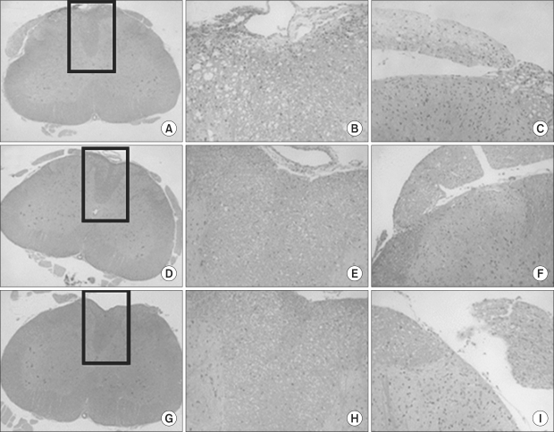
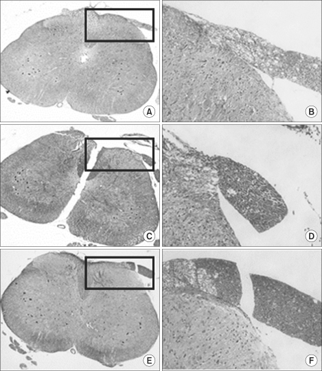
No significantly abnormal histopathologic findings were observed in the P and N groups. However, in group A, various histopathological forms of neurological deficit, such as local neuritis, myelin loss, and meningeal inflammation occurred at each time point of drug administration (P < 0.05, Table 2, Fig. 1, 2).

DISCUSSION

Bisphosphonates show specific, powerful binding to the inorganic matrix of bone, and thereby suppress the maturation and activity of osteoclasts, ultimately causing apoptosis of osteoclasts [17,18]. One of the bisphosphonates, pamidronate, strongly inhibits bone resorption [1,2]. It is a drug that treats hypercalcemia, osteolytic bone metastasis, and Paget's disease due to malignant tumors by inhibiting bone resorption by osteoclasts.
Pamidronate has been reported to have excellent efficacy in the treatment of chronic lower back pain due to osteoporotic fracture, alleviating pain due to bone metastasis of breast cancer, lowering severe bone pain in Paget's disease, improving the pain of cancer metastasis, and controlling the pain in complex regional pain syndrome [1,19-22].
Bonabello et al. [3] reported that the antinociceptive effects of bisphosphonates occurred with no respect to peripheral opioid receptors in both the central and peripheral nervous systems, with no association in bone lesions. It has also been reported that bisphosphonates modify such substances as prostaglandin E2, sensitizing anti - inerleukine (IL)-1, anti - IL - 6, and anti-tumor necrosis factor - alpha (TNF - alpha) effects, as well as the traumatic pain receptor, and thereby have an analgesic effect, in addition to inhibiting osteolclastic activity [23]. Another study reported that the afferent nerve terminal suppresses the release of various neuropeptides following the onset of trauma in patients with complex regional pain syndrome [22]. In addition, such effects as inhibition of protein prenylation, inhibition of neoangiogenesis, and immune modulation have an influence on the alleviation of pain [24].
Currently, pamidronate is administered via oral route or intravenous infusion. It can be administered up to a maximal dose of 300 mg/d for oral administration and 90 mg/d for intravenous infusion [24]. However, there are a few limitations to administration via the oral route, and disadvantages to the oral administration of bisphosphonate are as follows: 1) a lower rate of intestinal absorption (1-10%) [25]; 2) affection by food ingestion; 3) obligation to administer it during the fasting state; and 4) gastrointestinal disturbances, such as nausea or abdominal pain. Due to these disadvantages, patient compliance has been reported to be lower [25], and injection methods are favored. Intravenous injection has many advantages in terms of cost-effectiveness and patient compliance. A persistent intravenous infusion conversely induces osteochondrosis, and also produces such adverse effects as phlebitis, febrile sensation, chilling sensation, myalgia, malaise, thrombophlebitis, and hypophosphatemia [24,25]. In the early stage of administration, adverse effects, such as increased bone pain, transient leukocytopenia [26], and necrosis of the jaw bone [27,28], used to evolve. In addition, drugs are expensive and hospitalization is needed for an intravenous infusion, which can also be problematic.
From these findings, we considered the epidural treatment of bisphosphonate as an alternative for non-bone-related pain. An epidural approach is currently used most prevalently to control pain, and it is more effective than other administration routes in cases in which the spinal cord is the target of drug administration [29]. Even a minimal dose can produce the same effect, and it is considered that systemic adverse effects and risk of neurotoxicity can be reduced. It is also advantageous in that inhibition of the necessary spinal level can be selective. Pamidronate is also expected to have an excellent effectiveness when directly administered to the area adjacent to the spinal cord. However, administration of any drug into the spinal cord or an epidural space should be evaluated for potential occurrence of neurotoxicity in the spinal cord [10,29]. Although the addictives that can cause neurotoxicity with higher probability, such as anti-oxidants, anti-disinfectants, or excipients, could be ruled out, direct administration of drugs to the area adjacent to the spinal cord can cause direct contact between a high-dose of drugs to the nerve when compared with other administration routes. Therefore, neurological safety must first be demonstrated.
The usual dose of pamidronate recommended for pain control is 15-60 mg/d in humans. In the current study, the dose was 50 times higher than the usual dose for an intravenous infusion based on the weight of the rats, which corresponds to 750 mg in humans. Because 1/30 of the dose for oral administration is administered via epidural route, it is assumed that 22,500 mg is sufficient for a toxicity trial if it is converted to the dosage for oral administration.
Determination of drug volume has been known to correspond with an epidural infusion of 10-15 ml in humans and 0.1 ml in SD rats. In SD rats weighing 250-350 g, a contrast medium of 0.3 ml in volume is distributed across 10-11 segments of the spinal cord. It can therefore be predicted that most drugs with a viscosity lower than that of the contrast medium would be distributed in more extensive areas. It can therefore be inferred that the dosage is sufficient for examination of sensory and motor change in the lower limbs, including the 3-6th lumbar nerve root [30].
Although statistically significant neurotoxicity findings or ambulatory disorder were not observed on light microscopy, an ambulatory disorder was observed in a rat in the P group on day 2 (Grade 4). Because no special abnormal findings were observed on light microscopy, it can therefore be inferred that physical injury caused by catheter use, migration of the catheter into the subarachnoid space, or intravascular migration of the catheter would be a factor responsible for the presence of abnormal findings, rather than drug-induced basic changes in nerves.
In conclusion, epidural administration of pamidronate 12.5 mg/kg did not produce significant neurotoxic adverse effects on behavioral observation and histopathologic examination in SD rats. Further studies in other animal species are warranted to examine the clinical effects of an epidural infusion of pamidronate that could lead to clinical application.

ACKNOWLEDGEMENTS

This work was supported by the research grant of the Jeju National University in 2007.

References

1. Gangji V, Appelboom T. Analgesic effect of intravenous pamidronate on chronic back pain due to osteoporotic vertebral fractures. Clin Rheumatol. 1999; 18:266–267. PMID: 11206358.
crossref
2. Reid IR. Bisphosphonates: new indications and methods of administration. Curr Opin Rheumatol. 2003; 15:458–463. PMID: 12819475.
crossref
3. Bonabello A, Galmozzi MR, Bruzzese T, Zara GP. Analgesic effect of bisphosphonates in mice. Pain. 2001; 91:269–275. PMID: 11275384.
crossref
4. Bonabello A, Galmozzi MR, Canaparo R, Serpe L, Zara GP. Long-term analgesic effect of clodronate in rodents. Bone. 2003; 33:567–574. PMID: 14555260.
crossref
5. Cui JG, Holmin S, Mathiesen T, Meyerson BA, Linderoth B. Possible role of inflammatory mediators in tactile hypersensitivity in rat models of mononeuropathy. Pain. 2000; 88:239–248. PMID: 11068111.
crossref
6. Kawabata A, Kawao N, Hironaka Y, Ishiki T, Matsunami M, Sekiguchi F. Antiallodynic effect of etidronate, a bisphosphonate, in rats with adjuvant-induced arthritis: involvement of ATP-sensitive K+ channels. Neuropharmacology. 2006; 51:182–190. PMID: 16678221.
crossref
7. Sevcik MA, Luger NM, Mach DB, Sabino MA, Peters CM, Ghilardi JR, et al. Bone cancer pain: the effects of the bisphosphonate alendronate on pain, skeletal remodeling, tumor growth and tumor necrosis. Pain. 2004; 111:169–180. PMID: 15327821.
crossref
8. Walker K, Medhurst SJ, Kidd BL, Glatt M, Bowes M, Patel S, et al. Disease modifying and anti-nociceptive effects of the bisphosphonate, zoledronic acid in a model of bone cancer pain. Pain. 2002; 100:219–229. PMID: 12467993.
crossref
9. Yanow J, Pappagallo M, Pillai L. Complex regional pain syndrome (CRPS/RSD) and neuropathic pain: role of intravenous bisphosphonates as analgesics. ScientificWorld-Journal. 2008; 8:229–236. PMID: 18661047.
crossref
10. Hassenbusch SJ, Portenoy RK, Cousins M, Buchser E, Deer TR, Du Pen SL, et al. Polyanalgesic Consensus Conference 2003: an update on the management of pain by intraspinal drug delivery-- report of an expert panel. J Pain Symptom Manage. 2004; 27:540–563. PMID: 15165652.
crossref
11. Hayashi N, Weinstein JN, Meller ST, Lee HM, Spratt KF, Gebhart GF. The effect of epidural injection of betamethasone or bupivacaine in a rat model of lumbar radiculopathy. Spine. 1998; 23:877–885. PMID: 9580954.
crossref
12. Kawakami M, Matsumoto T, Hashizume H, Kuribayashi K, Tamaki T. Epidural injection of cyclooxygenase-2 inhibitor attenuates pain-related behavior following application of nucleus pulposus to the nerve root in the rat. J Orthop Res. 2002; 20:376–381. PMID: 11918320.
crossref
13. Bajrović F, Sketelj J. Extent of nociceptive dermatomes in adult rats is not primarily maintained by axonal competition. Exp Neurol. 1998; 150:115–121. PMID: 9514823.
crossref
14. Ochi T, Ohkubo Y, Mutoh S. FR143166 attenuates spinal pain transmission through activation of the serotonergic system. Eur J Pharmacol. 2002; 452:319–324. PMID: 12359273.
crossref
15. Sinnott CJ, Cogswell LP III, Johnson A, Strichartz GR. On the mechanism by which epinephrine potentiates lidocaine's peripheral nerve block. Anesthesiology. 2003; 98:181–188. PMID: 12502995.
crossref
16. Chatani K, Kawakami M, Weinstein JN, Meller ST, Gebhart GF. Characterization of thermal hyperalgesia, c-fos expression, and alterations in neuropeptides after mechanical irritation of the dorsal root ganglion. Spine. 1995; 20:277–289. PMID: 7537391.
crossref
17. Benford HL, McGowan NW, Helfrich MH, Nuttall ME, Rogers MJ. Visualization of bisphosphonate-induced caspase-3 activity in apoptotic osteoclasts in vitro. Bone. 2001; 28:465–473. PMID: 11344045.
crossref
18. Body JJ, Diel IJ, Lichinitser MR, Kreuser ED, Dornoff W, Gorbunova VA, et al. Intravenous ibandronate reduces the incidence of skeletal complications in patients with breast cancer and bone metastases. Ann Oncol. 2003; 14:1399–1405. PMID: 12954579.
crossref
19. Conte PF, Latreille J, Mauriac L, Calabresi F, Santos R, Campos D, et al. Delay in progression of bone metastases in breast cancer patients treated with intravenous pamidronate: results from a multinational randomized controlled trial. The Aredia Multinational Cooperative Group. J Clin Oncol. 1996; 14:2552–2559. PMID: 8823335.
crossref
20. Diener KM. Bisphosphonates for controlling pain from metastatic bone disease. Am J Health Syst Pharm. 1996; 53:1917–1927. PMID: 8862204.
crossref
21. Kubalek I, Fain O, Paries J, Kettaneh A, Thomas M. Treatment of reflex sympathetic dystrophy with pamidronate: 29 cases. Rheumatology (Oxford). 2001; 40:1394–1397. PMID: 11752511.
crossref
22. Robinson JN, Sandom J, Chapman PT. Efficacy of pamidronate in complex regional pain syndrome type I. Pain Med. 2004; 5:276–280. PMID: 15367305.
crossref
23. Abildgaard N, Glerup H, Rungby J, Bendix-Hansen K, Kassem M, Brixen K, et al. Biochemical markers of bone metabolism reflect osteoclastic and osteoblastic activity in multiple myeloma. Eur J Haematol. 2000; 64:121–129. PMID: 10997332.
crossref
24. Gallacher SJ, Ralston SH, Patel U, Boyle IT. Side-effects of pamidronate. Lancet. 1989; 2:42–43. PMID: 2567810.
crossref
25. Dodwell DJ, Howell A, Ford J. Reduction in calcium excretion in women with breast cancer and bone metastases using the oral bisphosphonate pamidronate. Br J Cancer. 1990; 61:123–125. PMID: 2297483.
crossref
26. Morton AR, Howell A. Bisphosphonates and bone metastases. Br J Cancer. 1988; 58:556–557. PMID: 3064797.
crossref
27. Harinck HI, Papapoulos SE, Blanksma HJ, Moolenaar AJ, Vermeij P, Bijvoet OL. Paget's disease of bone: early and late responses to three different modes of treatment with aminohydroxypropylidene bisphosphonate (APD). Br Med J (Clin Res Ed). 1987; 295:1301–1305.
crossref
28. Ralston SH, Gardner MD, Dryburgh FJ, Jenkins AS, Cowan RA, Boyle IT. Comparison of aminohydroxypropylidene diphosphonate, mithramycin, and corticosteroids/calcitonin in treatment of cancer-associated hypercalcaemia. Lancet. 1985; 2:907–910. PMID: 2865417.
29. Yaksh TL, Collins JG. Studies in animals should precede human use of spinally administered drugs. Anesthesiology. 1989; 70:4–6. PMID: 2912314.
crossref
30. Kim YC, Lim YJ, Lee SC. Spreading pattern of epidurally administered contrast medium in rabbits. Acta Anaesthesiol Scand. 1998; 42:1092–1095. PMID: 9809094.
crossref
Fig. 1
Microscopic findings in spinal cord and nerve on the 2nd day (A, D, and G), 7th day (B, E, and H), and 21st days (C, F, and I) following epidural injection of alcohol (A-C), pamidronate (D-F), and normal saline (G-I). Hematoxylin and eosin stain. Figures in middle and right columns (×200) show the high power view of the adjacent left side column (×40) (A, D, and G). In the epidural alcohol group, marked vaculolar change of posterior funicules (B) and lymphocytic infiltration in the spinal cord (C) are visible. In the epidurally-injected pamidronate and normal saline groups, no vacuolation or inflammation are visible.
kjp-23-116-g001
Fig. 2
Microscopic findings in spinal cord and nerve following epidural injection of alcohol (A, B), pamidronate (C, D), and normal saline (E, F). Luxol fast blue stain. Right-side figures (×200) (B, D, and F) show the high power view of the adjacent left side column (×40) (A, C, and E). In the epidural alcohol group, pale and diminished myelin is visible, and presented as a purple color (B). In the epidurally-injected pamidronate or normal saline groups, no definite abnormal morphologic findings of myelin are visible.
kjp-23-116-g002
Table 1
Evaluation of Pinch-Toe Test and Motor Deficit Following Epidural Drug Injection
kjp-23-116-i001

Values are expressed as number (%) of abnormal rats. *P < 0.05 versus corresponding data of Groups N and P. Group P: 0.3 ml (3.75 mg) of epidural pamidronate and Groups A and N: the same volume of epidural alcohol and normal saline, respectively. Grade 1: normal gait with no evidence of motor paresis, Grade 2: normal gait with slight hind paw deformity, such as plantar flexion of toes, Grade 3: slight gait disturbance with motor weakness and/or an inverted hindpaw, Grade 4: prominent limping gait with a dropped hindpaw. Rats with a motor disturbance of grade 2 or above were considered to have a motor deficit.

Table 2
Neuropathologic Findings of Spinal Cordand Nerve Under Light Microscopic Examination Following Epidural Drug Injection
kjp-23-116-i002

Values are expressed as number of positive animals. Group P: 0.3 ml (3.75 mg) of epidural pamidronate and Groups A and N: the same volume of epidural alcohol and normal saline, respectively. The 2nd, 7th, and 21st: days after epidural injection of test drugs. *P < 0.05 versus corresponding data of Groups N and P.

TOOLS
Similar articles