

 PDF
PDF Citation
 Print
Print


INTRODUCTION
Pain can be pathophysiological classified into the categories: nociceptive, neuropathic, and “other pain” (that not involve apparent damage, like fibromyalgia) [1]. The spared nerve injury (SNI), used to model neuropathic pain, results in injury and inflammation of a peripheral nerve model [2]. Intraarticular injection of 3% carrageenan, used to model arthritis, induces acute and chronic inflammation [3]. Repeated intramuscular acid injections, used to model chronic widespread muscle pain, is non-inflammatory, with hyperalgesia maintained by the central mechanisms [4]. The neuropathic pain and inflammatory pain models result in sensitization of nociceptors, and sensitization of central pathways in the dorsal horn of the spinal cord and supraspinal sites [5], while the non-inflammatory pain model is maintained by sensitization of central pathways in the dorsal horn and supraspinal sites [4,6].
Several non-pharmacological forms of analgesic treatments are available for effective treatment of pain. Transcutaneous electrical nerve stimulation (TENS) delivers electrical current through the skin to treat pain, and is effective in several clinical conditions including those associated with inflammatory, neuropathic, and non-inflammatory pain [7]. Spinal cord stimulation (SCS), which delivers electrical current to the dorsal columns through implanted electrodes, is commonly used for the treatment of neuropathic pain. Clinical studies in patients and animal models of neuropathic pain showed that SCS reduces pain and allodynia [8-10]. Manual acupuncture (MA) is effective for treatment of several types of pain conditions in animal models and humans, including neuropathic and inflammatory pain conditions [11,12]. Both TENS and SCS produce analgesia through similar mechanisms, including the release of inhibitory neurotransmitters gamma-aminobutyric acid (GABA), serotonin, and opioids in the central nervous system, and a reduction of central neuron sensitization [10,13-15]. Similarly, MA produces analgesia through release of serotonin, noradrenaline, and opioids, and reduces not only central sensitization [12,16-18], but also inflammation [19].
We hypothesized that TENS and SCS would have similar effects on pain behaviors in 3 different types of pain: inflammatory, neuropathic, and non-inflammation; but that acupuncture would show a different pattern. Thus, we compared the effects of TENS, SCS, and MA on neuropathic, inflammatory, and non-inflammatory pain models.
Go to : 
MATERIALS AND METHODS
All experiments were approved by the University of Iowa Animal Care and Use Committee and were carried out according to the guidelines of the National Institutes of Health (approval No. 1208181). Two researchers were trained to perform the experiment. One researcher was responsible for the surgeries and treatment of the animals. The other one did the behavior assessment, and he was blinded about the surgeries and treatment.
1. Animals
Male Sprague-Dawley rats weighing 225-300 g (n = 54) were used in these studies. The animals were kept in a 12-hour dark-light cycle with free access to standard rat food and water.
2. Nerve injury model (n = 18)
Rats were anesthetized with 2-3% isoflurane via a nose cone, the tibial and common peroneal nerves on one limb were tightly ligated with 4-0 silk, and the sural nerve was kept intact, as previously described [2]. The overlying muscle was sutured with 4-0 silk, and the tissue was sutured with 3-0 silk. Local anesthetic was applied to the incision.
3. Induction of inflammation (n = 18)
Rats were deeply anesthetized with 2%-3% isoflurane via a nose cone. A solution of 3% carrageenan (0.1 mL, pH 7.4) in sterile saline was then injected into the left knee joint to induce inflammation.
4. Acid saline-induced chronic muscle pain (n = 18)
Rats were anesthetized with 2%-3% isoflurane a via nose cone and 20 µL of sterile saline (pH 4.0, adjusted with HCl) was injected in the left gastrocnemius muscle. This procedure was performed again 5 days after the first injection [4].
5. Behavior tests
All behavioral tests were done with the experimenter blinded to treatment and experimental group. A separate experimenter applied the intervention. After that, she helped the other experimenter to assess, covering the behavioral test. Behavioral tests were performed in groups of 6 animals with two animals receiving acupuncture, two receiving TENS, and two receiving SCS. Each group of 6 animals received the same injury: neuropathic pain, joint inflammation, or chronic muscle pain.
6. Mechanical withdrawal thresholds
All rats were acclimated to the room for 30 minutes, and to the plastic testing cage placed on an elevated wire mesh floor for 15 minutes. To test for the mechanical withdrawal thresholds of the paw, calibrated von Frey filaments (Touch test sensory evaluator, kit of 20; Stoelting Co., Wood Dale, IL) with bending forces ranging from 1 to 402 mN were applied to the plantar surface of the ipsilateral paw for the inflammatory and non-inflammatory models, as previously described [20-22]. For the SNI model, filaments were applied to the area innervated by the sural nerve, on the ipsilateral paw as previously described [10]. The lowest withdrawal force that produced a withdrawal was recorded as the threshold. A decrease in the mechanical withdrawal threshold of the paw is interpreted as cutaneous hyperalgesia of the paw in this study.
7. Muscle or joint withdrawal thresholds
Animals were acclimated to the testing room and procedures were performed 2 times per day for two days. For acclimation, rats were restrained in a gardener’s glove for 5 minutes and the hind limb gently extended. To test withdrawal thresholds, the experimenter extended one hind limb, and the knee joint or gastrocnemius muscle was compressed using a pair of calibrated forceps until the animal withdrew the limb [23,24]. The tip of the modified forceps (30 mm2) was used for compression. The maximum compression force applied at withdrawal was recorded as the threshold. Three trials spaced five minutes apart were averaged to obtain one reading at each time point. A decrease in the mechanical withdrawal threshold of the paw is interpreted as muscle hyperalgesia in this study.
8. Application of TENS
TENS 60 Hz, 200 μs, 90% motor threshold (MT) was administered after animals were anesthetized with 2-3% isoflurane. TENS was applied 15 minutes daily for 4 days in all three-pain models. Every day, before the application of TENS, the animals were shaved, and their skin was cleaned with 70% alcohol. Pre-gelled electrodes (1.2 cm in diameter) were placed on the medial and lateral aspects of the inflamed knee joint, over the injected gastrocnemius muscle, or over the lumbar paravertebral muscles unilaterally for those with SNI. Following fifteen min of administration of TENS, rats were removed from anesthesia, the use of TENS was discontinued, and the pre-gelled electrodes were removed.
There were three TENS treatment groups: 1) the SNI group, in which all rats were anesthetized, and 4 electrodes were placed diagonally on their shaved paravertebral muscles in the lumbar region; 2) the joint inflammation group, in which all rats were anesthetized, and 2 electrodes were placed on the shaved knee joint; and 3) the chronic muscle pain group, in which all rats were anesthetized, and 2 electrodes were placed over the gastrocnemius muscle. Amplitude was determined by increasing the intensity until a visible motor contraction was elicited, defined as the MT, and then decreased to 90% of MT.
9. Implantation of the SCS electrode
One week before induction of the model, an epidural lead and neurostimulator were placed in the animal while the animal was deeply anesthetized with 2%-4% isoflurane. A small laminectomy was performed at the level of T13, which corresponds to the upper lumbar spinal cord region after nerve injury. The lead was then inserted epidurally in the rostral direction, and the neurostimulator was placed between the muscle and the skin on the left flank of the animal for connection to a neurostimulator (InterStim iCon; model 3058; Medtronic Inc., Minneapolis, MN). This allowed us to program the stimulator externally and have the animals remain in their home cages for treatment (model 8840; Medtronic Inc.) previously described [10].
A neurostimulator was placed between the muscle and the skin on the left flank. SCS was applied at 60 Hz frequency at an intensity of 90% of the MT and pulse width of 0.25 ms for 15 minutes each day. We previously showed analgesic effects with 60 Hz SCS at 90% MT in animals with SNI and in animals with non-inflammatory muscle pain [10,25]. All parameters of stimulation were programmed into the stimulator immediately prior to the start of stimulation. The animals received SCS while awake and freely moving in their home cages.
10. MA treatment
Animals were acclimated to the testing room and procedures 2 times per day for two days in a gardener’s glove. The experimenter extended the ipsilateral hind limb to expose the limb for needling. MA stimulation was performed by inserting a stainless-steel needle (0.17 × 7 mm) to a depth of about 4-5 mm at the ipsilateral Sanyinjiao (SP6) and Zusanli (ST36) acupoints [12,19]. The needle was then rotated at a rate of two spins per second for 15 seconds each, with a total of 30 spins, and then the animals could rest with the needles still inserted for an additional 15 minutes in a transparent acrylic box (approximately 9 × 7 × 11 cm). During this period, the animals were not restrained, and no anesthetic was applied. The animals remained awake and still during the treatment, and no signs of distress were observed. Acupoints SP6 and ST36 in the rats were located as described previously [26]. Behavioral measurements were conducted 30 minutes after needle withdrawal [12].
11. Experimental design
Table 1 shows the experimental design for each model and treatment. Briefly, animals were tested behaviorally before and after induction of the model, and before and after treatment daily for 4-days. Treatments were in separate animals starting 2-weeks after SNI, 24 hours after joint inflammation, or 24 hours after induction of non-inflammatory pain.
Table 1
Experimental Design Showing Treatment Parameters for Each Modality in Each Model
![]()
12. Data analysis
For the mechanical withdrawal threshold of the paw, and for the mechanical withdrawal thresholds of the muscle, differences between groups (different pain models and different treatments) were tested with a repeated measures ANOVA with dependent variables of time (before the pain models, before and after treatment each day) and side (ipsilateral and contralateral), and with the independent variable of intervention (MA, TENS, and SCS).
The parametric paired t-test was used to analyze changes in the mechanical withdrawal threshold of the hind paw (all pain models) and the calibrated forceps (knee joint withdrawal threshold and muscle withdrawal threshold) at each time point (before and after treatment on the same day). Post hoc testing between groups was performed with a Tukey’s test. A P value of < 0.05 was considered significant. Analysis of the data was performed using SPSS Statistics ver. 19.0 (IBM Corp., Armonk, NY).
Go to :
RESULTS
When compared to the baseline, there was a decrease in the paw withdrawal threshold two weeks after the nerve injury, the paw and joint withdrawal thresholds 2-4 hours after joint inflammation, and the paw and muscle withdrawal thresholds decreased 24 hours after the second acid injection (Figs. 1, 2). There were no changes in withdrawal thresholds contralaterally in the neuropathic or joint inflammation model (data not shown); significant decreases from the baseline occurred for the withdrawal threshold contralaterally in the non-inflammatory pain model.
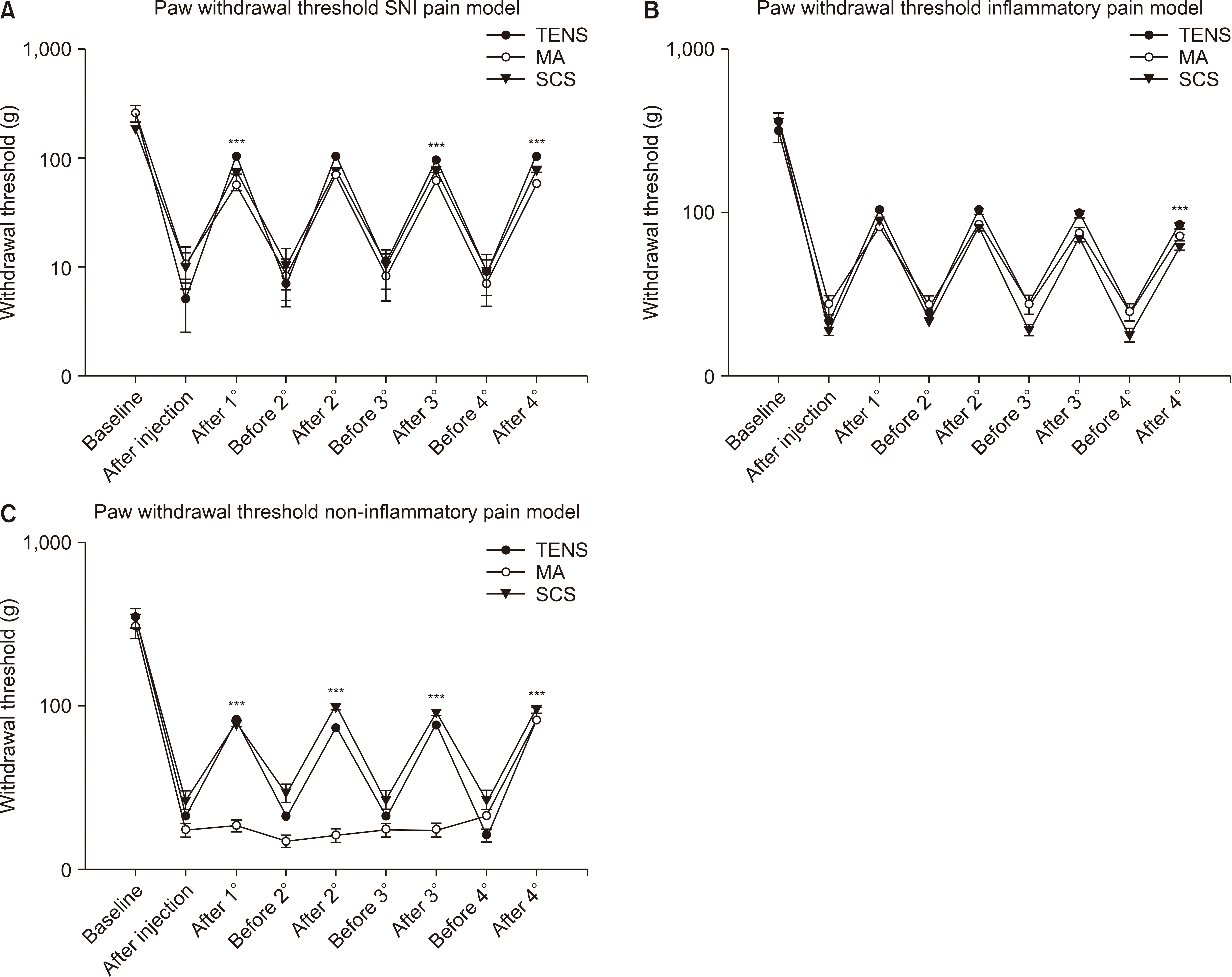 | Fig. 1Average paw withdrawal thresholds in the SNI group (A), joint inflammation group (B), and the non-inflammatory muscle pain group (C). Each graph shows the effects before and after induction of the model, and before and after treatment on each day. A significant decrease in withdrawal thresholds occurred after induction of the models, and this reduced withdrawal thresholds was increased after treatment on each day for the SNI and the joint inflammation model. For the acid model, a significant decrease occurred in all groups, and this decreased threshold was reversed by TENS and SCS but not by MA. SNI: spared nerve injury, TENS: transcutaneous electrical nerve stimulation, SCS: spinal cord stimulation, MA: manual acupuncture. ***P < 0.001.
|
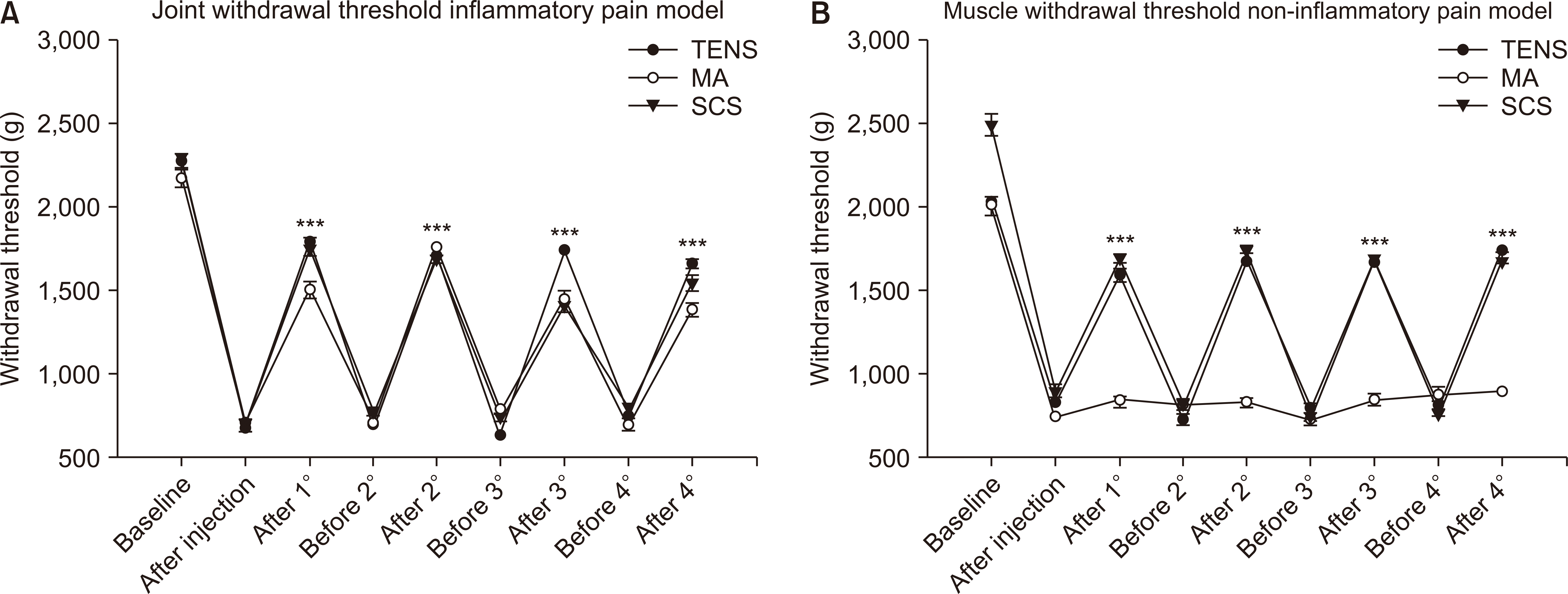 | Fig. 2(A) Average joint withdrawal thresholds before (baseline) and after induction of knee joint inflammation, and before and after treatment on each day with TENS, MA, or SCS. Significant increases occurred after treatment on each day. TENS was significantly higher than SCS and MA. (B) Average muscle withdrawal thresholds before (baseline) and after the second injection of acidic saline (pH 4.0), and before and after treatment on each day with TENS, MA, SCS. Significant increases occurred after treatment on each day. SCS and TENS were significantly greater than MA. TENS: transcutaneous electrical nerve stimulation, MA: manual acupuncture, SCS: spinal cord stimulation. ***P < 0.001.
|
1. Effects of treatment in neuropathic pain
In the SNI model (Table 2; lines SNI/left and right paw), the decreased withdrawal thresholds of the paw ipsilaterally were significantly increased by TENS, SCS, and MA immediately after treatment on each day (time effect; F8, 120 = 15.0, P < 0.001). The effect of TENS, SCS, and MA on the withdrawal thresholds of the paw was lost 24 hours after treatment prior to the next treatment. A significant effect occurred for the side, with the ipsilateral side showing significant decreases in withdrawal threshold compared to the contralateral side (side effect; F1, 120 = 276, P < 0.001). There was no difference for intervention, i.e., between SCS, TENS and MA (Fig. 1A).
Table 2
Average Paw Withdrawal Thresholds in the Groups Before and After Induction of the Model, and Before and After Treatment on Each Day
| Group | Baseline (g) | Day 1 | Day 2 | Day 3 | Day 4 | ||||
|---|---|---|---|---|---|---|---|---|---|
|
|
|
|
|
||||||
| Pre (g) | Post (g) | Pre (g) | Post (g) | Pre (g) | Post (g) | Pre (g) | Post (g) | ||
| Left paw | |||||||||
| SNI-TENS | 259 ± 45 | 5 ± 3a | 103 ± 4ab | 7 ± 3a | 103 ± 4ab | 11 ± 2a | 95 ± 5ab | 9 ± 3a | 103 ± 4ab |
| SNI-SCS | 188 ± 0 | 10 ± 3a | 75 ± 4ab | 11 ± 4a | 75 ± 4ab | 11 ± 4a | 78 ± 3ab | 9 ± 4a | 78 ± 3ab |
| SNI-MA | 259 ± 45 | 11 ± 4a | 56 ± 5ab | 8 ± 3a | 70 ± 3ab | 8 ± 3a | 61 ± 4ab | 7 ± 3a | 58 ± 4ab |
| Right paw | |||||||||
| SNI-TENS | 366 ± 66 | 402 ± 0 | 402 ± 0 | 402 ± 0 | 366 ± 36 | 366 ± 36 | 366 ± 36 | 366 ± 36 | 366 ± 36 |
| SNI-SCS | 259 ± 45 | 366 ± 36 | 400 ± 0 | 331 ± 45 | 366 ± 36 | 331 ± 45 | 331 ± 45 | 331 ± 45 | 390 ± 47 |
| SNI-MA | 331 ± 45 | 331 ± 45 | 331 ± 45 | 331 ± 45 | 366 ± 36 | 331 ± 45 | 331 ± 45 | 295 ± 48 | 295 ± 48 |
| Left paw | |||||||||
| Inf-TENS | 295 ± 48 | 20 ± 0a | 95 ± 5ab | 23 ± 3b | 95 ± 5ab | 25 ± 3a | 91 ± 5ab | 23 ± 3a | 78 ± 3abc |
| Inf-SCS | 331 ± 45 | 18 ± 1a | 75 ± 4ab | 20 ± 0b | 69 ± 5ab | 18 ± 1a | 61 ± 4ab | 17 ± 2a | 58 ± 4ab |
| Inf-MA | 331 ± 45 | 25 ± 3a | 75 ± 4ab | 25 ± 3b | 78 ± 3ab | 25 ± 3a | 69 ± 5ab | 23 ± 3a | 66 ± 6ab |
| Right paw | |||||||||
| Inf-TENS | 331 ± 45 | 402 ± 0 | 402 ± 0 | 366 ± 36 | 366 ± 36 | 402 ± 0 | 402 ± 0 | 402 ± 0 | 402 ± 0 |
| Inf-SCS | 366 ± 36 | 366 ± 36 | 402 ± 0 | 40 ± 0 | 402 ± 0 | 402 ± 0 | 402 ± 0 | 402 ± 0 | 402 ± 0 |
| Inf-MA | 366 ± 36 | 331 ± 45 | 402 ± 0 | 402 ± 0 | 402 ± 0 | 402 ± 0 | 402 ± 0 | 402 ± 0 | 402 ± 0 |
| Left paw | |||||||||
| NInf-TENS | 331 ± 45 | 20 ± 0a | 78 ± 3ab | 18 ± 1a | 70 ± 3ab | 20 ± 0a | 72 ± 3ab | 15 ± 1a | 78 ± 3ab |
| NInf-SCS | 331 ± 45 | 25 ± 3a | 75 ± 4ab | 28 ± 4a | 95 ± 5ab | 25 ± 3a | 87 ± 4ab | 5 ± 3ac | 91 ± 5ab |
| NInf-MA | 295 ± 48 | 17 ± 2a | 20 ± 0a | 14 ± 1a | 15 ± 1a | 17 ± 2a | 17 ± 2a | 20 ± 0a | 20 ± 0a |
| Right paw | |||||||||
| NInf-TENS | 402 ± 0 | 259 ± 45 | 295 ± 48 | 224 ± 36 | 224 ± 36 | 188 ± 0 | 188 ± 0 | 188 ± 0 | 188 ± 0 |
| NInf-SCS | 331 ± 45 | 331 ± 45 | 331 ± 45 | 331 ± 45 | 331 ± 45 | 331 ± 45 | 331 ± 45 | 331 ± 45 | 331 ± 45 |
| NInf-MA | 331 ± 45 | 224 ± 36 | 224 ± 36 | 188 ± 0 | 188 ± 0 | 188 ± 0 | 188 ± 0 | 188 ± 0 | 188 ± 0 |
![]()
2. Effects of treatment in joint inflammation
In the joint inflammation model, there were significant differences for time (F8, 120 = 31.6, P < 0.001), for side (F1, 120 = 3,014, P < 0.001), and for the withdrawal thresholds (Table 2; lines Inf/left and right paw) of the paw. Joint inflammation reduced withdrawal thresholds ipsilaterally, and these reduced paw withdrawal thresholds were increased significantly by TENS, SCS, and MA after the first treatment; similar increases were observed after each treatment (P < 0.001). No significant difference between interventions was found for the withdrawal threshold of the paw (Fig. 1B).
For the joint withdrawal thresholds on the knee (Table 3; lines Inf/left and right paw), there were significant effects for time (F8, 120 = 275, P < 0.001), for side (F1, 120 = 8,677, P < 0.001), and for intervention (F1, 115 = 275, P < 0.001). The threshold for the ipsilateral paw was significantly less than the contralateral paw. Induction of joint inflammation reduced the withdrawal thresholds of the paw ipsilaterally, and these reduced withdrawal thresholds were increased by TENS, SCS, and MA (P < 0.001). There was a significant interaction between time, side, and group (F1, 15 = 10.3286, P = 0.001). Specifically, the withdrawal thresholds after treatment with TENS were significantly greater than those after SCS (P = 0.01) and after MA (P < 0.001); there were no differences between SCS and MA (Fig. 2A).
Table 3
Average Joint (Inflammatory Model) and Muscle (Non-inflammatory Model) Withdrawal Joint Inflammation, and the Non-inflammatory Muscle Pain Group Before and After Induction of the Model, and Before and After Treatment on Each Day
| Group | Baseline (g) | Day 1 | Day 2 | Day 3 | Day 4 | ||||
|---|---|---|---|---|---|---|---|---|---|
|
|
|
|
|
||||||
| Pre (g) | Post (g) | Pre (g) | Post (g) | Pre (g) | Post (g) | Pre (g) | Post (g) | ||
| Left leg | |||||||||
| Inf-TENS | 2,251 ± 41 | 590 ± 33a | 1,739 ± 31ab | 605 ± 28a | 1,651 ± 21ab | 535 ± 22a | 1,688 ± 15ab | 667 ± 29 | 1,405 ± 21ab |
| Inf-SCS | 2,224 ± 24 | 609 ± 16a | 1,694 ± 37ab | 684 ± 25a | 1,636 ± 26ab | 644 ± 22a | 1,317 ± 21ab | 707 ± 19 | 1,487 ± 50ab |
| Inf-MA | 2,139 ± 51 | 616 ± 28a | 1,443 ± 51ab | 615 ± 14a | 1,709 ± 34abc | 699 ± 12a | 1,384 ± 139ab | 601 ± 39 | 1,219 ± 15ab |
| Right leg | |||||||||
| Inf-TENS | 2,372 ± 60 | 2,437 ± 16 | 2,305 ± 24 | 2,215 ± 25 | 2,313 ± 51 | 2,264 ± 15 | 2,380 ± 94 | 2,157 ± 28 | 2,200 ± 37 |
| Inf-SCS | 2,250 ± 47 | 2,207 ± 25 | 2,262 ± 29 | 2,113 ± 40 | 2,137 ± 98 | 2,201 ± 43 | 2,131 ± 28 | 2,057 ± 26 | 2,150 ± 108 |
| Inf-MA | 2,526 ± 93 | 2,450 ± 61 | 2,520 ± 63 | 2,193 ± 39 | 2,174 ± 26 | 2,300 ± 42 | 2,204 ± 49 | 2,024 ± 157 | 2,179 ± 25 |
| Left leg | |||||||||
| NInf-TENS | 1,993 ± 12 | 744 ± 18a | 1,536 ± 40ab | 636 ± 31a | 1,624 ± 18ab | 700 ± 35a | 1,612 ± 15ab | 728 ± 24 | 1,474 ± 20abc |
| NInf-SCS | 2,474 ± 70 | 812 ± 42a | 1,642 ± 25ab | 733 ± 8a | 1,687 ± 13ab | 652 ± 24a | 1,635 ± 20ab | 676 ± 14 | 1,479 ± 10abc |
| NInf-MA | 1,968 ± 56 | 655 ± 12a | 752 ± 40a | 726 ± 30a | 740 ± 30a | 638 ± 40a | 758 ± 31a | 790 ± 41 | 807 ± 16a |
| Right leg | |||||||||
| NInf-TENS | 2,045 ± 65 | 2,142 ± 38 | 2,043 ± 21 | 2,137 ± 18 | 2,086 ± 13 | 2,030 ± 22 | 2,095 ± 24 | 2,039 ± 25 | 2,060 ± 19 |
| NInf-SCS | 364 ± 71 | 1,975 ± 15 | 1,991 ± 7 | 1,963 ± 12 | 1,983 ± 4 | 2,011 ± 8 | 1,994 ± 6 | 1,964 ± 13 | 1,993 ± 17 |
| NInf-MA | 2,107 ± 63 | 2,187 ± 28 | 2,141 ± 34 | 2,112 ± 30 | 2,056 ± 32 | 2,048 ± 22 | 1,977 ± 48 | 2,028 ± 31 | 2,060 ± 28 |
![]()
3. Effects of treatment in non-inflammatory muscle pain
In chronic muscle pain model, there were significant decreases in withdrawal thresholds (Table 2; lines NInf/left and right paw) of the paw after induction of the model. Both TENS and SCS significantly reversed this decrease (time effect; F8, 120 = 84, P < 0.001). There were significant effects related to the side, with the ipsilateral side showing greater decreases than the contralateral side (F1, 120 = 189, P < 0.001). A significant effect for intervention occurred (group effect; F2, 15 = 6.5, P = 0.005), and there was a significant interaction between time, side, and intervention (F16, 120 = 2.2, P = 0.009). There was no effect of MA on paw withdrawal thresholds in the non-inflammatory pain model (Fig. 1C), with the MA group showing lower withdrawal thresholds than the TENS (P = 0.093) and the SCS (P = 0.008) groups.
For the muscle withdrawal thresholds (Table 3; lines NInf/left and right paw), there were significant effects for time (F8, 120 = 387, P < 0.001), for side (F1, 120 = 22.505, P = 0.001), and a significant interaction between time, side, and group (F16, 120 = 31.6, P < 0.001). Specifically, the withdrawal thresholds decreased ipsilaterally after induction of the model, and these withdrawal thresholds were reversed by TENS and SCS, but not by MA (Fig. 2B). The muscle withdrawal thresholds of the paw were lower in the MA groups than those of the TENS (P < 0.001) and SCS (P < 0.001) groups; MA had no effect on the withdrawal thresholds after repeated acid injections into the muscle.
Go to :
DISCUSSION
The current study compared the effects of three different types of non-pharmacological treatments (TENS, SCS, MA) for three different types of pain (neuropathic, inflammatory, and non-inflammatory). MA was only effective in the neuropathic and inflammatory models, while TENS and SCS were effective in all 3 models. The analgesic effects of TENS, SCS, and MA involve complex neuronal processes that utilize multiple neurotransmitters and modulators (see Table 4 for a summary), including opioid peptides and serotonin. The differences between interventions found in the current study could be explained by the different mechanisms of action used by each type of treatment.
Table 4
Summary of Mechanisms for SCS, TENS, and MA
![]()
It is known that TENS activates central inhibitory pathways to reduce hyperalgesia [14,27]. Prior studies have shown the good effects of TENS in models of joint, paw, and muscle inflammation [13,27,28], analgesia lasting for 12-24 hours [21], and repetitive use producing analgesic tolerance at the central opioid receptors. Specifically, TENS reduces central neuron sensitization, and activates opioid, serotonin, GABA, and muscarinic receptors in the spinal cord and supraspinal pathways [13,14,21,27,28]. In experimental studies on neuropathic pain, TENS reduces allodynia and hyperalgesia, and there is a reduction in central neuron sensitization and glial cell activity [29]. However, little is known about the effects of TENS in the non-inflammatory model. It is thought that pain may be alleviated by using electrical stimulation directly over the area of pain (inflammatory or non-inflammatory). This peripheral stimulation induces electrical activity which inhibits the brain’s perception of pain. The ‘gate control theory’ of Wall and Melzack is based on the principle that there is a gateway in the dorsal horn of the spinal cord, which somehow controls or regulates the flow of pain messages that are then sent to (ascending) and from (descending) higher levels of the brain for central processing, thus reducing the perception of pain [30,31]. Other postulated mechanisms of the pain relief mediated by TENS include the promotion of endorphin release in the brain [32] and local dilatation of blood vessels in injured tissue [33]. Clinically, TENS is effective for pain conditions associated with neuropathic pain, inflammatory pain, and non-inflammatory pain such as reducing phantom pain [34], diabetic peripheral neuropathy [35], postoperative pain [36], spinal nerve injury [37], trigeminal neuralgia [38], osteoarthritis [39], chronic musculoskeletal pain [40], and fibromyalgia [7]. Thus, TENS is effective for a variety of pain conditions with different underlying tissue pathologies.
The field of SCS also owes its inception to the concept of gate control theory, which proposed that “control of pain may be achieved by selectively activating the large, rapidly conducting fibers” [41] and can be used in inflammatory pain, although chronic pain is the main target. Conventional SCS activates large Aβ dorsal column axons. This activation can be measured as action potentials propagated antidromically in the peripheral nerves [42]. Electrical stimulation alters the membrane potential of neurons and other cell types exposed to electric fields, thereby altering the electrochemical properties of the segments affected [43]. Electrophysiology and molecular biology have provided a view of the effect of SCS on neurotransmitters and their receptors, which have led to the formulation of segmental and supraspinal mechanisms. The literature supports the involvement of glial cells in chronic pain and their characteristic response to electrical fields [43].
The current study showed that MA reduces hyperalgesia in the neuropathic and inflammatory pain models. However, it did not reduce hyperalgesia in the non-inflammatory pain model used in the current study. Prior work shows that MA using the SP6 and ST36 acupoints has an analgesic effect that lasts up to two hours [12], produces a cumulative effect [19], and reduces hyperalgesia in neuropathic [44] and inflammatory pain models [19,26]. Neurophysiological mechanisms, by which MA exerts its analgesic effects, show activation of both peripheral and central mechanisms. Centrally, MA activates the descending inhibitory systems [18,45] using opioids and serotonin in both inflammatory and neuropathic pain models [12,16,17]. Peripherally, MA releases adenosine triphosphate which converts to adenosine and activates the adenosine A1 receptor in inflammatory pain [46]. MA, however, also promotes the resolution of inflammation. Specifically, our prior studies show that MA reduces muscle inflammation, measured by reduced inflammatory cell infiltration, vascular permeability, neutrophilic activity, and edema [19,26]. Further, MA has a direct effect on the immune system, increasing release of the anti-inflammatory cytokine interleukin (IL)-10 and producing a phenotypic switch in the macrophage phenotype, to an increased M2 phenotype (IL-10 source) and a reduced M1 phenotype in the inflamed muscle [26]. Thus, the lack of effect on the non-inflammatory pain model may be related to the actions of MA on inflammation observed after nerve injury and injection of carrageenan. Surprisingly, however, there was not a sustained effect on nociceptive behaviors.
We speculate that while inflammation was reduced, it was not eliminated, and thus, inflammatory mediators could continue to activate nociceptors to produce nociceptive behaviors. Clinically, MA is effective in a variety of pain conditions, including neuropathic pain [47], knee osteoarthritis [45,48,49], acute and chronic back pain [49], and fibromyalgia [50]; however, there is a large variability in the acupoints used. Thus, it is possible that a different protocol of MA would be more effective in each of these different models.
Potential limitations of our study were the methods of the pain models. The variability of these methods, although not big, may be avoided in the future by using most precise ones. Also, we suggest that the protocols using different parameters of frequency, and alternating currents investigated with other methodologies should be included. Future basic science studies need to understand the different mechanisms between treatments in different models, and future clinical studies need to confirm animal data to provide a solid evidence base for the use of SCS, TENS, and MA.
The present study examined the efficacy of TENS, SCS, and MA on the neuropathic, inflammatory, and non-inflammatory models of pain. While all 3 interventions were successful on minimizing pain on neuropathic and inflammatory models, only TENS and SCS were effective with the non-inflammatory pain model. These data suggest that MA may not be useful for non-inflammatory pain, while all 3 interventions could be used for neuropathic and inflammatory pain.
Go to :
 XML Download