Journal List > Int J Stem Cells > v.11(1) > 1159121

Jeong, Yim, Park, Cho, Hong, Kim, and Oh: Mesenchymal Stem Cell Therapy for Ischemic Heart Disease: Systematic Review and Meta-analysis

Abstract

Background and Objectives

Mesenchymal stem cells (MSC) have emerged as breakthrough treatments for myocardial infarction. However, the efficacy of MSC remains unclear. The aim of the study was to evaluate treatment effect of MSC in terms of mechanical, regenerative, and clinical outcomes for patients with myocardial infarction (MI) using meta-analysis.

Methods

A systematic search and critical review of MEDLINE, EMBASE, and Cochrane database literature published from inception through December 2017 was performed. The inclusion criteria were randomized controlled trials, studies on patients with myocardial infarction, and studies compared with placebo as a control group.

Results

A total of 950 patients from 14 randomized placebo controlled trials were included in the final meta-analysis. MSC treatment showed benefits for mechanical, regenerative, and clinical outcomes. In terms of mechanical outcomes, the LVEF of the MSC treatment group increased by 3.84% (95% CI: 2.32~5.35, I2=43) and the effect was maintained for up to 24 months. Regenerative outcomes were measured by scar mass and WMSI. Scar mass was reduced by −1.13 (95% CI: −1.80 to −0.46, I2=71) and WMSI was reduced by −0.05 (95% CI: −0.07 to −0.03, I2=45) at 6 months after MSC treatment. Mortality rate and incidence of re-hospitalization for HF in MSC group patients trended toward reduced incidence compared to the control group, although this was not statistically significant because of the low event rate.

Conclusions

The findings of this meta-analysis indicate that MSCs can be beneficial in improving heart function in the treatment of MI. However, the efficacy of MSCs must be further explored through large randomized controlled trials based on rigorous research design.

Introduction

Several meta-analyses demonstrated that bone marrow-derived mononuclear cell (BM-MNC) therapy for patients with ischemic heart disease showed slightly but significantly increased left ventricular ejection fraction (LVEF) from 2.51 to 4.77% compared to controls (14). However, the results of individual patient data including 1,252 patients from 12 randomized clinical trials showed no changes in LVEF (mean difference: 0.96%, 95% confidence interval: −0.2 to 2.1) compared to controls (5). BM-MNC was not associated with reduced all-cause mortality and morbidity compared to standard therapy for patients with acute myocardial infarction (AMI) (6). Conversely, BM-MNC reduced mortality, periprocedural adverse events were infrequent, and the incidence of non-fatal myocardial infarction and arrhythmias was reduced in patients with chronic myocardial infarction (CMI) (7).
Mesenchymal stem cells (MSC) and cardiac stem cells have emerged as breakthrough treatments of myocardial infarction. MSC can be safely obtained from adult bone marrow and enriched and expanded by in vitro culturing. MSC can be safely administered without the need for immunosuppressant agents, and they are less prone to genetic abnormalities and malignant transformation during multiple passages in vitro (810). Studies in animal models of myocardial infarction have demonstrated the ability of transplanted MSC to engraft and differentiate into cardiomyocytes and vascular cells (1113). MSC have been used in humans for approximately 10 years to repair or regenerate injured heart, either directly or indirectly (through paracrine effects). Clinical trials suggest that intracoronary administration of autologous bone marrow-derived MSC can improve left ventricular function in patients with myocardial infarction (14). Also, clinical feasibility and safety trials have been published using bone marrow allogeneic mesenchymal cells in patients with myocardial infarction (15, 16).
Mortality is currently regarded as the most important endpoint to evaluate efficacy of heart failure (HF) drugs in regulatory perspectives (17). Since 2002 (18), numerous clinical trials on efficacy of stem cell therapy for heart disease have been performed but most of the efficacy have evaluated using surrogate endpoints such as LVEF and infarct size. A previous study reported that improved LVEF (mechanical parameter) and reduced infarct size (regenerative parameter) have correlated with long-term outcomes in heart disease (19). In addition, the employment of sophisticated imaging like MRI has played a role in accurately detecting regeneration of myocardium in the assessment of stem cell efficacy (20).
However, no consensus has been developed in MSC treatment of ischemic heart disease focusing on mechanical, regenerative, and clinical outcomes. We conducted a systematic review and meta-analysis based on the current evidence of prospective randomized controlled trials on treatment with MSC in terms of mechanical, regenerative, and clinical outcomes for patients with MI.

Materials and Methods

Search strategy and study selection

This systematic review was performed according to the Preferred Reporting Items for Systematic Reviews and Meta-Analyses (PRISMA) guidelines (http://www.prisma-statement.org/). A systematic search and critical review of the literature published from inception through December 2017 was performed of the MEDLINE, EMBASE, and Cochrane databases for clinical studies written in English of MSC transplantation in patients with MI using the following terms: “Mesenchymal Stromal Cell”, “Mesenchymal Stem cell”, “Mesenchymal Progenitor Cell”, “Multipotent Mesenchymal Stromal Cells”, “Myocardial Infarction” “Cardiovascular Stroke” and “Heart Failure”. The complete search strategy was provided in the Supplementary appendix. The inclusion criteria were restricted to the following studies to evaluate the efficacy of MSC treatment: randomized controlled trials (RCT), studies of patients with acute myocardial infarction (AMI) or chronic myocardial infarction (CMI), and studies comparing placebo as a control group. The administration route, origin of MSC, and MSC differentiation were not restricted. Exclusion criteria were in vivo or in vitro studies; injection of cells other than MSC; studies other than RCT, secondary report, poster presentation, review, or editorial; and studies not written in English. Article selection procedures were independently conducted by two authors with standard methods. Two investigators independently screened all titles and abstracts to identify extracted relevant studies that met the inclusion and exclusion criteria and then independently assessed a full text review for relevant studies. Disagreements were resolved by consensus.

Data extraction

Two investigators extracted data using customized data extraction forms. The following data were extracted: year of publication, country of patient enrollment, numbers of patients allocated to stem cell treatment and control groups, route of stem cell administration, MSC cell type, cell origin, injection dose, follow-up period (months), and whether or not cell differentiation was performed. The efficacy of MSC therapy was assessed in terms of mechanical, regenerative, and clinical outcomes. Mechanical outcomes were evaluated by left ventricular ejection fraction (LVEF), left ventricular end-systolic volume (LVESV), left ventricular end-diastolic volume (LVEDV), 6-minute walking distance (6MWD), and wall motion score index (WMSI). Regenerative outcome was assessed by reduced infarct size. Clinical outcomes were evaluated based on all-cause mortality, non-fatal myocardial infarction (MI), arrhythmia, re-hospitalization for HF, and revisualization. Outcome data were extracted up to the maximum follow-up period to evaluate MSC treatment effect. The time point farthest from cell transplantation was used because this was the best predictor of clinical outcome. Mechanical outcome data, including LVEF, LVESV, and LVEDV, were extracted at baseline and at 3, 6, 12, and 24 months to identify changes over time and evaluate length of MSC treatment. Mean changes of LVEF, LVESV, and LVEDV were evaluated at 3, 6, 12, and 24 months from baseline. The trials were assessed for efficacy outcomes at each follow-up point.
For dichotomous outcomes, the number of events was recorded. For continuous outcomes, the mean, standard deviation, and total sample size were recorded for both treatment and control groups. Outcome data were extracted for the maximum follow-up period, and MSC treatment effects were evaluated based on difference between treatment and control groups. If follow-up data were not present in the original article, changes (Δ) from baseline to final time point were included in the meta-analysis. All data were extracted in accordance with criteria based on the Cochrane Handbook for Systematic Reviews of Interventions (21).

Quality assessment

Two investigators independently assessed the risk of bias for randomized controlled trials using criteria based on the Cochrane Handbook for Systematic Reviews of Interventions (21). Each study was examined for sequence generation, allocation concealment, masking, performance bias, detection bias, incomplete outcome data, selective outcome reporting, and other bias that might affect the study outcome. Judgments were expressed simply as “low risk of bias,” “high risk of bias,” or “unclear risk of bias.” Overall quality was divided into low risk of bias and high risk of bias. Low risk of bias in overall quality was defined as all of the quality assessment domains evaluated as other than “high risk of bias.” Quality assessments were independently conducted by two authors. Disagreements were resolved by discussion between the two authors.

Statistical analyses

Outcome data were analyzed using Review Manager 5.3 (Cochrane collaboration). For dichotomous data, MSC treatment effects were calculated as a risk ratio (RR) with 95% confidence intervals (CI) using Mantel-Haenszel methods. For continuous data, MSC treatment effects were presented as weighted mean difference (WMD) if outcomes were measured in the same way across trials. For outcomes measured using different methods, treatment effect data were combined using the standardized mean difference (SMD). Mechanical and regeneration outcome data were pooled using the DerSimonian-Laird random-effects model because of the high degree of heterogeneity. Clinical outcome data were pooled using the fixed effect model because they showed low heterogeneity. The existence of heterogeneity among effect sizes of individual studies was analyzed with the I2 statistic. Heterogeneity was defined as low (25% to 50%), moderate (50% to 75%), or high (>75%) (22).
Because of different disease characteristics with AMI and CMI, MSC treatment effect differed due to the disease itself. Several previous meta-analyses of BM-MNC injection in patients with MI revealed that treatment effect varied depending on route of administration and measurement tools (23). A previous meta-analysis suggested that randomized controlled trials testing efficacy should be appropriately designed and rigorously applied to avoid bias (4). Therefore, preplanned subgroup analyses were conducted based on patient characteristics (AMI or CMI), route of delivery (intracoronary, intravenous, or intramyocardial), measurement tools (single-photon emission computed tomography (SPECT), magnetic resonance imaging (MRI), or echocardiography), whether cell differentiation was conducted or not, and overall risk of bias (low risk or high risk) to explore the source of heterogeneity. The likelihood of publication bias was tested graphically and quantitatively using funnel plot and Egger test.

Results

Search results

The initial search identified 2,272 articles, of which 2,113 references were excluded in the first screening. Of the remaining 159 potentially relevant references, 104 were excluded for being experimental studies, reviews, editorials, or comments. Fifty-five articles were reviews with full text. Fourteen randomized placebo controlled trials of 35 references were eligible for final analysis (1416, 2434). A summary of the study classification is presented in a PRISMA flow diagram (Fig. 1).

Characteristics of included studies

A total of 950 patients (MSC injection group 509, control group 441) from 14 randomized placebo controlled trials were included in the final meta-analysis. Publication years were from 2004 to 2017. Most of the included studies (11 out of 14) were published from 2013 to 2017. Of 12 studies, 6 included acute MI patients (14, 15, 24, 2729), and the other 8 included chronic MI patients (16, 25, 26, 3034). The baseline LVEF of AMI patients ranged from 43.1 to 52.0, and that of CMI patients was between 17.5 and 35.7. MSC was infused into intracoronary (5 studies) (24, 2730), intramyocardial (5 studies) (16, 25, 26, 31, 32), and intravenous (2 studies) (14, 15) locations. Total injected cell doses ranged from 1 to 600×106. Follow-up duration was from 3 to 24 months. In 4 of 14 studies, MSC were differentiated into cardiopoietic stem cells or mesenchymal precursor cells (Table 1) (16, 25, 26, 34).

Risk of bias assessment of included studies

A summary of the risk of bias is presented in Fig. 2. Of 14 randomized placebo controlled trials, 12 used computer-generated random sequence numbers to allocate participants. Nine studies mentioned allocation concealment (1416, 25, 26, 3134). Eight studies made an effort to maintain double blindness to reduce performance or detection bias (1416, 26, 3134). Eleven studies were evaluated as low risk of bias in terms of incomplete outcome data (1416, 25, 26, 28, 29, 3134). Nine studies were assessed as low risk of bias in selective outcome reporting compared to protocols from the protocol registration site (1416, 26, 28, 3134). Eight studies were evaluated as low risk of bias across all quality assessment domains (1416, 26, 3134) that were considered “low risk of bias” in the overall risk of bias assessment (Table 1).

Efficacy of MSC

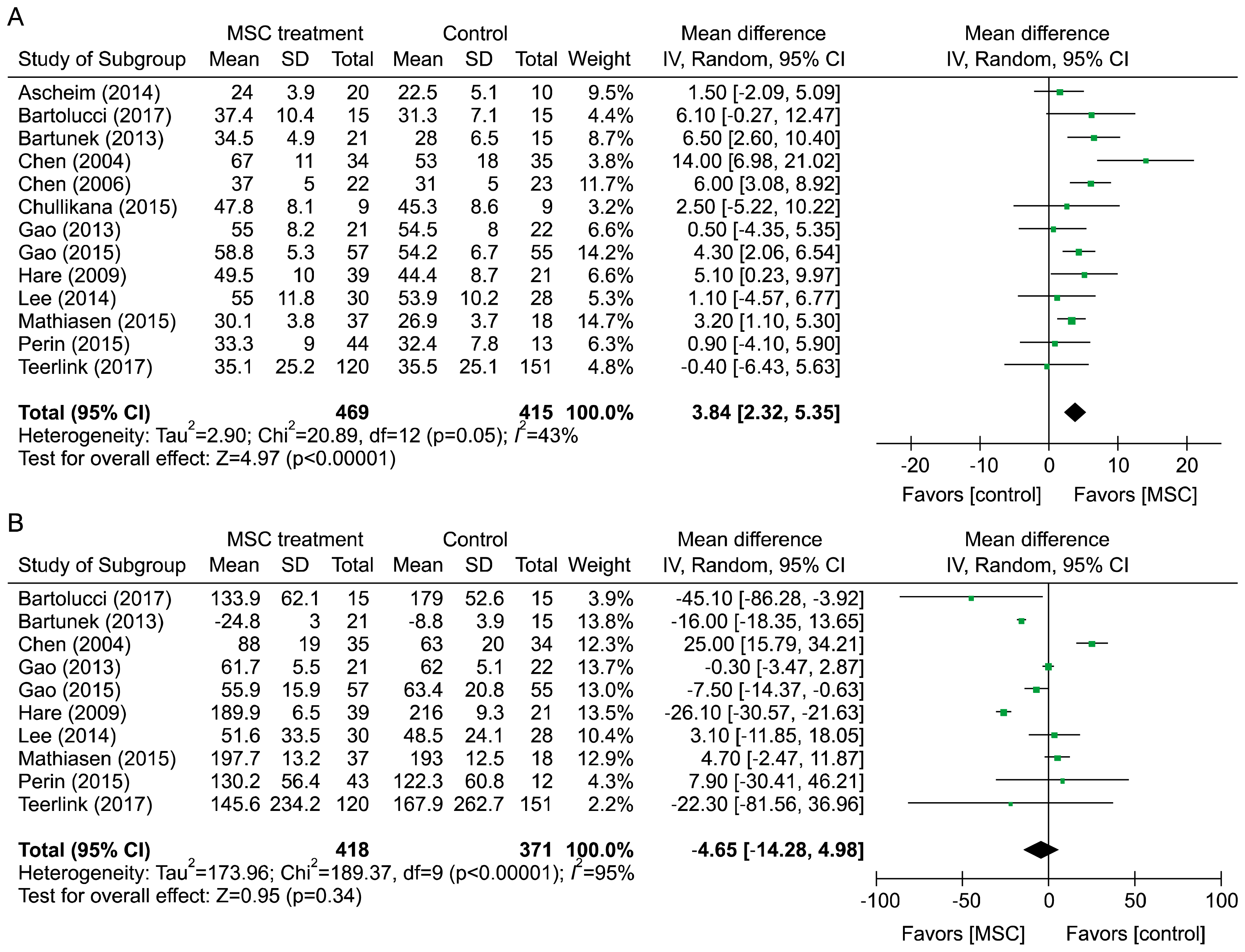
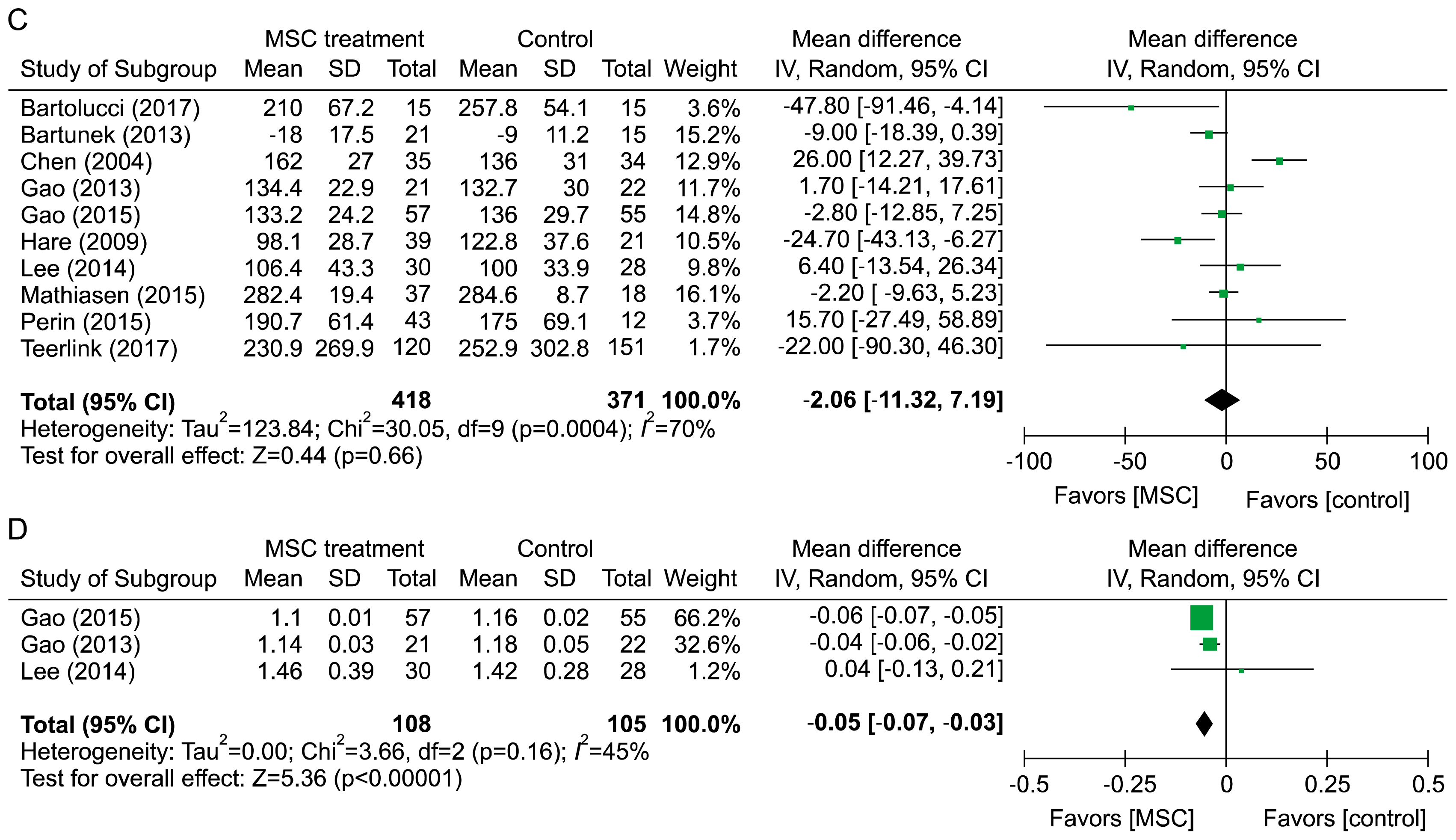
In terms of mechanical outcomes, all but one study reported the efficacy of MSC using LV function (31). Mechanical outcomes were evaluated by LVEF, LVESV, LVEDV, and WMSI. WMSI was calculated by dividing the sum of the wall motion score by the number of visualized segments; a normal WMSI was 1. Compared to the control group, LVEF of the MSC treatment group increased by 3.84% (95% CI: 2.32~5.35, I2=43), LVESV decreased by 4.65 ml (95% CI: −14.25 to 4.98, I2=95), LVEDV decreased by 2.06 ml (95% CI: −11.32 to 7.19, I2=70), and WMSI decreased by 0.05 (95% CI: −0.07 to −0.03) (Fig. 2).
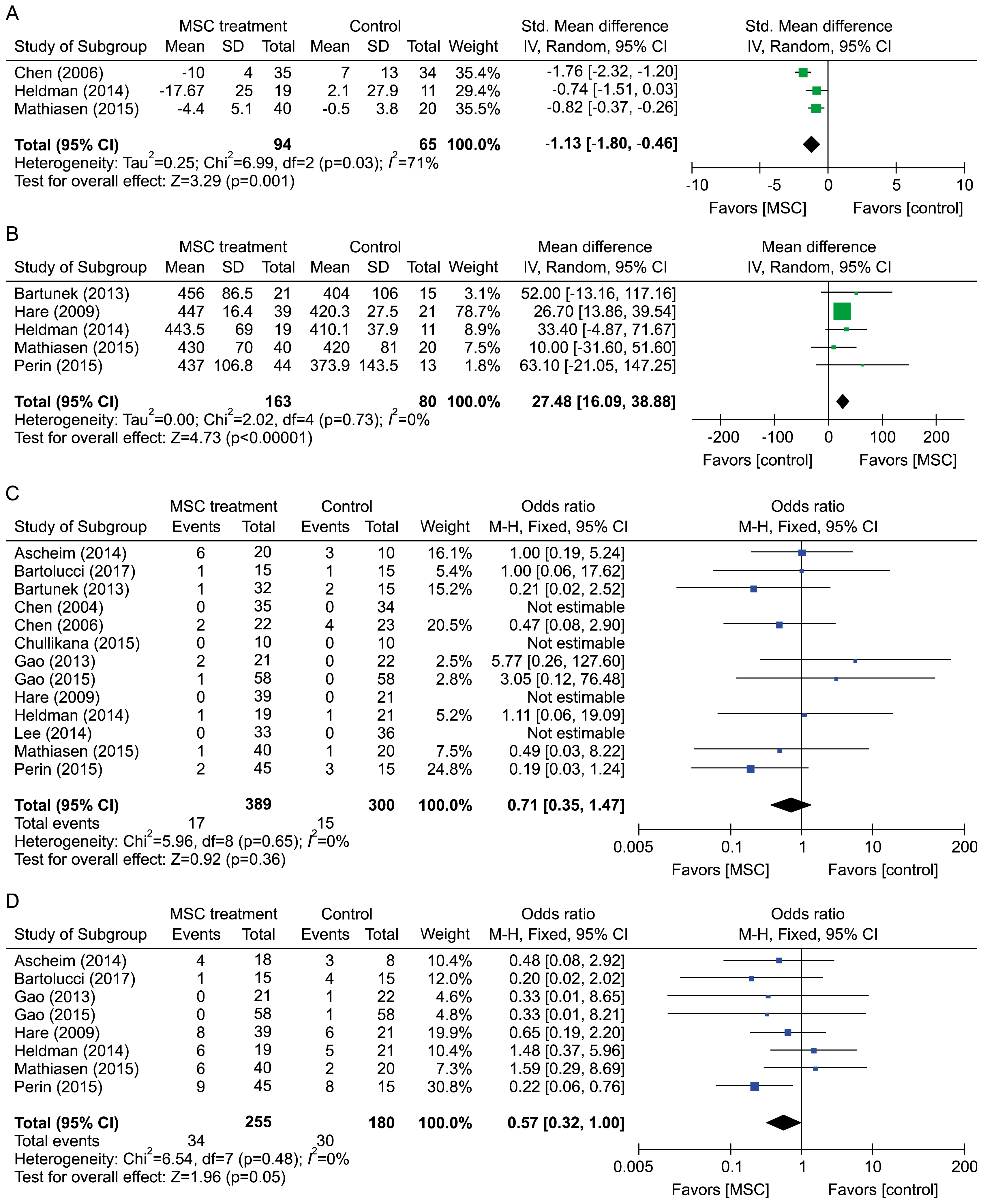
Regenerative outcomes were measured by scar mass. Scar mass was reduced by 1.13 (95% CI: −1.80 to −0.46, I2=71) at 6 months after MSC treatment. Regarding clinical outcomes, all of the included studies reported all-cause mortality. The mortality rate in the MSC group was lower than that of control group (OR=0.71, 95% CI: 0.35~1.47, I2=0%). Incidences of arrhythmia, re-hospitalization for HF, and revascularization in MSC group patients were lower than those of control groups (OR=0.37, 95% CI: 0.14~1.00, I2=14%; OR=0.57, 95% CI: 0.32~1.00, I2=0%; OR=0.37, 95% CI: 0.17~2.61, I2=0%, respectively). However, incidence of non-fatal MI in MSC patients was higher than that of control patients (OR=2.71, 95% CI: 0.32~22.97, I2=0%). Though differences in clinical outcomes were not statistically significant, point estimates showed the beneficial effect of MSC on clinical outcome. 6MWD improved by 27.48 m (95% CI: 16.09 to 38.88, I2=0) compared to the control group. MSC treatment showed benefits on mechanical, regenerative, and clinical outcomes (Fig. 3).

Subgroup analyses

Subgroup analysis showed that improvements in LV function were statistically significant in CMI patients. When studies were compared based on the autologous or allergenic origin of MSC, there was greater improvement in autologous origin MSC (4.62, 95% CI: 1.16 to 8.08 vs. 2.78, 95% CI: 0.55 to 5.01, respectively). Regarding route of injection, intracoronary injection was associated with greater reduction in LVEF. Regarding LVEF measurement tools, there was greater improvement in SPECT than MRI or echocardiography. Studies with a high risk of bias showed slightly greater improvement than those with low risk of bias. Heterogeneity disappeared with autologous cell origin, intravenous administration, MRI assessment, and low risk of bias in methodological flaws based on the subgroup analysis (Table 2). The likelihood of publication bias was tested using a funnel plot and Egger test for LVEF. The funnel plot was symmetric and the Egger test was not significant (p=0.20), suggesting low susceptibility to publication bias (funnel plot not shown).

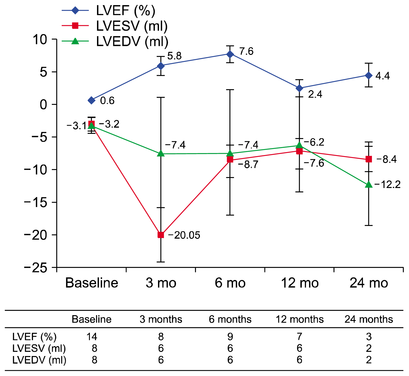
Changes in cardiac function times after transfusion of MSC

Compared to controls, the MSC treatment group LVEF improved by 5.8%, 7.6%, 2.4%, and 4.4% at 3, 6, 12, and 24 months, respectively, and LVESV decreased by 20.1 ml, 8.7 ml, 7.6 ml, and 8.4 ml, respectively. The MSC treatment group showed greater decreases in LVEDV than the control group at each follow-up point, with decreases of 7.4 ml, 7.4 ml, 6.2 ml, and 12.2 ml, respectively. Trials were assessed for efficacy outcomes at each follow-up point. The results show that LVEF increased after injection of MSC, and this effect was maintained for up to 24 months (Fig. 4).

Discussion

MSC treatment showed benefits on mechanical, regenerative, and clinical outcomes. In terms of mechanical outcomes, the LVEF of the MSC treatment group increased by 3.84% (95% CI: 2.32~5.35, I2=43). Cardiac function assessed by LVEF after injection of MSC was evaluated at 3, 6, 12, and 24 months from baseline. The results show that LVEF was increased, an effect that was maintained for up to 24 months. Regenerative outcomes were measured by scar mass and WMSI. Scar mass was reduced by 1.13 (95% CI: −1.80 to −0.46, I2=71), and WMSI was reduced by 0.05 (95% CI: −0.07 to −0.03) at 6 months after MSC treatment. The mortality rate and incidence of re-hospitalization for HF in MSC patients showed a trend toward reduction compared to the control group, although the differences were not statistically significant because of the low event rate. Six minute walking distance improved by 27.48 m (95% CI: 16.09 to 38.88, I2=0) in the MSC treatment group compared to the control group. Based on the present evidence, MSC might have a regenerative effect to reduce infarct size and wall motion in the myocardium. Preclinical evidence revealed that differentiation of MSCs to cardiomyocytes does not occur to a significant extent in vivo. Instead, the “paracrine effect” derived from secretion of MSCs is believed to be the dominant mechanism. MSC injection results in secretion of a range of growth factors, cytokines, and chemokines, which could help repair adverse remodeling in persistent ischemia of heart failure (35).
According to a meta-analysis of BMC treatment effect in ischemic heart failure with similar inclusion criteria as this study, BMC therapy affected mechanical, regenerative, and clinical outcomes. BMC transplantation resulted in improved LVEF (2.92%, 95% CI: 1.91~3.92), reduced infarct size (−2.25%, 95% CI: −3.55 to −0.95), and significantly lower incidence of all-cause mortality (OR= 0.55, 95% CI: 0.34~0.89) (36).
A recent in vivo study compared MSC and BMC cell delivery in a porcine model of chronic ischemic heart disease. This study showed that MSC was more effective than BMC in improving global heart function. Cell therapy was not associated with increased mortality (37). A preclinical meta-analysis also revealed that MSC were more beneficial in ischemic heart disease than BMC (38).
The exact mechanisms by which MSC act remain incompletely described. It is likely that there are myriad effects from these versatile stem cells, rather than a single mechanism of action. Research has shown that a number of factors, both in vitro and in vivo, contribute to the salutary effects of MSC therapy after MI. Possible mechanisms include: 1) MSC differentiate into new cardiomyocytes or other cell types (39), 2) MSC enhance the neovascularization response after myocardial infarction (40), 3) MSC act as reservoirs for paracrine mediators that are the actual effectors of observed cardiac repair (41), 4) MSC stimulate endogenous cardiac precursors to repair the damaged tissue (42). Further research into the precise mechanism might facilitate improved techniques to achieve even more functional repair for damaged hearts, which will benefit patients in the long run.
MI leads to significant cell loss and scar tissue formation. The remaining cardiomyocytes cannot reconstitute necrotic tissue, and cardiac function deteriorates during the ensuing course. The efficacy of MSC therapy in myocardial infarction might contribute to multiple biological mechanisms, including cardiac regeneration, neovascularization, paracrine effect, and immune regulation. MSC isolated from adult bone marrow differentiated into cardiomyocytes in large animal models (4345). Transplantation of MSCs by injection into the myocardium showed positive cardiac markers in infarcted myocardium (43, 44). MSCs can also achieve engraftment and long-term survival in scarred myocardium (35).
Though MSC have great potential as a new therapy to treat damaged myocardium, it is crucial to address safety concerns in clinical applications. Several reports have raised concerns about tumor formation with the use of BM-derived MSC and malignant tumors when transplanted in vivo (46, 47). Although malignant transformation and tumor formation with MSC are not yet reported in clinical trials, concerns related to the tumorgenicity of MSC were raised by preclinical studies demonstrating increased tumor progression (48). A clinical study reported that intracoronary infusion of MSC was involved in acute coronary artery occlusion during the BMSC injection procedure (27).

Conclusions

The findings of this meta-analysis indicate that BM-MSC can be beneficial in improving heart function in the treatment of MI. However, the efficacy of MSC must be further explored through large randomized controlled trials based on rigorous research design. MSC therapy appears to be safe. However, the safety of MSC must be further explored through clinical studies based on a research design that allows systematic monitoring and reporting of safety to establish the appropriate safety profile of MSC. In addition, potential AEs of MSC must be verified by long-term follow-up studies with continuous caution.

Acknowledgements

This research was supported by a grant (18172MFDS182) from the Ministry of Food & Drug Safety in 2018. The Ministry of Food & Drug Safety had no further role in areas of study design, collection, analysis, and interpretation of data, report writing, or in the decision to submit the paper for publication.

Notes

Ethical Approval and Consent to participate

This study has been granted an exemption from requiring ethics approval by the Institutional Review Board of The Catholic University of Korea (Approval number: MC17ZESE0055). Consent to participate is not applicable.

Consent for publication

All authors consent for publication

Availability of supporting data

Not applicable

Funding

This research was supported by a grant (18172MFDS182) from the Ministry of Food & Drug Safety in 2018.

Author contributions

HJ participated in eligibility screening, data extraction, protocol preparation, analysis, data interpretation, and writing the manuscript; HWY participated in the scientific concept, eligibility screen, data extraction, quality assessment data analysis, analysis and interpretation of data, protocol preparation, scientific guidance, and critical review of the manuscript; YC and HH participated in design and implementation of search strategies, initial eligibility screening, data verification, and critical review of the manuscript; HJP and IO participated in clinical content, comments on protocol, and critical review of the manuscript; NK participated in design and implementation of search strategies, data search, and validation. All authors read and approved the final manuscript.

Potential conflict of interest

The authors have no conflicting financial interest.

References

1. Lipinski MJ, Biondi-Zoccai GG, Abbate A, Khianey R, Sheiban I, Bartunek J, Vanderheyden M, Kim HS, Kang HJ, Strauer BE, Vetrovec GW. Impact of intracoronary cell therapy on left ventricular function in the setting of acute myocardial infarction: a collaborative systematic review and meta-analysis of controlled clinical trials. J Am Coll Cardiol. 2007; 50:1761–1767. DOI: 10.1016/j.jacc.2007.07.041. PMID: 17964040.
crossref
2. Martin-Rendon E, Brunskill SJ, Hyde CJ, Stanworth SJ, Mathur A, Watt SM. Autologous bone marrow stem cells to treat acute myocardial infarction: a systematic review. Eur Heart J. 2008; 29:1807–1818. DOI: 10.1093/eurheartj/ehn220. PMID: 18523058.
crossref
3. Zhang SN, Sun AJ, Ge JB, Yao K, Huang ZY, Wang KQ, Zou YZ. Intracoronary autologous bone marrow stem cells transfer for patients with acute myocardial infarction: a meta-analysis of randomised controlled trials. Int J Cardiol. 2009; 136:178–185. DOI: 10.1016/j.ijcard.2008.04.071.
crossref
4. Jeong H, Yim HW, Cho Y, Park HJ, Jeong S, Kim HB, Hong W, Kim H. The effect of rigorous study design in the research of autologous bone marrow-derived mononuclear cell transfer in patients with acute myocardial infarction. Stem Cell Res Ther. 2013; 4:82. DOI: 10.1186/scrt233. PMID: 23849537. PMCID: 3854784.
crossref
5. Gyöngyösi M, Wojakowski W, Lemarchand P, Lunde K, Tendera M, Bartunek J, Marban E, Assmus B, Henry TD, Traverse JH, Moyé LA, Sürder D, Corti R, Huikuri H, Miettinen J, Wöhrle J, Obradovic S, Roncalli J, Malliaras K, Pokushalov E, Romanov A, Kastrup J, Bergmann MW, Atsma DE, Diederichsen A, Edes I, Benedek I, Benedek T, Pejkov H, Nyolczas N, Pavo N, Bergler-Klein J, Pavo IJ, Sylven C, Berti S, Navarese EP, Maurer G. ACCRUE Investigators. Meta-Analysis of Cell-based CaRdiac stUdiEs (ACCRUE) in patients with acute myocardial infarction based on individual patient data. Circ Res. 2015; 116:1346–1360. DOI: 10.1161/CIRCRESAHA.116.304346. PMID: 25700037. PMCID: 4509791.
crossref
6. Fisher SA, Zhang H, Doree C, Mathur A, Martin-Rendon E. Stem cell treatment for acute myocardial infarction. Cochrane Database Syst Rev. 2015; (9):CD006536. PMID: 26419913.
crossref
7. Fisher SA, Doree C, Mathur A, Taggart DP, Martin-Rendon E. Stem cell therapy for chronic ischaemic heart disease and congestive heart failure. Cochrane Database Syst Rev. 2016; 12:CD007888. PMID: 28012165.
crossref
8. Izadpanah R, Trygg C, Patel B, Kriedt C, Dufour J, Gimble JM, Bunnell BA. Biologic properties of mesenchymal stem cells derived from bone marrow and adipose tissue. J Cell Biochem. 2006; 99:1285–1297. DOI: 10.1002/jcb.20904. PMID: 16795045. PMCID: 4048742.
crossref
9. Draper JS, Moore HD, Ruban LN, Gokhale PJ, Andrews PW. Culture and characterization of human embryonic stem cells. Stem Cells Dev. 2004; 13:325–336. DOI: 10.1089/scd.2004.13.325. PMID: 15345125.
crossref
10. Lee K, Majumdar MK, Buyaner D, Hendricks JK, Pittenger MF, Mosca JD. Human mesenchymal stem cells maintain transgene expression during expansion and differentiation. Mol Ther. 2001; 3:857–866. DOI: 10.1006/mthe.2001.0327. PMID: 11407899.
crossref
11. Toma C, Pittenger MF, Cahill KS, Byrne BJ, Kessler PD. Human mesenchymal stem cells differentiate to a cardiomyocyte phenotype in the adult murine heart. Circulation. 2002; 105:93–98. DOI: 10.1161/hc0102.101442. PMID: 11772882.
crossref
12. Ikegami Y, Miyoshi S, Nishiyama N, Hida N, Okamoto K, Miyado K, Segawa K, Ogawa S, Umezawa A. Serum-independent cardiomyogenic transdifferentiation in human endometrium-derived mesenchymal cells. Artif Organs. 2010; 34:280–288. DOI: 10.1111/j.1525-1594.2009.00859.x. PMID: 20420609.
crossref
13. Xu W, Zhang X, Qian H, Zhu W, Sun X, Hu J, Zhou H, Chen Y. Mesenchymal stem cells from adult human bone marrow differentiate into a cardiomyocyte phenotype in vitro. Exp Biol Med (Maywood). 2004; 229:623–631. DOI: 10.1177/153537020422900706.
crossref
14. Chullikana A, Majumdar AS, Gottipamula S, Krishnamurthy S, Kumar AS, Prakash VS, Gupta PK. Randomized, double-blind, phase I/II study of intravenous allogeneic mesenchymal stromal cells in acute myocardial infarction. Cytotherapy. 2015; 17:250–261. DOI: 10.1016/j.jcyt.2014.10.009.
crossref
15. Hare JM, Traverse JH, Henry TD, Dib N, Strumpf RK, Schulman SP, Gerstenblith G, DeMaria AN, Denktas AE, Gammon RS, Hermiller JB Jr, Reisman MA, Schaer GL, Sherman W. A randomized, double-blind, placebo-controlled, dose-escalation study of intravenous adult human mesenchymal stem cells (prochymal) after acute myocardial infarction. J Am Coll Cardiol. 2009; 54:2277–2286. DOI: 10.1016/j.jacc.2009.06.055. PMID: 19958962. PMCID: 3580848.
crossref
16. Perin EC, Borow KM, Silva GV, DeMaria AN, Marroquin OC, Huang PP, Traverse JH, Krum H, Skerrett D, Zheng Y, Willerson JT, Itescu S, Henry TD. A phase II dose-escalation study of allogeneic mesenchymal precursor cells in patients with ischemic or nonischemic heart failure. Circ Res. 2015; 117:576–584. DOI: 10.1161/CIRCRESAHA.115.306332. PMID: 26148930.
crossref
17. FDA. Guidance for Industry Cellular Therapy for Cardiac Disease [Internet]. [cited 2013 May 15]. Available from: http://www.fda.gov/downloads/BiologicsBloodVaccines/GuidanceComplianceRegulatoryInformation/Guidances/CellularandGeneTherapy/UCM164345.pdf.
18. Strauer BE, Brehm M, Zeus T, Köstering M, Hernandez A, Sorg RV, Kögler G, Wernet P. Repair of infarcted myocardium by autologous intracoronary mononuclear bone marrow cell transplantation in humans. Circulation. 2002; 106:1913–1918. DOI: 10.1161/01.CIR.0000034046.87607.1C. PMID: 12370212.
crossref
19. Cintron G, Johnson G, Francis G, Cobb F, Cohn JN. Prognostic significance of serial changes in left ventricular ejection fraction in patients with congestive heart failure. The V-HeFT VA Cooperative Studies Group. Circulation. 1993; 87(6 Suppl):VI17–V23. PMID: 8500235.
20. Karantalis V, Hare JM. Use of mesenchymal stem cells for therapy of cardiac disease. Circ Res. 2015; 116:1413–1430. DOI: 10.1161/CIRCRESAHA.116.303614. PMID: 25858066. PMCID: 4429294.
crossref
21. Higgins JPT, AD , Sterne JAC. Assessing risk of bias in included studies. Higgins JPT, Green S, editors. Cochrane Handbook for Systematic Reviews of Interventions. Version 5.1.0 (updated March 2011). Cochrane Collaboration;2011.
crossref
22. Huedo-Medina TB, Sánchez-Meca J, Marín-Martínez F, Botella J. Assessing heterogeneity in meta-analysis: Q statistic or I2 index? Psychol Methods. 2006; 11:193–206. DOI: 10.1037/1082-989X.11.2.193. PMID: 16784338.
23. de Jong R, Houtgraaf JH, Samiei S, Boersma E, Duckers HJ. Intracoronary stem cell infusion after acute myocardial infarction: a meta-analysis and update on clinical trials. Circ Cardiovasc Interv. 2014; 7:156–167. DOI: 10.1161/CIRCINTERVENTIONS.113.001009. PMID: 24668227.
24. Lee JW, Lee SH, Youn YJ, Ahn MS, Kim JY, Yoo BS, Yoon J, Kwon W, Hong IS, Lee K, Kwan J, Park KS, Choi D, Jang YS, Hong MK. A randomized, open-label, multicenter trial for the safety and efficacy of adult mesenchymal stem cells after acute myocardial infarction. J Korean Med Sci. 2014; 29:23–31. DOI: 10.3346/jkms.2014.29.1.23. PMID: 24431901. PMCID: 3890472.
crossref
25. Ascheim DD, Gelijns AC, Goldstein D, Moye LA, Smedira N, Lee S, Klodell CT, Szady A, Parides MK, Jeffries NO, Skerrett D, Taylor DA, Rame JE, Milano C, Rogers JG, Lynch J, Dewey T, Eichhorn E, Sun B, Feldman D, Simari R, O’Gara PT, Taddei-Peters WC, Miller MA, Naka Y, Bagiella E, Rose EA, Woo YJ. Mesenchymal precursor cells as adjunctive therapy in recipients of contemporary left ventricular assist devices. Circulation. 2014; 129:2287–2296. DOI: 10.1161/CIRCULATIONAHA.113.007412. PMID: 24682346. PMCID: 4243683.
crossref
26. Bartunek J, Behfar A, Dolatabadi D, Vanderheyden M, Ostojic M, Dens J, El Nakadi B, Banovic M, Beleslin B, Vrolix M, Legrand V, Vrints C, Vanoverschelde JL, Crespo-Diaz R, Homsy C, Tendera M, Waldman S, Wijns W, Terzic A. Cardiopoietic stem cell therapy in heart failure: the C-CURE (Cardiopoietic stem Cell therapy in heart failURE) multicenter randomized trial with lineage- specified biologics. J Am Coll Cardiol. 2013; 61:2329–2338. DOI: 10.1016/j.jacc.2013.02.071. PMID: 23583246.
27. Gao LR, Pei XT, Ding QA, Chen Y, Zhang NK, Chen HY, Wang ZG, Wang YF, Zhu ZM, Li TC, Liu HL, Tong ZC, Yang Y, Nan X, Guo F, Shen JL, Shen YH, Zhang JJ, Fei YX, Xu HT, Wang LH, Tian HT, Liu DQ, Yang Y. A critical challenge: dosage-related efficacy and acute complication intracoronary injection of autologous bone marrow mesenchymal stem cells in acute myocardial infarction. Int J Cardiol. 2013; 168:3191–3199. DOI: 10.1016/j.ijcard.2013.04.112. PMID: 23651816.
crossref
28. Gao LR, Chen Y, Zhang NK, Yang XL, Liu HL, Wang ZG, Yan XY, Wang Y, Zhu ZM, Li TC, Wang LH, Chen HY, Chen YD, Huang CL, Qu P, Yao C, Wang B, Chen GH, Wang ZM, Xu ZY, Bai J, Lu D, Shen YH, Guo F, Liu MY, Yang Y, Ding YC, Yang Y, Tian HT, Ding QA, Li LN, Yang XC, Hu X. Intracoronary infusion of Wharton’s jelly-derived mesenchymal stem cells in acute myocardial infarction: double-blind, randomized controlled trial. BMC Med. 2015; 13:162. DOI: 10.1186/s12916-015-0399-z.
crossref
29. Chen SL, Fang WW, Ye F, Liu YH, Qian J, Shan SJ, Zhang JJ, Chunhua RZ, Liao LM, Lin S, Sun JP. Effect on left ventricular function of intracoronary transplantation of autologous bone marrow mesenchymal stem cell in patients with acute myocardial infarction. Am J Cardiol. 2004; 94:92–95. DOI: 10.1016/j.amjcard.2004.03.034. PMID: 15219514.
crossref
30. Chen S, Liu Z, Tian N, Zhang J, Yei F, Duan B, Zhu Z, Lin S, Kwan TW. Intracoronary transplantation of autologous bone marrow mesenchymal stem cells for ischemic cardiomyopathy due to isolated chronic occluded left anterior descending artery. J Invasive Cardiol. 2006; 18:552–556. PMID: 17090821.
31. Heldman AW, DiFede DL, Fishman JE, Zambrano JP, Trachtenberg BH, Karantalis V, Mushtaq M, Williams AR, Suncion VY, McNiece IK, Ghersin E, Soto V, Lopera G, Miki R, Willens H, Hendel R, Mitrani R, Pattany P, Feigenbaum G, Oskouei B, Byrnes J, Lowery MH, Sierra J, Pujol MV, Delgado C, Gonzalez PJ, Rodriguez JE, Bagno LL, Rouy D, Altman P, Foo CW, da Silva J, Anderson E, Schwarz R, Mendizabal A, Hare JM. Transendocardial mesenchymal stem cells and mononuclear bone marrow cells for ischemic cardiomyopathy: the TAC-HFT randomized trial. JAMA. 2014; 311:62–73. DOI: 10.1001/jama.2013.282909. PMCID: 4111133.
crossref
32. Mathiasen AB, Qayyum AA, Jørgensen E, Helqvist S, Fischer-Nielsen A, Kofoed KF, Haack-Sørensen M, Ekblond A, Kastrup J. Bone marrow-derived mesenchymal stromal cell treatment in patients with severe ischaemic heart failure: a randomized placebo-controlled trial (MSC-HF trial). Eur Heart J. 2015; 36:1744–1753. DOI: 10.1093/eurheartj/ehv136. PMID: 25926562.
crossref
33. Bartolucci J, Verdugo FJ, González PL, Larrea RE, Abarzua E, Goset C, Rojo P, Palma I, Lamich R, Pedreros PA, Valdivia G, Lopez VM, Nazzal C, Alcayaga-Miranda F, Cuenca J, Brobeck MJ, Patel AN, Figueroa FE, Khoury M. Safety and efficacy of the intravenous infusion of umbilical cord mesenchymal stem cells in patients with heart failure: a phase 1/2 randomized controlled trial (RIMECARD Trial [Randomized Clinical Trial of Intravenous Infusion Umbilical Cord Mesenchymal Stem Cells on Cardiopathy]). Circ Res. 2017; 121:1192–1204. DOI: 10.1161/CIRCRESAHA.117.310712. PMID: 28974553.
crossref
34. Teerlink JR, Metra M, Filippatos GS, Davison BA, Bartunek J, Terzic A, Gersh BJ, Povsic TJ, Henry TD, Alexandre B, Homsy C, Edwards C, Seron A, Wijns W, Cotter G. CHART Investigators. Benefit of cardiopoietic mesenchymal stem cell therapy on left ventricular remodelling: results from the Congestive Heart Failure Cardiopoietic Regenerative Therapy (CHART-1) study. Eur J Heart Fail. 2017; 19:1520–1529. DOI: 10.1002/ejhf.898. PMID: 28560782.
crossref
35. Quevedo HC, Hatzistergos KE, Oskouei BN, Feigenbaum GS, Rodriguez JE, Valdes D, Pattany PM, Zambrano JP, Hu Q, McNiece I, Heldman AW, Hare JM. Allogeneic mesenchymal stem cells restore cardiac function in chronic ischemic cardiomyopathy via trilineage differentiating capacity. Proc Natl Acad Sci U S A. 2009; 106:14022–14027. DOI: 10.1073/pnas.0903201106. PMID: 19666564. PMCID: 2729013.
crossref
36. Afzal MR, Samanta A, Shah ZI, Jeevanantham V, Abdel-Latif A, Zuba-Surma EK, Dawn B. Adult bone marrow cell therapy for ischemic heart disease: evidence and insights from randomized controlled trials. Circ Res. 2015; 117:558–575. DOI: 10.1161/CIRCRESAHA.114.304792. PMID: 26160853. PMCID: 4553075.
crossref
37. van der Spoel TI, Gathier WA, Koudstaal S, van Slochteren F, Of Lorkeers SJ, Sluijter JP, Hoefer IE, Steendijk P, Cramer MJ, Doevendans PA, van Belle E, Chamuleau SA. Autologous mesenchymal stem cells show more benefit on systolic function compared to bone marrow mononuclear cells in a porcine model of chronic myocardial infarction. J Cardiovasc Transl Res. 2015; 8:393–403. DOI: 10.1007/s12265-015-9643-3. PMID: 26382088. PMCID: 4623074.
crossref
38. van der Spoel TI, Jansen of Lorkeers SJ, Agostoni P, van Belle E, Gyöngyösi M, Sluijter JP, Cramer MJ, Doevendans PA, Chamuleau SA. Human relevance of pre-clinical studies in stem cell therapy: systematic review and meta-analysis of large animal models of ischaemic heart disease. Cardiovasc Res. 2011; 91:649–658. DOI: 10.1093/cvr/cvr113. PMID: 21498423.
crossref
39. Oh H, Bradfute SB, Gallardo TD, Nakamura T, Gaussin V, Mishina Y, Pocius J, Michael LH, Behringer RR, Garry DJ, Entman ML, Schneider MD. Cardiac progenitor cells from adult myocardium: homing, differentiation, and fusion after infarction. Proc Natl Acad Sci U S A. 2003; 100:12313–12318. DOI: 10.1073/pnas.2132126100.
crossref
40. Kocher AA, Schuster MD, Szabolcs MJ, Takuma S, Burkhoff D, Wang J, Homma S, Edwards NM, Itescu S. Neovascularization of ischemic myocardium by human bone-marrow-derived angioblasts prevents cardiomyocyte apoptosis, reduces remodeling and improves cardiac function. Nat Med. 2001; 7:430–436. DOI: 10.1038/86498. PMID: 11283669.
crossref
41. Gnecchi M, Zhang Z, Ni A, Dzau VJ. Paracrine mechanisms in adult stem cell signaling and therapy. Circ Res. 2008; 103:1204–1219. DOI: 10.1161/CIRCRESAHA.108.176826. PMID: 19028920. PMCID: 2667788.
crossref
42. Boyle AJ, McNiece IK, Hare JM. Mesenchymal stem cell therapy for cardiac repair. Methods Mol Biol. 2010; 660:65–84. DOI: 10.1007/978-1-60761-705-1_5. PMID: 20680813.
crossref
43. Piao H, Youn TJ, Kwon JS, Kim YH, Bae JW, Bora-Sohn , Kim DW, Cho MC, Lee MM, Park YB. Effects of bone marrow derived mesenchymal stem cells transplantation in acutely infarcting myocardium. Eur J Heart Fail. 2005; 7:730–738. DOI: 10.1016/j.ejheart.2004.09.019. PMID: 16158494.
crossref
44. Katritsis DG, Sotiropoulou PA, Karvouni E, Karabinos I, Korovesis S, Perez SA, Voridis EM, Papamichail M. Transcoronary transplantation of autologous mesenchymal stem cells and endothelial progenitors into infarcted human myocardium. Catheter Cardiovasc Interv. 2005; 65:321–329. DOI: 10.1002/ccd.20406. PMID: 15954106.
crossref
45. Katritsis DG, Sotiropoulou P, Giazitzoglou E, Karvouni E, Papamichail M. Electrophysiological effects of intracoronary transplantation of autologous mesenchymal and endothelial progenitor cells. Europace. 2007; 9:167–171. DOI: 10.1093/europace/eul184. PMID: 17272327.
crossref
46. Jeong JO, Han JW, Kim JM, Cho HJ, Park C, Lee N, Kim DW, Yoon YS. Malignant tumor formation after transplantation of short-term cultured bone marrow mesenchymal stem cells in experimental myocardial infarction and diabetic neuropathy. Circ Res. 2011; 108:1340–1347. DOI: 10.1161/CIRCRESAHA.110.239848. PMID: 21493893. PMCID: 3109741.
crossref
47. Miura M, Miura Y, Padilla-Nash HM, Molinolo AA, Fu B, Patel V, Seo BM, Sonoyama W, Zheng JJ, Baker CC, Chen W, Ried T, Shi S. Accumulated chromosomal instability in murine bone marrow mesenchymal stem cells leads to malignant transformation. Stem Cells. 2006; 24:1095–1103. DOI: 10.1634/stemcells.2005-0403.
crossref
48. Djouad F, Plence P, Bony C, Tropel P, Apparailly F, Sany J, Noël D, Jorgensen C. Immunosuppressive effect of mesenchymal stem cells favors tumor growth in allogeneic animals. Blood. 2003; 102:3837–3844. DOI: 10.1182/blood-2003-04-1193. PMID: 12881305.
crossref

Fig. 1
Flow diagram of study classification in this review.
ijsc-11-001f1.tif
Fig. 2
MSC effect on mechanical outcomes. (A) LVEF (left ventricular ejection fraction); (B) LVESV (left ventricular end-systolic volume); (C) LVEDV (left ventricular end-diastolic volume); (D) WMSI (wall motion score index).
ijsc-11-001f2a.gifijsc-11-001f2b.tif
Fig. 3
MSC effect on regenerative and clinical outcomes. (A) Scar mass; (B) 6MWD (6-minute walking distance); (C) all-cause mortality; (D) re-hospitalization for heart failure (HF).
ijsc-11-001f3.tif
Fig. 4
Mean changes of cardiac function with MSC compared to control at baseline and at 3, 6, 12, and 24-month follow-up.
*Numbers in the table indicate the numbers of trials including analysis at each follow-up point. LVEF: left ventricle ejection fraction; LVEDV: left ventricle end-diastolic volume; LVESV: left ventricle end-systolic volume. Error bars indicated 95% Confidence Interval.
ijsc-11-001f4.tif
Table 1
Characteristics of included studies
Study Country of Pts’ enrollment Disease N Baseline LVEF (Mean) Route of delivery MSC origin Cell origin Cell dose (×106) Measurement tool MSC differentiation F/U (mo) RoB


Tx Co Tx Co
Ascheim (2014) USA CMI 20 10 17.5 19.3 IM BM Allo 25 Echo Yes 3 High
Bartolucci (2017) Chile CMI 15 15 32.6 29.6 IV UC Allo 1 MRI No 12 Low
Bartunek (2013) Belgium, Serbia, Switzerland CMI 32 15 27.5 27.8 IM BM Auto 2 Echo Yes 6 Low
Chen (2004) China AMI 35 34 49.0 48.0 IC BM Auto 5 SPECT No 6 High
Chen (2006) China CMI 22 23 26.0 23.0 IC BM Auto 3.1 SPECT No 12 High
Chullikana (2015) India AMI 10 10 43.1 43.4 IV BM Allo 4 Echo No 6 Low
Gao (2013) China AMI 21 22 50.8 51.4 IC BM Auto 2 Echo No 6 High
Gao (2015) China AMI 58 58 52.0 51.1 IC WJ Auto 6 SPECT No 18 High
Hare (2009) USA AMI 39 21 47.3 45.1 IV BM Allo 1 MRI No 6 Low
Heldman (2014) USA CMI 19 11 35.7 28.1 IM BM Allo 1.5 MRI No 12 Low
Lee (2014) Korea AMI 33 36 49.0 52.3 IC BM Auto 72 SPECT No 6 High
Mathiasen (2015) Denmark CMI 40 20 28.2 25.1 IM BM Auto 2~3* MRI No 6 Low
Perin (2015) USA CMI 45 15 31.3 34.6 IM BM Allo 25~150 Echo Yes 12 Low
Teerlink (2017) Hungary, Poland, Belgium, Israel, Italy, Spain, Switzerland, Bulgaria, Serbia CMI 120 151 27.0 28.0 IM BM Auto 600 Echo Yes 12 Low

* unit=ml;

unit=mol/L;

Tx: treatment, Co: control, LVEF: left ventricular ejection fraction, AMI: acute myocardial infarction, CMI: chronic myocardial infarction, IC: intracoronary, IV: intravenous, IM: intramyocardial, Echo: echocardiography, SPECT: single-photon emission computed tomography, MRI: magnetic resonance imaging, FU: follow-up, BM: bone marrow, WJ: Wharton’s jelly, Auto: autologous, Allo: allogeneic, RoB: risk of bias, Low: low risk of bias, High: high risk of bias.

Table 2
Subgroup analysis to explore source of heterogeneity on changes in LVEF
Subgroup No. of trials (No. of pts) Mean difference (95% CI) I2
Patients’ characteristics
 AMI 6 (524) 4.73 (−0.16 to 9.62) 90.8
 CMI 7 (223) 3.66 (1.85 to 5.46) 74.7
Cell origin
 Autologous 8 (689) 4.62 (1.16 to 8.08) 90.1
 Allogeneic 5 (195) 2.78 (0.55 to 5.01) 0
Route of delivery
 Intracoronary 5 (327) 5.30 (0.43 to 10.17) 92.4
 Intravenous 3 (108) 4.87 (0.43 to 8.33) 0
 Intramyocardial 5 (449) 2.96 (1.45 to 4.47) 29.7
Measurement tool
 SPECT 4 (284) 6.40 (1.16 to 11.64) 92.1
 MRI 3 (145) 3.72 (1.87 to 5.56) 0
 Echocardiography 6 (455) 2.34 (0.41 to 4.26) 20
Cell differentiation
 Yes 4 (394) 2.48 (−0.58 to 5.55) 48.3
 No 9 (490) 4.91 (1.66 to 8.17) 87
Overall risk of bias
 Low 5 (468) 3.09 (1.43 to 4.76) 0
 High 8 (411) 6.93 (5.83 to 8.04) 89.1

AMI: acute myocardial infarction; CMI: chronic myocardial infarction; SPECT: single-photon emission computed tomography; MRI: magnetic resonance imaging.

TOOLS
Similar articles