

 PDF
PDF Citation
 Print
Print


Introduction
Prostate cancer is the second most frequent cancer and the fifth leading cause of cancer death in men worldwide (1). Approximately 80% of prostate cancer is identified as clinically localized and treated with radical prostatectomy (RP) (2). RP was the first treatment for localized prostate cancer that benefits overall survival and cancer-specific survival (3). Furthermore, the introduction of pioneering nerve-sparing robot-assisted laparoscopic radical prostatectomy has significantly improved the potency rate after RP (4). Despite surgical technical advancements, many patients experience erectile dysfunction (ED) after prostatectomy because of cavernous nerve injury (CNI) (5). Type-5 phosphodiesterase inhibitors (PDE5Is) are the first line drugs for ED, but PDE5Is provide only symptomatic relief of ED and do not offer a cure for the disease. Therefore there is a growing interest in developing therapies, including stem cell therapy, that offer a cure (6).
Stem cells are considered a potential therapy for restoring the injured cavernous nerve due to their paracrine effect on surrounding penile tissue and differentiation into smooth muscle, endothelium, and neuronal tissues (7). Mesenchymal stem cells (MSC) have capacity for self-renewal and differentiation into multiple lineages, including muscle, cartilage, bone, and fat (8). The paracrine effect leads to immunomodulation through the secretion of cytokines and growth factors that reduce inflammation and accelerate healing (9–11). The practical use of stem cells, such as embryonic stem cells and bone marrow derived-stem cells, is restricted due to ethical considerations, painful procedures, and the necessity of spinal anesthesia. Adipose tissue-derived stem cells (ADSCs) are a mesenchymal stem cell source that can be easily isolated from adipose tissue. ADSCs have abundant sources that are localized in subcutaneous adipose tissue throughout the body. Using minimally invasive liposuction, it is easier to obtain a large amount of ADSCs than other stem cells. In addition, ADSCs can be used for autologous or allogeneic transplantation in the body safely with less implant migration and foreign body reaction (12).
Up until now, two meta-analyses have been reported on the efficacy of stem cell therapy in the ED rat model (13, 14). Hou et al. (13) had analyzed the effect of ADSCs on ED induced by various causes, including diabetes mellitus, CNI, cigarette smoking, tunica albuginea, and radiation. Shan et al. (14) had shown that different sources of stem cell, such as neural embryonic stem cells, skeletal muscle derived stem cells, bone marrow derived stem cells, and ADSCs, recovered ED in CNI rat models. They had reported the efficacy of stem cell therapy in the ED rat model, but they did not evaluate the efficacy of stem cell therapy in consideration of methodological quality assessment. In clinical studies, randomization controlled trial (RCT) is a rigorous study design for evaluating intervention efficacy. Few studies were evaluated the effect of blindness on outcome in animal models, though a potential effect of blinding has been reported in clinical trials. Since the animal backgrounds were homogeneous, most of the studies did not mention the randomization procedure. It may not be necessary for animals to be blinded because they do not have treatment preference. However, blinding of the outcome assessor is important in animal studies because researchers may anticipate positive results. As in human RCTs, animal studies should also utilize high quality study design and analysis to ensure that the efficacy of intervention is not affected by conscious or unconscious bias (15). Therefore, we analyzed the effect of ADSCs on CNI-induced ED in rats. Moreover, we explored how proper blinding of the outcome assessor affected outcomes.
Materials and Methods
The review methodology was specified in advance and documented using the SYRCLE (Systematic Review Centre for Laboratory Animal Experimentation) systematic review protocol for animal intervention studies (Supplementary File S1). The review question was: what is the effect of ADSCs on ED in experimental CNI?
Eligibility criteria
The studies were included if they satisfied the following criteria: included a CNI animal model, was a randomized controlled experiment using ADSCs, and the article was written in English. Studies were excluded if they were a conference abstract or review article or had not determined the ICP/MAP ratio.
Search methods
We searched through PubMed, EMBASE, Cochrane and Web of Science databases for relevant studies published before January 28, 2019 using the following terms: “Cavernous nerve injury,” “Prostatectomy,” “Erectile dysfunction,” and “Adipose tissue-derived stem cell.” The full search procedure was documented in the Supplementary File S2. References were transferred to Endnote to eliminate duplicates. Two investigators (HJP and HJ) independently extracted the references by using the titles and abstracts as the first screening step. Then, full-text manuscripts were reviewed to select the final eligible studies. We also included animal studies to evaluate the effects of ADSCs with or without additional materials. Disagreements between the two investigators were discussed with a third reviewer (JYL) to reach a resolution.
Data extraction
Two investigators extracted study characteristics with a structured data extraction form. For each study, we extracted study characteristic data as bibliographic information that included the name of the first author, year of publication, rat species, rat age, origin of the cell, cell number, modification which was contained 2 or more combined treatments biologically, chemically or physically such as growth factor, drug or shock wave, the site of cell injection, and follow-up period. If studies evaluated multiple treatment groups in comparison to control groups, they were considered separate experiments. Our primary outcome was erectile function measured as an ICP/MAP ratio. Secondary outcomes were structural changes, such as neuronal nitric oxide synthase (nNOS) levels, the cavernous smooth muscle and collagen (CSM/collagen) ratio, and cyclic guanosine monophosphate (cGMP). All outcome data were extracted as the mean, standard deviation (SD), and the number of animals in both the intervention and control groups. If some studies expressed outcome data with the standard error (SE), we changed the data from SE to SD. When data were presented graphically, we extracted the numerical data from the graph using a WebPlotDigitizer version 3.8 (https://automeris.io/WebPlotDigitizer). If we could not find available outcome data from the journal, we attempted contacting the authors to inquire the raw data.
Quality assessment
Two investigators independently assessed the risk of bias for the studies based on the SYRCLE Risk of Bias tool for animal studies (16), which is derived from the Cochrane Collaboration tool for assessing risk of bias in randomized controlled trials. Each study was assessed for sequence generation, baseline characteristics, allocation concealment, random housing, performance blinding, random outcome assessment, blinding of outcome assessment, and incomplete outcome data that might influence the study outcome. Selective outcome reporting was not assessed because all of the studies did not report the use of a study protocol predefining primary and secondary outcomes. Judgements were expressed as “low risk of bias,” “high risk of bias”, or “unclear risk of bias.” When assessing baseline characteristics, the studies were considered low risk of bias if the rat species and age in both groups were described. If the method of randomization or allocation concealment was not described, we considered them as unclear. If random outcome assessment or blinding of outcome assessor were mentioned, we considered them as low risk of bias. Quality assessments were independently conducted by two investigators. Disagreements were resolved by discussion.
Statistical analysis
Outcome data were analyzed with Review Manager 5.3 (Cochrane collaboration). Differences between the intervention and control groups were expressed as standardized mean differences with 95% confidence intervals (CIs) for continuous variables. The heterogeneity was analyzed using I2 statistics and defined as low (25% to 50%), moderate (50%~75%), or high (>75%) (17). I2>50% indicated significant heterogeneity. The DerSimonian-Laird random-effect model was used when the I2 value was >50%.
To explore the source of heterogeneity, we conducted preplanned subgroup analysis based on blinding of the outcome assessment (blinded, unblinded), origin of ADSC (autologous, allogenic, human), follow-up period (<6 weeks, ≥6 weeks), route of administration (intracavernous, cavernous nerve, others) and co-intervention (yes, no). Publication bias was examined graphically with a contour enhanced funnel plot. If publication bias was suspected, we adjusted the estimate using the trim and fill method.
Results
Search and study selection
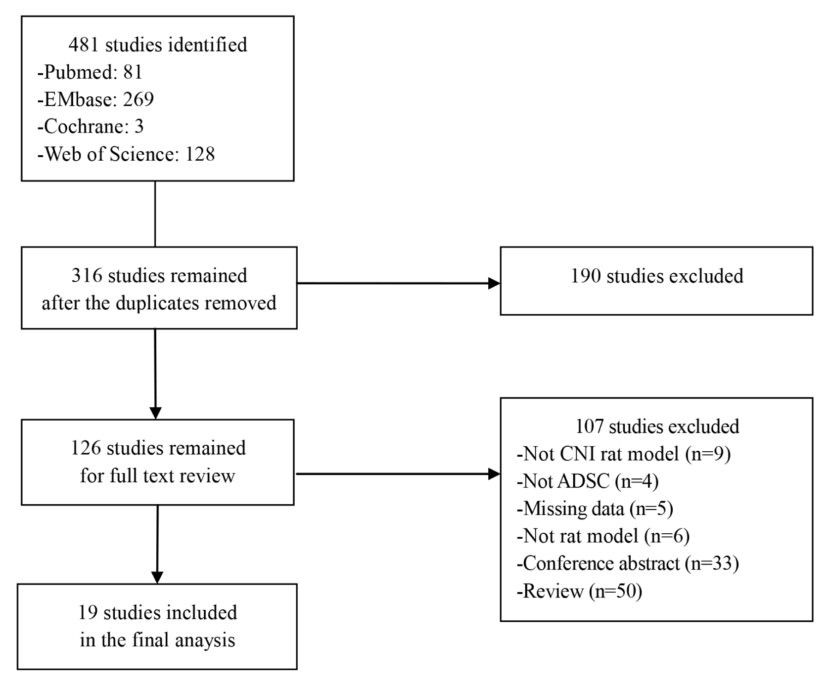
We identified 481 studies from PubMed (n=81), EMBASE (n=269), Cochrane (n=3) and Web of Science (n=128) databases. After removing duplicate articles, 316 studies remained. We excluded 190 studies after a first screening on title and abstract. Another 107 studies were excluded after assessing the full text manuscript. Finally, 19 studies were included in this meta-analysis (Fig. 1).
Study characteristics
Characteristics of the 19 included studies are summarized in Table 1. Eight studies reported on multiple treatments and were considered separate experiments (18–25). A total of 27 animal experiments from 19 studies were included in this meta-analysis. Publication years were from 2010 to 2018. All of the studies used Sprague Dawley (SD) rats. Allogenic, autologous, and human ADSCs were administered in 8 (25–32), 4 (18, 33–35), and 7 studies, respectively (19–24, 36). Injected cell doses were 1×10⁶ in 15 of the studies (19–26, 28–31, 33, 35, 36). Two studies used 2×10⁶ cells (18, 27). One study used 2×10⁵ cells (32). Cell doses in one study were not clear (34). Six studies reported on multiple treatment groups that contained 2 or more combined biological, chemical, or physical treatments, such as growth factor, drug, or shock wave, respectively (19–23, 25). Thirteen studies injected ADSCs into the intracavernous (IC) route (18, 22, 24–27, 29–33, 35, 36). Five studies injected ADSCs into the injured cavernous nerves (19–21, 23, 34). One study injected ADSCs into the grafted vein (28). Two studies evaluated the effect of ADSCs between different delivery routes (18, 24). One of these studies compared injections into the IC route and the dorsal penile perineural space (18); the other study compared periprostatic implantation to IC injection (24). The follow-up periods were from 4 to 12 weeks.
Table 1
Characteristics of the included studies
![]()
Risk of bias assessment in the included studies
The results of study quality and the risk of bias assessment are shown in Table 2. All of the studies mentioned randomization but failed to provide details of the randomization procedure. As a result, the risk of bias in terms of sequence generation and allocation concealment was unclear. All of the studies reported that the rats were Sprague Dawley (SD) rats of similar age and weight at baseline. As a result, the risk of bias for baseline characteristics was low. Three studies mentioned similar housing conditions (30, 33, 35) but did not report whether the rats were randomly housed during the experiment. As a result, the risk of bias related to random housing in the studies was unclear. All of the studies failed to report performance blinding. Five studies had mentioned that blinding of outcome assessment was applied during the experiment (20, 21, 23, 24, 35). However, this was only related to the outcome assessment of structural change. Despite insufficient descriptions, we considered the risk of bias to be low since the study authors had attempted to maintain blinding of the outcome assessors. Two studies had reported that the outcome assessment was randomly conducted. Therefore, random outcome assessment was considered a low bias risk (24, 35). Thirteen studies did not have missing number of rats in each group to analyze. This item was considered low for risk of bias. In one study, the risk of attrition bias was assessed as high because there were discrepancies between the number of rats that completed the study and at baseline (23). In five studies, the number of rats per group was mentioned only once at baseline, and the number of rats completing the study was not reported. Thus, the risk of attrition bias was unclear (19, 21, 28, 32, 33).
Table 2
Individual risk of bias and quality assessment using SYRCLE (Systematic Review Centre for Laboratory Animal Experimentation)
![]()
Efficacy of ADSC on ED
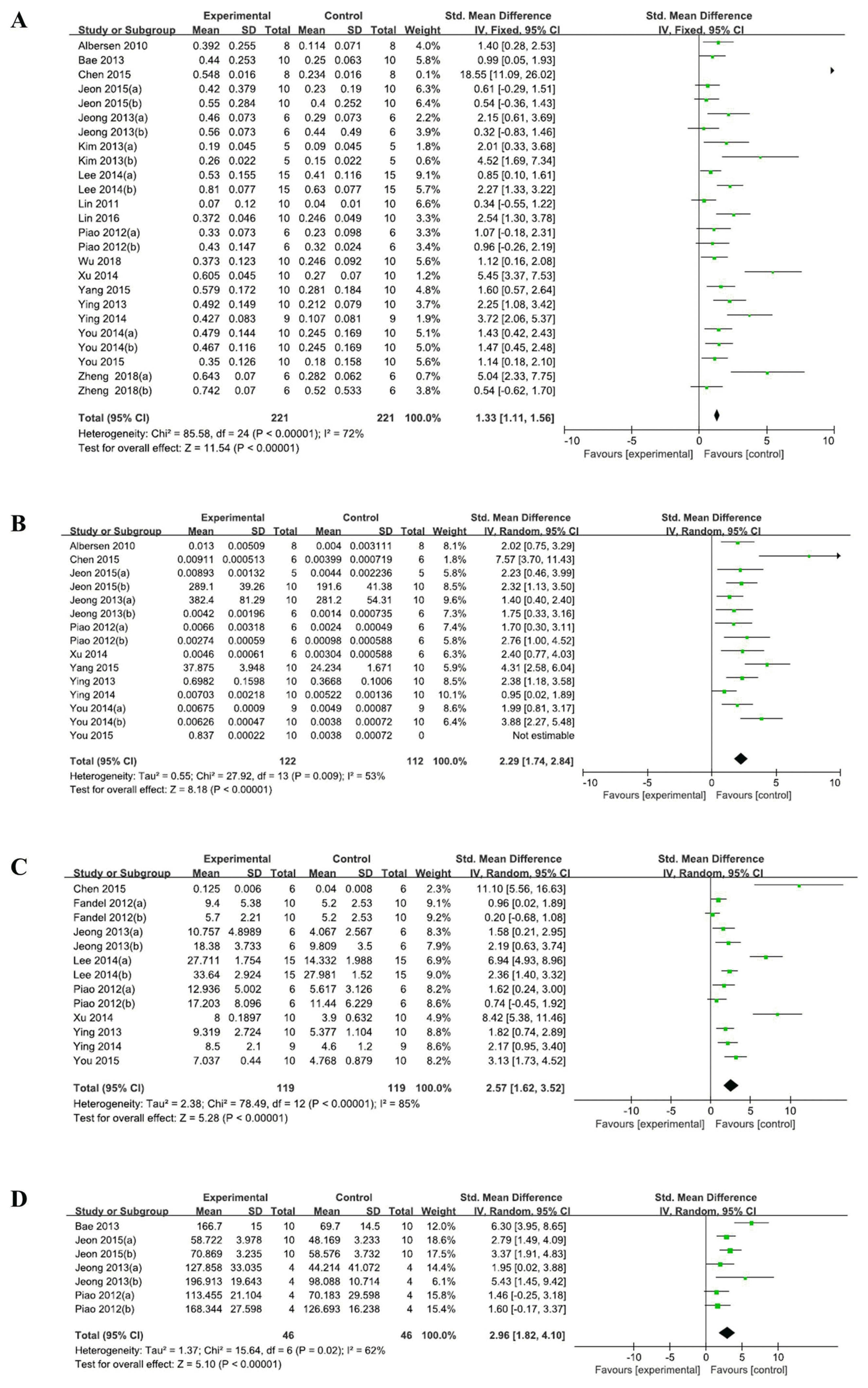
Eighteen out of the 19 studies had reported outcomes related to the ICP/MAP ratio. One study reported only the change in the ICP/MAP ratio from the baseline, and not the ICP/MAP ratio after treatment (18). Accordingly, we could not calculate the ICP/MAP ratio after the treatment from the reported data. We attempted to contact the corresponding author in order to obtain the raw data, but we received no response. Another co-author replied to email but he did not have the raw data. Thus, 18 studies were included in the quantitative analysis. As shown in Fig. 2, ADSC therapy significantly increased the ICP/MAP ratio (SMD 1.33, 95% CI, 1.11 to 1.56) as compared to the control group. Moderate heterogeneity was observed (I2=72%). The nNOS level (SMD 2.29, 95% CI 1.74~2.84; I2=75%), CSM/collagen (SMD 2.57, 95% CI 1.62~3.52; I2=85%), and cGMP (SMD 2.96, 95% CI 1.82~4.10; I2=62%) were increased compared to the control groups. A random-effects model was used for the overall analysis.
Subgroup analyses
Preplanned subgroup analysis was conducted to explore the source of heterogeneity. When studies were compared between the blinding of outcome assessment, the studies with a blinding of the outcome assessment had shown a lesser effect on erectile function (1.33, 95% CI: 0.86 to 1.80) than the studies that had not blinded the outcome assessment (1.81, 95% CI: 1.17 to 2.46). Heterogeneity in studies with outcome assessment blinding reduced by minimal level (I2=23%). When studies were compared according to the origin of ADSC, allogenic ADSCs (2.94, 95% CI: 1.72~4.17) resulted in greater improvement than autologous or human ADSCs (0.90, 95% CI: 0.26~1.54; 1.21, 95% CI: 0.81~1.61, respectively). A high degree of heterogeneity existed in allogenic ADSCs (I2=84%). Based on the follow-up periods, ADSCs showed a similar effect on erectile function between follow-up periods of <6 weeks and ≥6 weeks, and a high degree of heterogeneity remained. Substantial heterogeneity also existed in terms of both route of administration and whether co-intervention was applied or not (Table 3).
Table 3
Subgroup analysis to explore heterogeneity sources and changes in erectile dysfunction
| Subgroup | No. of trials (Tn/Cn) | SMD (95% CI) | I2 |
|---|---|---|---|
| Outcome assessment | |||
| Blinded | 5 (64/64) | 1.33 (0.86 to 1.80) | 23% |
| Unblinded | 13 (157/157) | 1.81 (1.17 to 2.46) | 80% |
| Cell origin | |||
| Autologous | 3 (28/28) | 0.90 (0.26 to 1.54) | 22% |
| Allogenic | 8 (79/79) | 2.94 (1.72 to 4.17) | 84% |
| Human | 7 (114/114) | 1.21 (0.81 to 1.61) | 41% |
| Follow-up period | |||
| <6 weeks | 14 (182/182) | 1.62 (1.12 to 2.12) | 72% |
| ≥6 weeks | 4 (39/39) | 1.73 (0.46 to 2.99) | 80% |
| Route of administration* | |||
| Intracavernous | 14 (138/138) | 1.91 (1.26 to 2.56) | 78% |
| Cavernous nerve | 9 (64/64) | 0.97 (0.43 to 1.50) | 43% |
| Others | 2 (19/19) | 2.49 (0.30 to 4.69) | 81% |
| Co-intervention* | |||
| No | 17 (149/149) | 1.67 (1.11 to 2.22) | 72% |
| Yes | 8 (72/72) | 1.63 (1.18 to 2.08) | 75% |
![]()
Publication bias
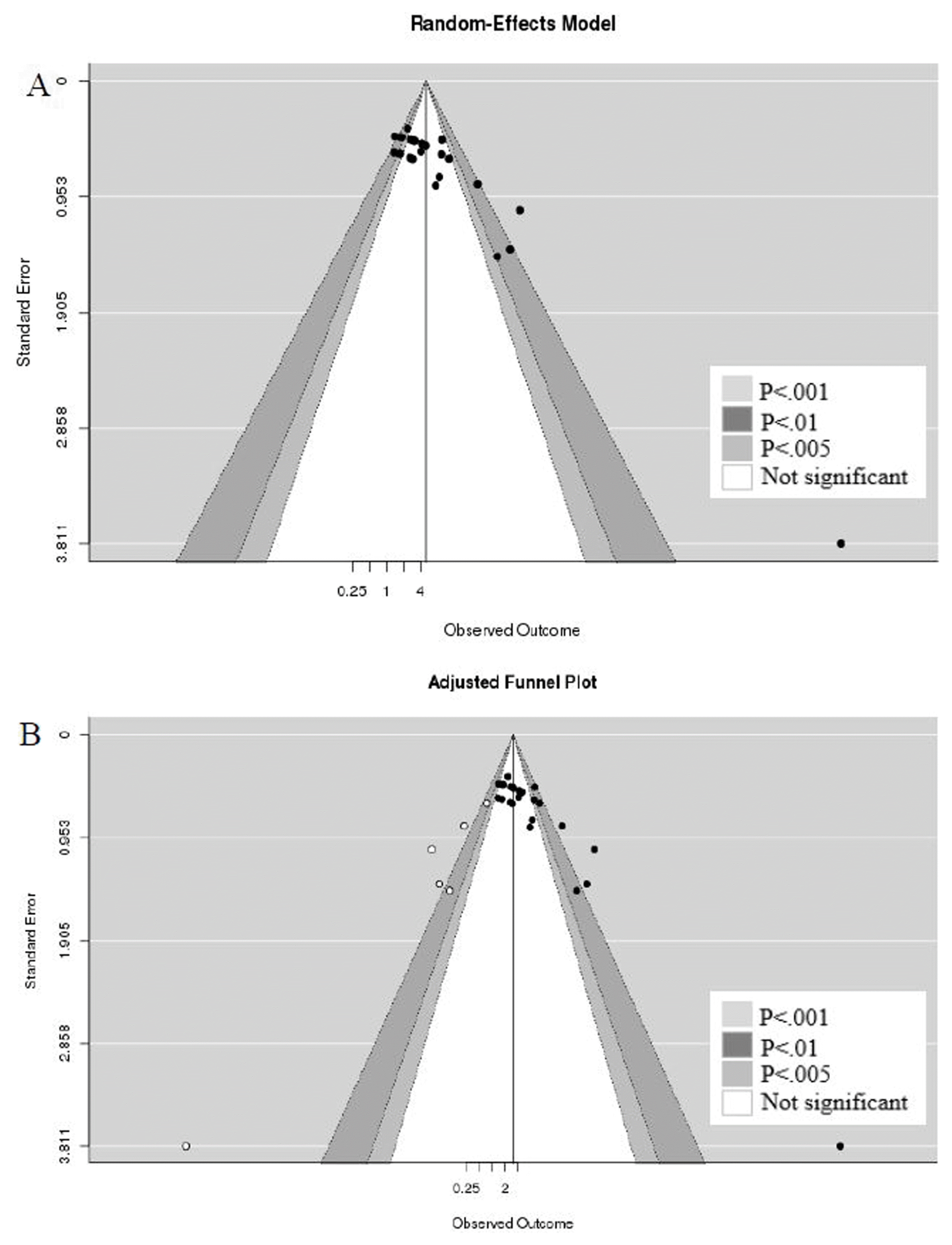
To interpret publication bias, contour enhanced funnel plot was applied. Conventional funnel plot is often interpreted as being caused by publication bias if asymmetry in the appearance of a funnel plot. On the other hand, contour enhanced funnel plot can be enhanced by adding contours of statistical significance to aid in interpreting the funnel plot. If studies appear to be missing in areas of low statistical significance, then it is possible that the asymmetry is due to publication bias. Publication bias was suspected when evaluated with a contour enhanced funnel plot (Fig. 3). We can re-display the funnel plot, taking into account the Trim and Fill adjustment. The trim and fill method is an iterative non-parametric technique to adjust for publication bias by imputing studies estimated to be missing from the funnel plot asymmetry (37). The five imputed studies are shown as white circles, and the imputed estimates of the ICP/MAP ratios were reduced from 1.63 (95% CI: 1.18~2.08) to 1.17 (95% CI 0.64~1.70) after adjustment of the publication bias.
Discussion
Our meta-analysis included 19 studies that investigated the effects of ADSCs in rats with CNI-induced ED. The primary findings showed that ADSC therapy improved erectile function through increased ICP/MAP ratios. ADSC therapy also improved structural changes in the corpus cavernosume, which was evaluated by nNOS levels, cGMP, and CSM/collagen, suggesting that ADSCs had regenerated damaged cavernous tissues.
The nitric oxide (NO)-cGMP pathway is a main modulator in the corpus cavernosum for penile erection. NO produced by nNOS in nervous tissue leads to an increase in the intracellular cGMP concentration, which results in smooth muscle relaxation to attain a penile erection (38). During radical prostatectomy, the incision, heat, and mechanical stress may damage the cavernous nerve. CNI induces neurapraxia, decreased NOS, smooth muscle apoptosis, and penile fibrosis, which result in erectile dysfunction. Although the exact mechanism of ADSC therapy remains unclear, ADSCs have shown a paracrine effect on surrounding smooth muscle, neurons, and endothelium to promote regeneration (10). To elucidate the mechanisms of ADSCs in improving erectile function, a study tracked ADSCs after injection in a CNI rat model using 5-ethynyl-2-deoxyuridine-labeled ADSCs. They had found that CNI upregulated stromal cell-derived factor-1 expression in the major pelvic ganglion (MPG), thereby attracting intracavernously injected ADSCs to the MPG to promote neuroregenerative effects on the cell bodies of injured nerves, resulting in enhanced erectile response (18).
There have been two previous meta-analysis studies on stem cells and ED. Hou et al. (13) had assessed 20 studies that reported that ADSCs significantly improved erectile function induced by various causes of ED in rat models. The nNOS levels, cGMP, and CSM/collagen were also improved in the ADSC group when compared to the control. DM and CNI rats were the most commonly used rat models in the studies; however, the different ED models had no influence on the ADSC efficiency. Moderate to high heterogeneity (I2=68%) was suspected in the meta-analysis though. Shan et al. (14) had used data from 12 studies to analyze the effect of diverse sources of stem cells on CNI-induced ED. The meta-analysis showed a significant difference in erectile function as assessed by the ICP/MAP ratio between the stem cell and control groups (SMD 1.701, 95% CI 1.245~2.158) with significant heterogeneity (I2=60.6%). The previous two studies had analyzed a variety of stem cells and ED models, but they did not evaluate the efficacy of stem cell treatments including the quality assessment of the studies.
To our knowledge, this is the first systematic review and meta-analysis to assess the effect of ADSCs on CNI-induced ED. We evaluated more homogenous sources of stem cells and ED rat models. Moreover, we performed subgroup analysis to identify sources of heterogeneity including quality assessment. Randomization is important in clinical trials in order to balance the distribution of confounding factors. However, randomization in animal studies might have less of an effect on the results than in human clinical trials since animal studies utilize the same species with similar age and weight. On the other hand, blinding the outcome assessor might be crucial in animal studies because researchers may anticipate positive outcomes in their studies. When observer bias is introduced into intervention results, the efficacy of the intervention might be overestimated. In the current study, five of the 19 studies attempted to blind the outcome assessor (20, 21, 23, 24, 35). Those studies showed a lesser effect on erectile function (1.33, 95% CI: 0.86 to 1.80) as compared to the non-blinded studies (1.81, 95% CI: 1.17 to 2.46). In addition, heterogeneity was lower when the outcome assessment was blinded (I2= 23%). Jeong et al. (39) had mentioned that the treatment effect may be overestimated when blinding was not maintained during the study. We assumed that the ADSC efficacy may be overestimated in studies a non-blinded outcome assessor. The cell origin subgroup showed that allogenic ADSCs were significantly more effective for erectile function than autologous or human ADSCs. None of the studies with allogenic ADSCs maintained blinding of outcome assessments.
This study had several limitations. The efficacy of ADSCs from this meta-analysis should be considered carefully because of the small number of studies. When outcome data were presented graphically in some studies, we extracted the numerical data from a graph using a program. Therefore, there might be discrepancies between the numerical data presented on the program and actual data. The quality assessment of studies was generally poor because the randomization method, allocation concealment, and blinding were not described. This poor methodological quality has weak internal validity. In addition, publication bias was also suspected. If the missing studies were systematically different, the results could be biased. Our results showed that the funnel plot was distributed asymmetrically around the mean effect size. Conradi et al. (40) had reported that published studies had more positive outcomes and statistically significant results than unpublished studies in animal research.
Conclusions
Our meta-analysis indicates that ADSCs can be effective in improving erectile function and structural change of CNI-induced ED. However, caution should be used when evaluating the efficacy of ADSCs due to low methodological quality. A non-blinded outcome assessor may cause detection bias and overestimate ADSC efficacy. Therefore, the efficacy of ADSCs in animal models must be further evaluated with a rigorous study design in order to avoid bias.
 XML Download