

 PDF
PDF Citation
 Print
Print


Introduction
Mesenchymal stem cells (MSCs) are multipotent stem cells with a self-renewing capacity, able to differentiate into multiple mesenchymal lineages, such as adipocytes, chondrocytes, osteocytes, smooth muscle cells, fibroblasts and hematopoietic stroma (1).
These cells were first observed between the late 60s and the early 70s by Friedenstein et al., who proved the existence of a non-hematopoietic cell population, fibroblast-like cells, plastic-adherent cells derived from bone marrow (1). MSCs have been documented to originate a wide range of cellular types such as adipocytes, chondrocytes, osteocytes, smooth muscle cells, fibroblasts and hematopoietic supportive stroma (1, 2). MSC-like cells can been collected from different tissues, such as skeletal muscle, adipose tissue, umbilical cord, synovium, dental pulp, amniotic fluid, as well as fetal blood, liver, bone marrow, lung and heart (2, 3).
Although MSCs derive from different tissues, they do share some common features: they do not display the peculiar markers that hematopoietic and endothelial cells express, like Cluster of Differentiation (CD)34, CD45, CD11b, CD11c, CD14, CD19, CD79α, CD86, and HLA class II molecules and they do express surface markers, in accordance with the commonly accepted minimal criteria of the International Society for Cellular Therapy (ISCT), such as CD90, CD105, CD44, CD73, CD9, and very low levels of CD80 (4).
MSCs can be divided into two classes: adult and fetal/perinatal MSCs, respectively derived from adult tissues, such as bone marrow (BM-MSCs) and adipose tissue (AD-MSCs) (5), and from fetal/perinatal tissues, that include cells obtained from the embryo/foetus itself and cells obtained from extra-embrionic tissues otherwise known as birth associated tissues, such as placenta umbilical cord, Wharton’s jelly mesenchymal stem cells (WJ-MSCs) and amniotic membrane (6). MSCs isolated from adult tissues are most commonly used for therapeutic purposes, despite a very limited proliferative capacity. Instead, MSCs derived from extra embryonic tissues, represent ideal choice for therapeutic use, as they share many characteristics with adult MSCs, like BM-MSCs. Although they retain several features of embryonic stem cells (ESCs) they do not form teratomas (7) and their research does not raise any 3ethical or legal issues related to their applications in regenerative medicine. During their short life, perinatal MSCs, such as WJ-MSCs, are spared from pro-aging factors. It has also been shown that the age of the donor tissue affects many properties of MSCs (8). All these features open perspectives for the diffusion and engineering of umbilical cord derived MSCs for cell-based therapies. Additionally, fetal MSCs possess an immune-privileged status which makes them a favourable choice for regenerative medical applications (9).
The first description of Wharton’s Jelly (WJ) by Thomas Wharton dates back to 1656. It is a mucous connective tissue of the umbilical cord located between the amniotic epithelium and the umbilical vessels. McElreavey et al. (10), 1991, was the first to isolate MSCs from the WJ portion of the umbilical cord.
Several studies indicate that WJ-MSCs can be used in many fields such as neurological disorders (11), kidney injury (12), lung injury (13), orthopaedic injury (14), liver injury (15), cancer therapy (16).
This review will first attempt to provide a summary of the biology of MSCs derived from WJ and then discuss their potential application for the treatment of immune-mediated disorders such as GvHD. Additionally, it aims to provide a panoramic view of the most recent clinical trials involving WJ-MSCs for therapeutic use.
This review includes papers identified through a MEDLINE and EMBASE search using combinations of the following subject heading terms: WJ-MSCs, Immunomodulation, Graft versus Host Disease (GvHD), Mesenchymal Stem Cells, Human umbilical cord blood.
Of the retrieved articles, 83 papers regarding WJ-MSCs, their biological properties and their therapeutic potential were carefully selected, and therefore added together to describe the therapeutic benefits of WJ-MSCs, in particular their role in the management of GvHD.
Go to : 
Immunomodulatory Property of WJ-MSCs
A mandatory requisite for allogeneic transplantation is low cellular immunogenicity. The therapeutic utility of the WJ-MSCs is strictly associated with their regenerative and immunomodulatory potential. WJ-MSCs are capable of immune suppression and immune avoidance, making them optimal candidates for cellular therapies in allogenic transplantation. Furthermore, WJ-MSCs exhibit very low expression of Human Leukocyte Antigen (HLA) class I and an absence of HLA-DR (17, 18). Additionally, it induces the expansion of regulatory T cells (Treg), which would contribute to the suppression of the effectors responses to alloantigens (19). WJ-MSCs are not capable of generating in vitro immune responses from allogeneic T cells (20), suggesting that WJ-MSCs possess a specific low immunogenicity and will be tolerated in allogeneic transplantation. Unlike BM-MSCs, WJ-MSCs produce large amounts of tolerogenic Interleukin (IL)-10, Transforming Growth Factor-β (TGF-β) (20–22) and they also express IL-6 and vascular endothelial growth factor (VEGF), which are important for the immunosuppressive capability of MSCs (20, 23). The immunosuppressive potential of WJ-MSCs on T lymphocytes (18) was investigated. Soluble factors, such as hepatocyte growth factor (HGF), prostaglandin E2 (PGE2), TGF-β1 and indoleamine 2 3-dioxygenase (IDO) (24) may mediate the immunomodulatory effects of MSCs on T cells (Fig. 1). WJ-MSCs, when cultured with CD14+ monocytes, inhibit their differentiation into mature DCs, both via direct contact and through soluble factors (Fig. 1) (25). The immunomodulatory effects of MSCs concern both the cell-mediated and humoral component of immune system. However, the impact of WJ-MSCs on B cells has been poorly investigated. Until today, only a few studies have been reported: in 2012 Che et al. (26), it was reported that WJ-MSCs, in vitro, are capable of inhibiting B cell proliferation, differentiation and antibody production; Other authors, however, proved that the WJ-MSCs have no influence on the activation of B cells (27). Further investigation is needed.
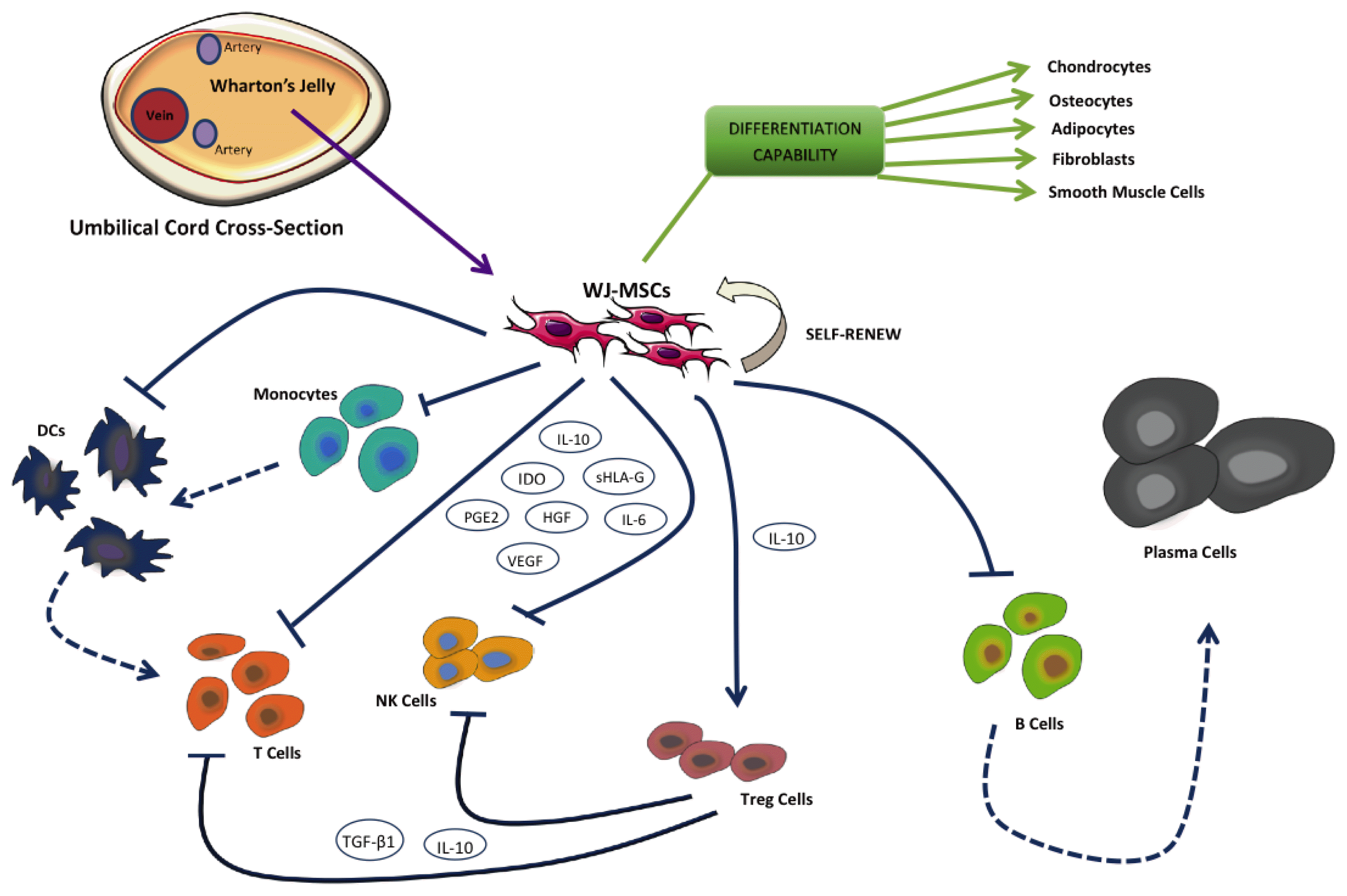 | Fig. 1MSCs mediate immunosuppression of B, T and Natural Killer (NK) cells via different mechanisms. Soluble factors secreted by MSCs such as IDO, PGE2, sHLA-G5 can suppress T and NK cell functions. In addition, MSCs can indirectly mediate immunosuppression by inhibiting dendritic cells (DCs) and inducing the expansion of regulatory T cells (Tregs).
|
In summary, immunomodulatory properties of WJ-MSCs, confers upon these cells the potential for therapeutic application.
Go to :
Phenotypic and Genetic Markers Expression of WJ-MSCs
WJ-MSCs are good candidates for therapeutic applications because of their primitive features (28), as they exhibit several characteristics of ESCs, such as ESC-like antigen Tra-1-60, Tra-1-81, SSEA-1 and SSEA-4 (29), and numerous pluripotency genes like: Oct-4, NANOG, and SOX-2 (28).
Nekanti et al. (30), in a comparative gene expression profiling study between WJ-MSCs and BM-MSCs, demonstrated that BM-MSCs express higher levels of NANOG, DNMT3B and GABRB3 and an increased expression of Brix, CD9, Gal, Kit and Rex1, a pluripotent/stem cell marker. Their expression is thought to reflect the more primitive nature of WJ-MSCs, compared to their adult counterparts in the bone marrow. These findings, combined with the fact that human WJ-MSCs express the primitive stem cell marker TERT (31) explain, at least in part, the observation that WJ-MSCs have a shorter doubling time (32) and a more extensive in vivo expansion capacity (7).
The most peculiar characteristic of WJ-MSCs is their ability to express the HLA-G6 isoform, implicated, as previously mentioned, in immune-modulation, an essential feature for promoting the use of WJ-MSCs in a cell-based therapy.
WJ provides a generous source of MSCs, and, although WJ-MSCs show some variations in terms of cell quality, it is still a very useful depot of MSCs, with a wide range of potential therapeutic applications.
Go to :
WJ-MSCs for the Management of Graft-Versus-Host Disease
Allogeneic hematopoietic stem cell transplantation (allo-HSCT), which consists in the infusion of hematopoietic stem cells (HSCs) from a matching donor, is a fundamental therapeutic option for several hematologic malignancies.
In about 50% of cases, the procedure is complicated by acute or chronic graft-versus-host disease (aGvHD and cGvHD, respectively) and clinical conditions occur when donor-derived T cells recognize the host cells as non-self and attack them (33).
aGvHD commonly involves the skin, intestinal tract and liver, frequently manifesting itself as a maculopapular rash, nausea, vomiting, diarrhea and hepatic cholestasis. cGvHD generally occurs after 100 days from the transplantation and leads to inflammation and fibrosis of involved organs, presenting as sicca syndrome-like, scleroderma-like skin, cytopenias, chronic pulmonary fibrosis, hepatic and intestinal diseases (34).
The main risk factor associated with the development and severity of GvHD is that the HLA may mismatch between donor and recipient.
The primary treatment for both aGvHD and cGvHD involves immunosuppression by glucocorticoids, with a response rate ranging from 30 to 50%. On the contrary, many immunosuppressive strategies have been studied for steroid-resistant aGvHD (such as mycophenolate mofetil, pentostatin, monoclonal antibodies directed against T lymphocytes, cytokines and their receptors, mTOR inhibitors and extracorporeal photopheresis) but none have proven to have a consistent effectiveness and safety level (35), so aGvHD is still characterized by a poor prognosis and a Treatment-related mortality (TRM) >50% reported in several studies (33). Therefore, alternative strategies are needed to treat GvHD.
Promising treatments for steroid-refractory aGvHD involve the infusion of ex vivo expanded MSCs. The majority of data published reported the use of the BM as standard source of MSCs for human clinical applications. However, to harvest the MSCs from BM a painful, invasive procedure with certain risks is required.
Kang-Hsi Wu et al. in 2011 reported the first case of Wharton’s Jelly derived MSCs used in a human clinical application, in two pediatric patients with severe steroid-resistant aGvHD (36).
The first patient was a 4-year-old boy who required an unrelated 6/6 HLA-matched allo-HSCT for severe aplastic anemia not responding to immunosuppression. On day 45, the patient developed an aGvHD with vomiting, hyperbilirubinemia, and maculopapular rash on both legs and arms. A steroid therapy was ineffective. One unit of WJ-MSCs 4/6 HLA-matched was obtained and expanded in vitro to 6.6×10⁷ cells (3.3×10⁶ cells/kg body weight) at passage 3 and then cryopreserved again. A normal karyotype and the absence of pathogenic contamination were confirmed and then, the WJ-MSCs were thawed, washed, and infused into the patient through a central venous catheter. Before the infusion, the grade of aGvHD was IV (gut staging: 4; skin staging: 3; liver staging: 3) according to standard aGvHD criteria proposed by Glucksberg et al. (37). Two days after infusing the WJ-MSCs, the severity of the diarrhea improved and subsequently subsided, the total bilirubin concentration decreased to a normal range in 6 days and the skin rash declined and disappeared in 7 days. On day 28 after the infusion, the recurrence of a III grade aGvHD (gut staging: 3; skin staging: 2; liver staging: 3) was treated with a 3/6 HLA-matched WJ-MSCs expanded to 7.2×10⁶ cells/kg at passage 3 with the complete disappearance of symptoms within 6 days. Despite continued cyclosporine administration, by day 25 after the second MSCs infusion, the IV aGvHD (gut staging: 4; skin staging: 3; liver staging: 2) flared up again and the patient received a third infusion of WJ-MSCs (8.0×10⁶ cells/kg at passage 4) matching 3/6 HLAs with total regression of manifestations 6 days after the WJ-MSCs infusion. Cyclosporine was slowly tapered 2 months after the third infusion, all immunosuppressive drugs were discontinued 12 months after HSCT and no chronic GvHD was observed.
The second patient was a 6-year-old boy transplanted with 2 units of umbilical cord blood 4 of 6 HLA matched for an acute lymphoblastic leukemia in second remission. On day 21, the patient developed hyperbilirubinemia and maculopapular rash.
He received a 3/6 matching HLA MSCs infusion expanded to 4.1×10⁶ kg at passage 3. Clinical manifestations of aGvHD intestinal, cutaneous or hepatic disappeared 5 days after the MSC infusion. Cyclosporine was tapered slowly 2 months after the MSCs infusion, all immunosuppressive drugs were discontinued 10 months after transplantation and no chronic GVHD was observed. No side effects and no severe infections incurred during or after each MSCs infusion in both patients.
In 2016, Boruczkowski et al. (38) described the use of WJ-MSCs for the treatment of 10 patients with steroid-resistant GvHD (7 patients diagnosed with aGvHD and 3 patients with cGvHD). After 1 to 3 WJ-MSC infusions at a median dose of 1.5×10⁶ cells/kg of recipient body weight, a complete or partial response was observed in 4 patients with aGvHD and in 2 patients with cGvHD. Five out of 6 patients (83.3%) from the responder group and only 1 out of 4 non-responders (25%) survived the follow-up. The deaths occurred in patients with aGvHD. In the responder group, 2 patients showed complete remission of GvHD while symptoms were alleviated in 4 patients and the intensity of immunosuppressive therapy could be reduced. No serious adverse effects were observed. Interestingly, it seems that the therapy outcome did not correlate with the number of infusions, while the timing of infusion is crucial for therapy outcomes: in fact, 2 cases of complete remission were observed in patients who received only 1 infusion, but those patients received MSCs infusions 22 and 24 days after the GvHD diagnosis, which were the earliest cell applications in the aGvHD group (38).
In 2015 a second author, Wu et al. (39), futher documented that MSCs could be used for clinical treatment of refractory extensive cGvHD or aGvHD graded II–III. Twenty-four patients were included in the study: 8 patients suffered from aGvHD (4 grade III, 2 grade IV, 2 grade II), 16 patients cGvHD. All the patients received umbilical cord blood-derived MSCs infusion once. The average MSC dose given was 0.6×10⁶/kg body weight (range 0.5~1.0×10⁶/kg body weight). After a mean follow-up period of 1.5 years (range 1 month~2 years), 22 patients were still alive and 2 patients died: one due to IV aGvHD and one during liver transplant surgery.
The clinical manifestations of GvHD improved significantly in about one month after MSC infusion, with skin and oral mucosa involvement improving remarkably at the effective rates of 55.6% and 100%, respectively. However, hepatic and pulmonary involvement underwent no significant improvements. At the last follow-up, the immunosuppressive agent therapy, after MSCs transfusion, was tapered and/or discontinued in 18 patients, while 2 patients with no remission and 2 patients, who achieved progressive disease (PD), maintained their basic therapies or required further increases of immunosuppressive agents. None of the surviving patients were found to have experienced a relapse of the primary disease, the incidence of infection or other transplant-related complications. No infusion-related toxicity was observed during or after the administration of MSCs. Table 1 provides an overview of the international literature regarding the management of Graft Versus Host Disease with WJ-MSCs infusions.
Table 1
Literature overview for the management of graft versus host disease with WJ-MSCs infusions
![]()
Go to :
Current Clinical Trials of WJ-MSCs
The current trials regarding WJ-MSCs were investigated by a study on the website clinicaltrials.gov. Nine results were obtained using the following research queries: “Wharton’s Jelly Mesenchymal Stem Cell”, “Wharton Jelly Stem Cell”, “WJMSC”, “WJ Mesenchymal Stem Cell” and “Wharton Jelly Cell”. Two of these trials are complete, one has an unknown status, one is enrolling by invitation, one is not yet recruiting and four are recruiting (Table 2).
Table 2
Ongoing trials on WJ-MSCs from clinicaltrial.gov
| Clinicaltrials.gov Identifier | Condition | First registration/phase | Treatment | Estimated enrollment | Sponsor | Final goal |
|---|---|---|---|---|---|---|
| NCT0129-1329 | Elevation myocardial infarction | February 2011/2 | Intracoronary infusion of WJ-MSCs or placebo medium into the infarct artery 4~7 days after successful reperfusion therapy | 160 | Navy General Hospital, Beijing | Investigate the efficacy of intracoronary WJ-MSCs |
| NCT0236-8587 | Ischemmic cardiomyophaty | March 2015/2 | Intracoronary infusion of WJ-MSCs or placebo in Patients with ischemic heart failure. | 160 | Navy General Hospital, Beijing | Investigate the therapeutic safety and efficacy of WJ-MSCs in patients with ischemic cardiomyopathy |
| Intravenous infusion of WJ-MSCs or placebo in Patients with ischemic heart failure | ||||||
| NCT0116-6776 | Varices of umbilical cord | June 2010 | Isolate Wharton’s jelly matrix; test its ability to support the growth and differentiation of transplanted | 64 | University of Kansas Medical Center | Identifie Wharton’s jelly matrix characteristics for used in tissue engineering |
| MSCs; perform preliminary animal test to study its ability to support bone tissue regeneration in vivo | ||||||
| NCT0164-9752 | Improving implantation rates | April 2015/1 | A differentiated stem cell therapy group; a undifferentiated stem cell therapy group; a control group | 60 | Kasr El Aini Hospital | Test the effect of placental derived MSC in improving implantation rates in selected patients |
| NCT0288-1476 | Amyotrophic lateral sclerosis | November 2015/1 | Group I - patients receiving intrathecally one application of WJ-MSCs | 30 | University of Warmia and Mazury | Investigate the tolerability of allogeneic WJ-MSCs |
| Group II - patients receiving intrathecally three applications of WJ-MSCs | ||||||
| NCT0294-5449 | Erectile dysfunction | January 2017/1,2 | Dose I: Three intracavernous injections of 30×106 WJ-MSCs | 15 | Sophia Al-Adwan | Treat the erectile dysfunction in diabetic patients |
| Dose II: Three intracavernous injections of 60×106 WJ-MSCs | ||||||
| Dose III: Three intracavernous injections of 90×106 WJ-MSCs | ||||||
| NCT0300-3364 | Chronic traumatic spinal cord injury | December 2016/1,2 | XCEL-UMC-BETA (initial treatment)/Placebo (month 6) | 10 | Banc de Sang i Teixits | Obtain efficacy data in intrathecal administration of expanded WJ-MSCs |
| Placebo (initial treatment)/XCEL-UMC-BETA (month 6) | ||||||
| NCT0296-3727 | Knee osteoarthrosis | January 2017/1 | Intra-articular injection of WJ-MSCs in 2 doses each of 50 million of cells | 10 | University of Jordan | Inject WJ-MSCs in patients with Knee osteoarthrosis |
| NCT0216-6294 | Diabetic foot ulcers | June 2014 | Pressure bandage; Standard of Care Cross over to NEOX. | 30 | Amniox Medical, Inc. | Evaluate the efficacy and safety of NEOX® CORD 1K in patients suffering from non-healing diabetic foot ulcers |
![]()
“Intracoronary Human Wharton’s Jelly-Derived Mesenchymal Stem Cells (WJ-MSCs) Transfer in patients with acute myocardial infarction (AMI)” (NCT01291329) (40) is a completed trial held by the Navy General Hospital of Beijing. The purpose of the trial was to investigate the efficacy and safety of the intracoronary transfer of WJMSCs in patients with ST-segment elevation myocardial infarction. The Navy General Hospital of Beijing is also the sponsor of the “Intracoronary or intravenous infusion human Wharton’ Jelly-derived Mesenchymal Stem Cells in patients with ischemic cardiomyopathy” (NCT-02368587) study, which, with similar objectives to the previous study, investigates the aptitude of WJ-MSCs to be differentiated into cardiomyocytes and endothelial cells and be integrated in vascular and ischemic cardiac tissue, improving heart function in patients with ischemic cardiomyopathy. To date, the study is not yet enrolling.
The other completed trial is “A research study looking at specific tissue of the umbilical cord” (NCT01166776), sponsored by the university of Kansas medical center. This study aims to investigate not the use of WJ-MSCs, but the WJ matrix without the cellular component as a scaffold for the implantation of MSCs with origins not otherwise specified.
The “Role of stem cells in improving implantation rates in Intracytoplasmic Sperm Injection ICSI patients” (NCT-01649752) study status is unknown, since the completion date has passed and the status has not been updated in over two years. This study, sponsored by Kasr El Aini hospital, aimed to improve endometrial receptivity before transferring good embryos, taking advantage of the regenerative properties of MSCs. Both placenta-derived MSCs and WJ-MSCs were induced to differentiate in endometrium. This target was achieved by the placenta-derived population, which was later deposited in the uterine cavity.
The study “Therapeutic treatment of amyotrophic lateral sclerosis” (ALS) (NCT02881476), carried out by university of Warmia and Mazury, is enrolling participants by invitation only. The WJ-MSCs are administrated intrathecally in one application for one group and in three applications for the second. The therapeutic outcome is hypothesized on the base of a neurocrine and paracrine effect of WJ-MSCs, which are investigated in terms of their capacity to influence the neurodegeneration present in ALS.
The study “Use of WJ in erectile dysfunction” (NCT-02945449), is being conducted by Sophia Al-Adwan, university of Jordan. This study, which is now enrolling patient, is designed to evaluate the effect of intracavernous WJ-MSCs injection in diabetic patients with erectile dysfunction. The patients are divided in three groups, each receiving a different dose of WJ-MSCs. The efficacy is evaluated with doppler ultrasound and a sexual health inventory for men/international index of erectile function/erection hardness score questionnaire.
Another study, which is now recruiting patients is “Intrathecal administration of expanded WJ-MSCs in chronic traumatic spinal cord injury” (NCT03003364) by Banc de Sang i Teixits. This randomized, double-blind, single-dose, placebo-controlled trial examines the advantages that intrathecal administration of WJ-MSCs can produce in patients with chronic traumatic spinal cord disease. These effects are evaluated with scales, questionnaires, electrophysiology, imaging techniques and laboratory findings.
“Use of WJMSCs for knee osteoarthrosis” (NCT02963727) is a trial that evaluates the efficacy of intra-articular WJ-MSCs injection in patients with knee osteoarthrosis. This trial, sponsored by the university of Jordan, is recruiting patients and assesses the efficacy of the treatment by MRI at 12 months.
The last clinical trial founded as a result of our research is the “NEOX CORD 1K vs standard of care in non-healing diabetic foot ulcers (CONDUCT I)” trial (NCT02166294), sponsored by Amniox Medical Inc., NEOX CORD 1K is a cryopreserved umbilical cord allograft, containing only WJ matrix but not cells. The trial compares the time for closure of the ulcer using NEOX CORD 1K versus the standard treatment, with pressure bandage.
Go to :
Conclusions
As seen above, WJ possess the essential requisites to express a huge immunomodulation potential, which is, in fact, proven by early clinical data regarding the treatment of GvHD. Actual clinical trials hypothesize that WJ-MSCs can be used to improve patients’ health or the outcome of life-threatening conditions.
Considering the increasing interest in the role of stem cells in regenerative medicine, the need for immunomodulatory treatment with lighter side effects and the spreading of allogenic transplantation as a therapy for consolidating haematological neoplasm as well as a saving therapy in several intractable diseases, WJ appears to play a leading role in the future of medicine and be the fulcrum of the newest research.
Go to :
 XML Download