

 PDF
PDF Citation
 Print
Print


Introduction
Anterior cruciate ligament (ACL) injuries are common in sports and it may result in symptoms such as movement dysfunction, knee instability, meniscus damage and early post-traumatic osteoarthritis (1, 2). Because of the poor regenerative potential of implanted graft, ACL reconstruction often results in the failure (3–5). The success of ACL reconstruction depends on many factors, including the initial mechanical properties of the graft (6, 7), positioning of the femoral and tibial tunnel (8, 9), fixation method (10, 11) and the postoperative rehabilitation. In addition, the biologic behavior of the graft is the one of the most important factors determining the mechanical properties of the grafts (12).
Improvement of early and secure graft integration to the bone can enable allow early and aggressive rehabilitation after ACL reconstruction. With the advanced knowledge of ACL healing and the development of biological strategies, many biologic agents have been introduced and aggressively developed in recent years. These include plate-let-rich plasma (13, 14) growth factors (13, 15), and stem cell therapy (16–19). Among these agents, MSCs derived from bone marrow (bMSCs) have demonstrated promising potential in promoting graft osteointegration and graft healing (16–18). As well, the clinical application of mesenchymal stem cells (MSCs) has been explored as a way of enhancing the tissue regeneration in many orthopedic fields. MSCs are multipotent cells that can differentiate into variable cell types, including osteoblasts, chondrocytes, adipocyte and tenocytes (20–22), and are being investigated extensively in the enhancement of healing of tendon injuries and improvement of ACL reconstruction (23, 24). Fibrin glue is a formulation of fibrinogen and thrombin that is used to create a fibrin clot (25). Fibrin glue promotes repair of various connective tissues including cartilage and plays essential role in carrier of stem cells in ACL reconstruction in animal models (26, 27).
This study investigated three groups (control, fibirin, and MSCs group) and we conducted radiologic assessment with Quantum GX μCT imaging system for exact measurement of bone density, tunnel diameters, and tunnel lengths. To our knowledge, there were no studies which investigated three groups for bone density, tunnel widening and length with micro-CT. The purpose of the present study was to investigate the effect of MSCs or fibrin glue on tunnel widening after ACL reconstruction compared with biologic-free control using radiologic and histologic evaluations in a rabbit model.
Go to : 
Materials and Methods
Animal and experimental design
Eighteen healthy, adult female New Zealand white rabbits weighing 3,000 to 3,500 g underwent bilateral ACL reconstruction using an extensor digitorum longus tendon autograft harvested from the same surgical site. The physical conditions of all knees were carefully monitored during the experimental period. All experimental animal procedures were performed in strict accordance with our Institutional Animal Care and Use Committee. All knees were distributed into three groups according to use the biologic agent; control group (n=12) without any biologic agent, fibrin group (n=12) and MSCs group (n=12) with stem cell and fibrin.
Preparation of MSCs
Bone marrow aspiration was carried out from the iliac crest of the rabbits in the control group as previously described (25, 26). Bone marrow (1 ml) was collected and put in complete Dulbecco’s Modified Eagle Medium (DMEM-lg, Gibco, USA) containing 10% fetal bovine serum and cephazolin l g/ml. The sample was washed twice with DMEM-lg and centrifuged at 2,000 rpm for 6 min. The supernatant was removed and the precipitating component was suspended in 10 ml of stem cell growth medium. The stem cells were counted and plated (1.0 to 1.5×10⁷ cells) in a 100 mm dish. Cultures were incubated at 37°C in an atmosphere 5% CO2. After three days, non-adherent cells were removed when the medium was replaced with fresh medium. Once the colonies of MSCs reached 70~80% confluence, adherent cells were obtained from the flask with 0.05% trypsin. Homogenous MSCs were obtained after 2 weeks of culture. Passage two MSCs (P-2) were used.
Surgical technique
Each animal was then anesthetized with ketamine (35 mg/kg) and positioned in supinen. Both knees of each rabbit were shaved, scrubbed with betadine and covered with a sterile drape exposing only the unilateral knee. After midline incision, a medial para-patellar approach was performed and then the native ACL and extensor digitorum longi (EDL) tendon were identified (28) and the length was measured (Fig. 1A). The EDL tendon was harvested to a length of 20 mm or more and was kept immersed in sterile saline until implantation (Fig. 1B). After excision of the native ACL at its femoral and tibial origins, a 2 mm-diameter bone tunnel was made at the footprints of the native ACL on the tibia and lateral femoral condyle (Fig. 1C, D). The reproducible and accurate tunnel positioning in relation to the native ACL footprint was necessary to achieve loading conditions similar to those of native knee joint. The free end of each EDL graft secured with 3-0 vicryl. In the fibrin group, 2 mm of fibrin glue (TISSEEL Kit, IMMUNO AG Vienna, Austria) was injected to the end of graft and MSCs embedded in fibrin glue by mixing was injected in the MSCs group. A total of 1×10⁶ bone marrow stem cell with 1 ml fibrin glue were injected in the end of graft and bone tunnel in MSCs groups. In fibrin group, a total of 2 ml fibrin glue was injected in graft and tunnel. The prepared graft was passed through the tibial and femoral tunnels (Fig. 1E). The gap between the graft and the tunnel was filled with each biologic agent. A total of 1×10⁷ stem cells with 1 ml fibrin glue were injected to the end of graft and filled into the tunnel in MSCs groups. After extra-osseous post-screws were inserted near the tunnel orifices, the end of each implanted graft was anchored to screws using sutures (Fig. 1F). The incision was closed in layers with the arthrotomy and subcutaneous layers. For initial stability, the postoperative knee was maintained in extension by an elastic bandage. Then the contralateral knee was operated on using the same method. All animals were euthanized at 12 weeks postoperatively for histologic analysis and for the evaluation of tunnel widening using micro-CT analysis.
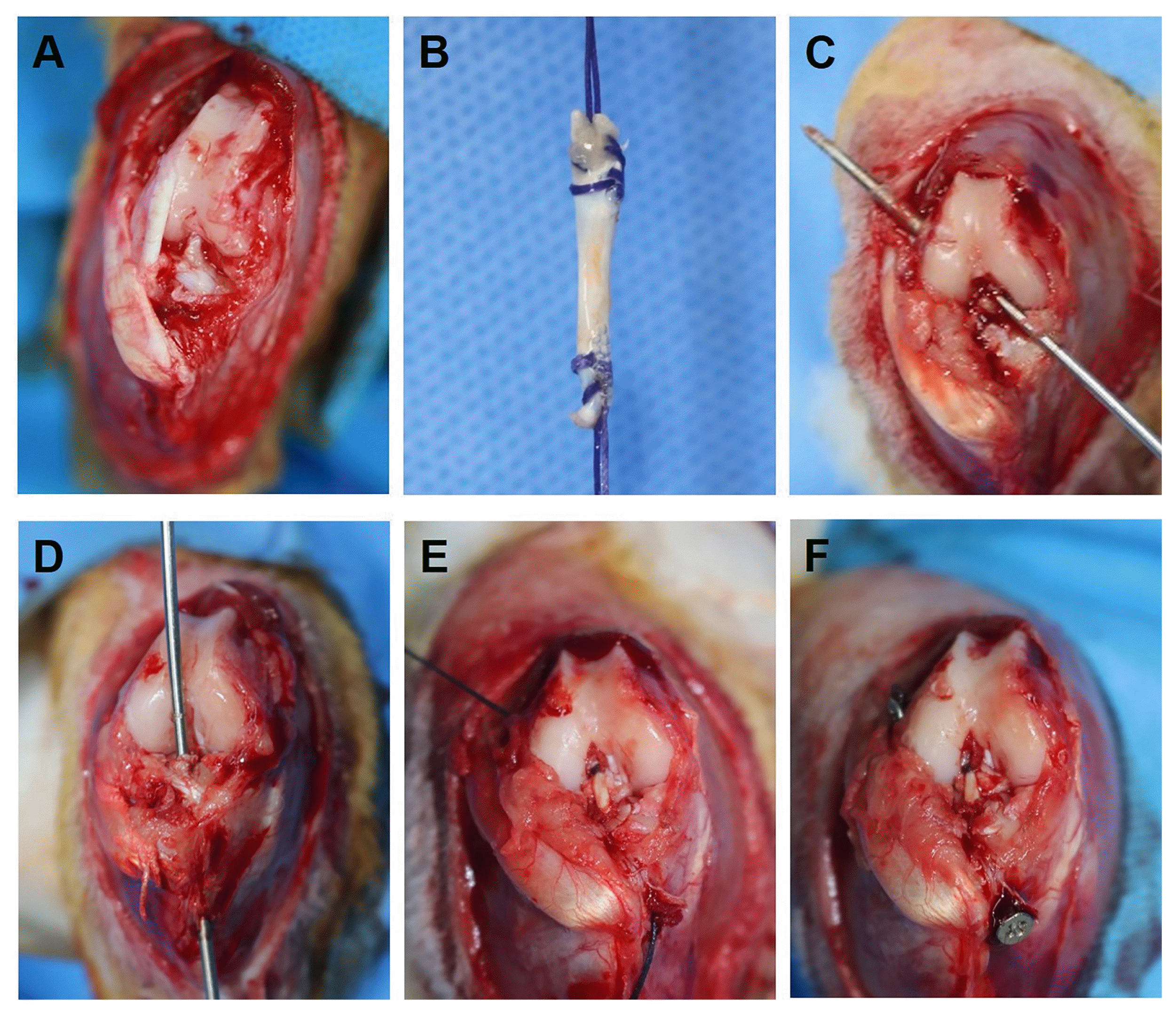 | Fig. 1Surgical process of ACL reconstruction in rabbit model. (A) The native ACL and extensor digitorum longus were identified after medial para-patellar arthrotomy. (B) The EDL tendon with 2 mm diameter and 25 mm length was harvested. (C, D) Femoral and Tibial tunnel were made using 2 mm drill at footprints of native ACL. (E) The harvested tendon was passed through the femoral and tibial tunnel and (F) was tethered with suture to the screw inserted into the bone.
|
Radiologic analysis
The knee joint was obtained by amputating at femoral and tibial shaft after euthanasia. CT imaging was performed using a Quantum GX μCT imaging system (PerkinElmer, Waltham, MA, USA). The X-ray source was set to levels of 90 kV and 88 mA with a field of view of 72 mm with a slice thickness of 0.144 mm. The scanning time was 14 min in a 360° rotation. A matrix size of 512×512×512 was then used to reconstruct using 3D Viewer, existing software within the Quantum GX. Following scanning, image segmentation was performed by Analyze software (AnalyzeDirect, Overland Park, KS, USA). To calculate the tunnel widening, the oblique-coronal and oblique-sagittal plane were obtained parallel to the tunnel (Fig. 2). The tunnel diameter was measured on 2 mm proximal and distal from the femoral and tibial articular surface. The tunnel widening was defined as difference between measured diameter on CT scanning and drilled hole diameter (2 mm).
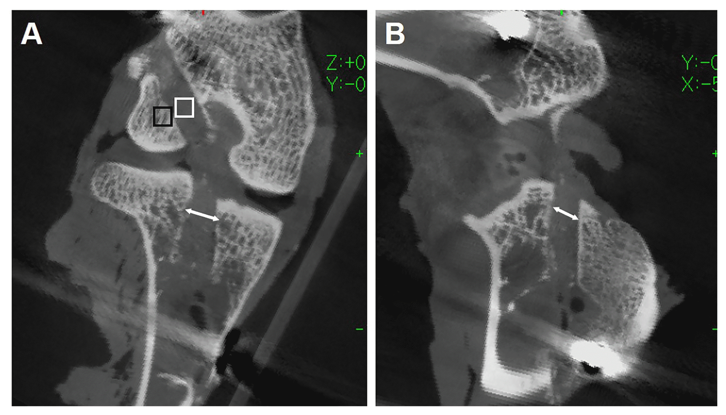 | Fig. 2CT image measuring tunnel widening and Hounsfield units (HUs). The tunnel diameter (white arrow) were calculated on the oblique-coronal (A) and oblique-sagittal image (B) 2 mm distal from the femoral and tibial articular surface. On the same distance from articular surface, HUs were measured in regions of interest (1.5 mm×1.5 mm squared) in the tunnel (white square) and the cancellous bone around the tunnel (black square).
|
Then, Raw CT values were converted to Hounsfield Unit (HU) using intensity values of dry air was −1,000 HU and that of a water was 0 HU. To define each 1.5 mm×1.5 mm squared specific regions of interest (ROI) within the rabbit knees, the oblique plane images of the bone tunnel 2 mm proximal and distal from the femoral and tibial articular surface were analyzed using Quantum GX software. HUs were measured in each ROI in the tunnel and around tunnel in cancellous bone. All radiographic measurements were performed by two blinded observers not participating in surgery.
Histologic analysis
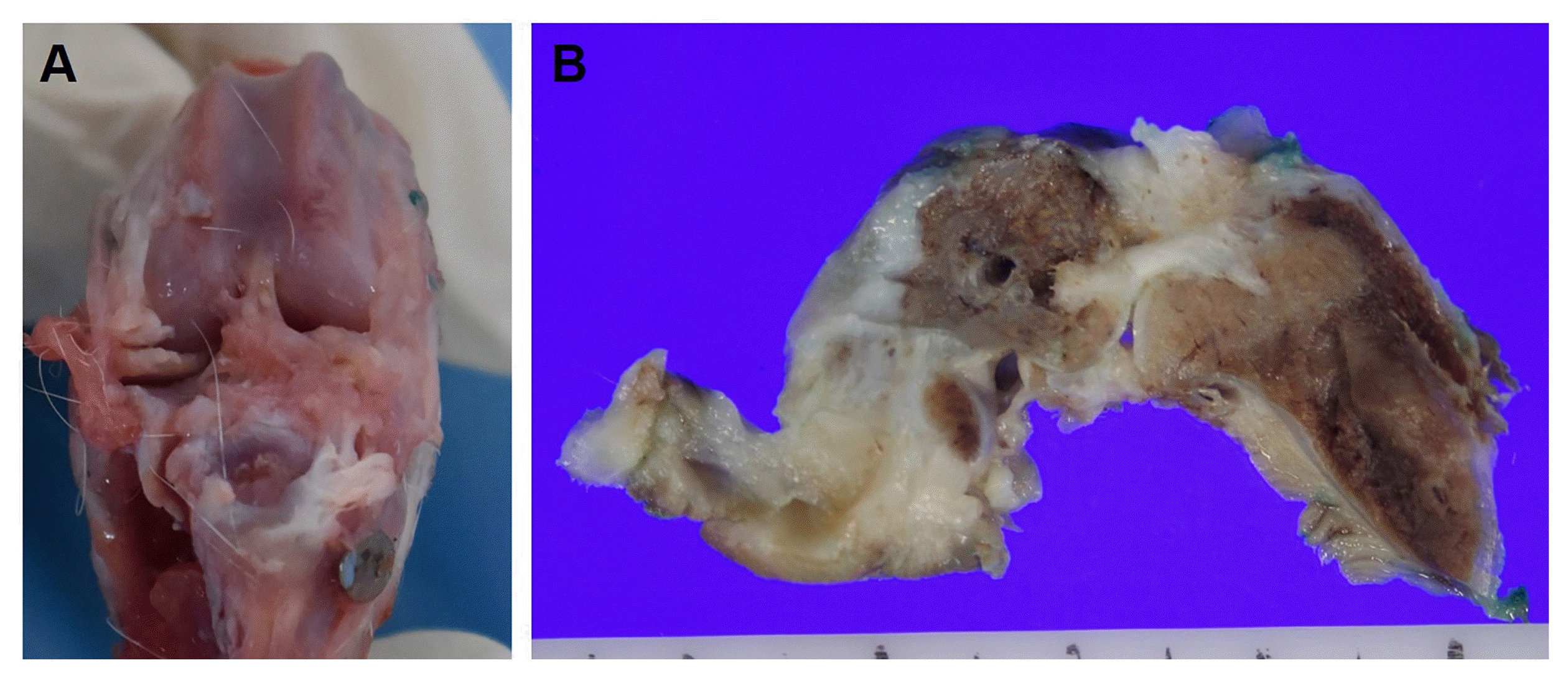
After euthanasia, each knee was carefully stripped from the surrounding soft tissues except from the autograft itself (Fig. 3). For histopathologic examination of the knee joints, each sample was fixed in 10% buffered formalin. The specimens were decalcified in a decalcifying agent (Calci-Clear Rapid, National Diagnostics, GA) for 12 hours, cut along the grafted tendon in the oblique-sagittal direction, and embedded in paraffin. The paraffin block were sectioned at a 3 μm thickness using a microtome (Leica Microsystems, Inc, Wetzlar, Germany) and each section was stained with Hematoxylin and Eosin (H & E). The stained slides of the knee joint sections were observed under a light microscope and were read by a pathologist.
Statistical analysis
Statistical evaluation was made using two ways repeated measures Kruskal-Wallis test to detect significant differences in the CT density scales and tunnel widening. When significant differences were found, post hoc comparisons with the Mann-Whitney test were made. The study results were evaluated with SPSS version 20.0 for Windows (SPSS Inc, Chicago, Illinois). Difference were considered statistically significant at p<0.05.
Go to :
Results
One rabbit in the control group died on postoperative 2 days and was excluded. One knee in the fibrin and one in the MSCs group had septic arthritis and were also excluded. The remaining 32 knees were evaluated radiologically and histologically. The average length of the grafts was 5.8 cm and all the grafts were intact at the time of dissection. Macroscopically, the implanted grafts were white in color with densely packed fiber bundles. All were cylindrical with a straight orientation.
Radiologic findings
The average combined length of the tunnels was 3.8 cm. There were no statistically significant differences among the three groups in terms of tunnel lengths (p>0.05).
Comparing the three groups, there are significant difference of femoral and tibial tunnel widening on the oblique-sagittal view (p=0.001, 0.018, respectively). The mean extent of widening of the femoral and tibial tunnel in oblique-sagittal image was significantly greater in the control than in the fibrin and MSCs groups (p<0.05) (Table 1). No significant differences were evident in terms of the mean tunnel widening on oblique-coronal views among the three groups (p>0.05).
Table 1
Tunnel widening following ACL reconstruction
| Control Group | fibrin Group | MSC Group | p-value* | |
|---|---|---|---|---|
| Femoral tunnel (mm) | ||||
| Oblique-coronal | 0.42±0.2 | 0.27±0.1 | 0.32±0.1 | 0.288 |
| Oblique-sagittal | 0.70±0.4 | 0.22±0.1 | 0.25±0.1 | 0.001 |
| Tibial tunnel (mm) | ||||
| Oblique-coronal | 0.65±0.3 | 0.36±0.1 | 0.44±0.3 | 0.092 |
| Oblique-sagittal | 0.76±0.5 | 0.27±0.1 | 0.29±0.2 | 0.018 |
![]()
HUs in the femoral tunnel (control : 95.06±38.6, fibrin : 103.69±21.8, MSCs : 105.75±13.2) and in the tibial tunnel (control : 98.43±57.8, fibrin : 102.77±32.4, MSCs : 101.93±57.9) were similar to tendon structures in previous study (29). In addition, HUs around the femoral tunnel (control : 424.88±70.0, fibrin : 407.54±29.5, MSCs : 387.30±50.1) and around the tibial tunnel (control : 405.85±55.2, fibrin : 401.68±31.4, MSCs : 421.08±42.7) were similar to prior values obtained from the cancellous bone of humans (29). HUs in and around the bone tunnel were not significantly different between the three groups (p>0.05) (Table 2).
Table 2
Hounsfield unit around the bone tunnel after ACL reconstruction
| Control Group | fibrin Group | MSC Group | p-value* | |
|---|---|---|---|---|
| Femoral tunnel | ||||
| In tunnel | 95.06±38.6 | 103.69±21.8 | 105.75±13.2 | 0.248 |
| Around tunnel | 424.88±70.0 | 407.54±29.5 | 387.30±50.1 | 0.593 |
| Tibial tunnel | ||||
| In tunnel | 98.43±57.8 | 102.77±32.4 | 101.93±57.9 | 0.797 |
| Around tunnel | 405.85±55.2 | 401.68±31.4 | 421.08±42.7 | 0.130 |
![]()
Histologic findings
Gross observation revealed intact intra-articular grafts in all cases. For evaluation of tendon-bone integration and graft architecture, the intra-osseous portion of each graft was obtained and analyzed. H & E staining 12 weeks after surgery revealed preservation of smooth bone to graft bonding and good architecture were observed in the control group. However, there were fewer fibrocytes (hypocellularity) and less compact collagen fiber compared with those of other groups (Fig. 4A, D). Focal areas of disorganized architecture and degenerative alterations were evident in the control group. In the fibrin group, although disorganized architecture or partial tear of the graft was observed, there are good cellularity and more compact collagen fiber compared with the control group (Fig. 4B, E). In the MSCs group, excellent restoration of the bone to graft bonding to the well-formed tunnel was observed. The good architecture with hypercellularity and compact collagen deposition was shown in compared with other groups (Fig. 4C, F).
 | Fig. 4Photomicrographs of H & E staining in 12-week ACL reconstruction. In the control group (A, D), the smooth tendon-bone integration (firm attachment fo the graft to the bone) were observed. However, hypocellularity and less compacted collagen fiber was observed. In fibrin group (B, E), although partial tear of the graft has been shown, there are good cellularity and more compact collagen fiber. In the MSCs groups (C, F), the good architecture with hypercellularity and compacted collagen deposit was shown in compared with other groups.
|
Go to :
Discussion
The main purpose of this study was to compare the effect of MSCs and fibrin glue on ACL reconstruction in terms of radiologic evaluation as well as histologic assessment. The most important finding of present study was that MSCs seemed intra-articular graft healing and prevented tunnel widening.
Tendon-bone integration and graft healing after ACL reconstruction is a complex process. Natural tendon-bone integration without any biologic agent usually results in fibrous scar tissue, which is of inferior property compared to native attachment (30, 31). In addition, grafts undergo the process of avascular necrosis and revascularization the first period after their implantation (32, 33). This process lasts from 3 months to 1 year or more, and the detrimental effects of avascular necrosis on the mechanical properties can prelude graft failure or unsatisfactory function of the knee joint (32, 34, 35).
For evaluation of the effect of MSCs on promoting graft osteointegration, Ouyang et al. (16) implanted bMSCs embedded within fibrin glue into the bone tunnel after ACL reconstruction in rabbit model. Four weeks later, Tendon–bone interface revealed more perpendicular collagen fiber formation and increased proliferation of cartilage-like cells. Similar positive results were reported in two other studies using a rabbit model. These studies investigated the effect of MSCs on osteointegration of autografts (17) and allografts (18) in the bone tunnel. A mature zone from bone to the graft was observed in the MSCs group at 8 weeks in both studies, while the controls showed mature scar tissue resembling Sharpey’s fibers spanning the tendon–bone interface.
The present experiment had demonstrated that administration of cultured MSCs caused an influx of cells into the ACL graft. Regarding the origin of these cells, one possibility is that the implanted MSCs proliferate and differentiate into ligament fibroblasts. Another possibility is that the cellular proliferation originates from cells recruited locally: injected MSCs may secrete a variety of growth factors to stimulate the activation and recruitment of local fibroblasts. An in vitro study on MSCs in graft healing reported that MSCs seemed to be the most suitable candidate for the development of tissue-engineered ligament, with the highest cell proliferation and highest collagen production (19).
Our study showed that the tunnel widening in the fibrin and MSCs groups was less than the control group after ACL reconstruction. Although whether tunnel widening influences clinical outcome is debatable, sagittal tunnel widening did positively affect on clinical outcome and anterior knee laxity after primary ACL reconstruction in one study (36).
There are several mechanical and biological factors for tunnel widening after ACL reconstruction. Biological factors include inflammatory and immune response to allografts, increased cytokine levels within knee joint and cell necrosis caused by drilling of the bone (37, 38). In our experimental model, we tried to control mechanical factors including motion of the graft within the tunnel, insecure fixation, improper graft placement and accelerated rehabilitation. Simultaneously, we used MSCs and fibrin glue to prevent tunnel widening.
There was no difference in HUs between each group in the micro-CT analysis. This finding indicates that there was no heterotopic ossification in and around tunnels caused by MSCs. MSCs is likely to cause ectopic bone formation because of the multi-potential to differentiate into osteoblasts, chondrocytes and adipocytes. Actually, in a horse model, MSCs stimulated bone formation in the cartilage defects (39). However, no ectopic ossification was developed in and around the tunnels in the present study.
The rabbit ACL model has been validated in previous reports. Still, there are some limitations concerning the experimental model used in this study. A rabbit is too small for surgery and so differs from the human situation. Most studies of stem cells therapy are based on small animal models, in which the graft osteointegration process occurs at a faster rate than in humans. Further studies to test the effectiveness of this application in large animal models are necessary to confirm its clinical feasibility. In addition, more accurate and reliable experiments such as biomechanical testing are needed with a large animal model to best judge how to enhance the process of ligament reconstruction in humans. Lastly, we did not identify the ligamentization of the tendon using ligament-specific antibody by immunohistochemistry.
Go to :
 XML Download