Journal List > Int J Stem Cells > v.12(1) > 1158993

Lee, Jung, Choi, Kim, Chae, and Han: Role of HIF1α Regulatory Factors in Stem Cells

Abstract

Hypoxia-inducible factor 1 (HIF1) is a master transcription factor that induces the transcription of genes involved in the metabolism and behavior of stem cells. HIF1-mediated adaptation to hypoxia is required to maintain the pluripotency and survival of stem cells under hypoxic conditions. HIF1 activity is well known to be tightly controlled by the alpha subunit of HIF1 (HIF1α). Understanding the regulatory mechanisms that control HIF1 activity in stem cells will provide novel insights into stem cell biology under hypoxia. Recent research has unraveled the mechanistic details of HIF1α regulating processes, suggesting new strategies for regulating stem cells. This review summarizes recent experimental studies on the role of several regulatory factors (including calcium, 2-oxoglutarate-dependent dioxygenase, microtubule network, importin, and coactivators) in regulating HIF1α activity in stem cells.

Introduction

Hypoxia and stem cells

Stem cells exposed to hypoxia must physiologically adapt to low oxygen conditions to maintain their pluripotency and survival. Metabolic switching during hypoxia is essential in decreasing the accumulation of oxidative metabolism-derived reactive oxygen species (ROS) and the availability of oxygen (13). Recent studies indicate that differences in metabolic profiles of pluripotent stem cells are closely related to self-renewal and initial cell fate decision of stem cells. Gu and colleagues reported that naive pluripotent stem cells in humans exhibit a high glycolytic flux and pentose phosphate pathway activity. However, human embryonic stem cells (hESCs) primed under feeder-free conditions exhibit a low glycolytic flux (4). Moreover, metabolic switching induced by hypoxia prevents the excessive generation of ROS and reduces the demand for ATP, both closely associated with maintaining the physiological functions and survival of stem cells (13). Despite the metabolic switching, chronic hypoxia-induced ROS accumulation leads to mitochondrial apoptosis in embryonic and mesenchymal stem cells (5, 6). Hypoxia-preconditioned mesenchymal stem cells (HP-MSCs) exhibit higher transplantation survival rates and therapeutic potential than normoxia-preconditioned mesenchymal stem cells (MSCs) (7, 8). Other study showed that HP-MSCs exhibit higher concentrations of fatty acids and synthase-mediated lipogenesis, which stimulates cell migration, proliferation, and survival (9, 10). Additionally, hypoxia induces the hexosamine biosynthesis pathway, thereby increasing O-linked-N-acetyl-glucosaminylation concentrations, which is critical for anti-apoptosis in mouse embryonic stem cells (mESCs) exposed to hypoxia (11).

Hypoxia-inducible factor 1 (HIF1)

Semenza et al. first reported that hypoxia-inducible factor 1 (HIF1) is a human erythropoietin gene inducer in ischemic liver and kidney tissues (12). Alpha (HIF1α) and beta (HIF1β) subunits of HIF1 are dimerized in the nucleus, which activates the heterodimeric transcription factor for the HIF1-target genes (13, 14). HIF1 subunits belong to the basic helix loop helix (bHLH) and PER-ARNT-SIM (PAS) domain protein family (Fig. 1). The bHLH and PAS domains within HIF are required for its dimerization and for binding it to DNA. After nuclear translocation, HIF1 binds to the E-box-like hypoxia-responsive element (HRE), which comprises the consensus sequence 5′-[A/G] CGTG-3′ (3). The HIF1α subunit has two transactivation domains (TADs): specifically, the N-terminal TAD (NTAD), comprising amino acids 531~575, and the C-terminal TAD (CTAD), comprising amino acids 786~826. Deletion of inhibitory domain (ID) sequences enhances the transcriptional activity of the HIF1-target genes (15). HIF1α also contains an oxygen-dependent degradation domain (ODDD), which includes prolyl hydroxylase (PHD)-targeted prolyl hydroxylation sites (P402 and P564) (15, 16). HIF1α is controlled by post-translational modifications, including ubiquitination mediated by PHD, which is followed by its stabilization. In contrast, CTAD comprises another hydroxylation site (N803) (17). The hydroxylation of CTAD, which is induced by factor-inhibiting HIF1 (FIH), inhibits the interaction between HIF1α and CREB-binding proteins (CBP/p300), which decrease gene transcriptional activity of HIF1 (18). However, HIF1β does not contain an ODDD, CTAD, or ID. Owing to structural differences between the two HIF1 subunits, HIF1α plays an important role in regulating gene transcription activity of HIF1. Therefore, a deeper understanding of HIF1α is required to comprehend the functional regulation of stem cells under hypoxia.
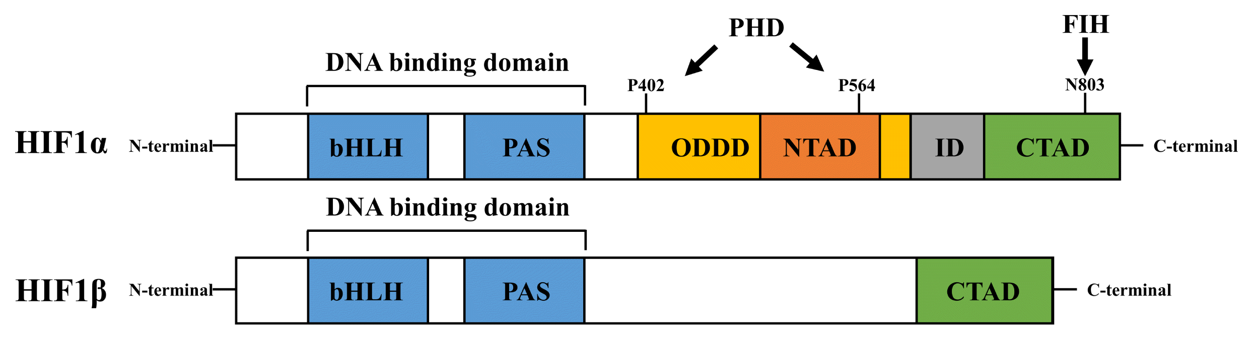
HIF1 stimulates the expression of various hypoxia-responsive genes, which in turn regulate various kinds of cellular physiology including metabolic reprogramming, anti-apoptosis, migration, proliferation, amyloid β production, and prion stabilization 3, 1923). Recent studies have reported the role of HIF1α in stem cell physiology. Von Hippel–Lindau (VHL)-HIF1 signaling induces metabolic compartmentalization in embryonic cells, which is important for cardiac development and functional maturation (24). Sustained HIF1 activation induces cardiac chamber defects and dysfunction when conduction system regulating genes are altered (24). HIF1α induced by the GRP78-Akt axis is critical in enhancing functions, including the proliferation and survival of MSCs under hypoxia (25, 26). In hematopoietic stem cells, treatment with the 4-prolyl hydroxylase inhibitor FG-4497 stabilizes the HIF1 transcription factor and enhances cell mobilization (27). Furthermore, midkine and arachidonic acid induces HIF1α expression, critical for anti-apoptosis, IL-6 production, and proliferation in mESCs under hypoxia (2830).

HIF1α regulation by hypoxia

HIF1α is tightly regulated by oxygen-dependent and oxygen-independent pathways (3133). Unlike HIF1α, HIF1β is constitutively expressed (34). The suppressive effect of hypoxia on global gene transcription is reportedly induced by inhibitions against recruitment of RNA polymerase III and tRNA gene transcription, whereas HIF1A gene transcription is regulated by Sp1 and NF-κB under hypoxia (3537). Tossato et al. showed that hypoxia treatment decreases HIF1A mRNA levels in patients with breast cancer (38). Moreover, Chamboredon et al. reported that hypoxia progressively decays HIF1A mRNA in endothelial cells via tristetraprolin, a mRNA-destabilizing protein (39). In stem cells, chronic hypoxia suppresses the expression of HIF1A mRNA in hESCs, but increases the expression of HIF1A mRNA and HIF1α protein in MSCs derived from rat bone marrow (40, 41). These findings suggest that the effect of hypoxia on HIF1A mRNA transcription genes differs relative to the type of stem cell impacted by hypoxia. In addition, previous studies have shown the regulatory role of the target of rapamycin (TOR) pathway in regulating HIF1 in mammals. Many studies have shown that activating the Akt/mammalian target of rapamycin (mTOR) pathway increases the cap-dependent mRNA translation of HIF1α (4244). In addition to the capacity of mTOR to translationally upregulate HIF1α, mTOR has been reported to increase the stabilization of HIF1α and gene transcription activity of HIF1. Mint3 phosphorylation induced by mTOR stabilizes HIF1α by inactivating FIH (45). In addition, a regulatory protein of mTOR (Raptor), a subunit of the mTOR complex 1 (mTORC1), directly interacts with an mTOR signaling motif of HIF1α when upregulating HIF1-target gene transcription activity (46). However, the effect of hypoxia on mTOR has been debated. It is well documented that mTOR activity is reduced under hypoxic conditions by the tuberous sclerosis protein 1 and 2 complex and REDD1, a protein that regulates development and DNA damage response 1 is inactivated under hypoxic conditions (9, 47, 48). In contrast, several studies have shown that hypoxia-activated Akt induces mTOR signaling by enhancing vascular cell proliferation and angiogenesis (49). In addition, silencing HIF1A or inhibiting fatty acid synthase halts the hypoxia-induced mTORC1 signaling pathway in UCB-MSCs, suggesting that HIF1α-induced lipogenesis is critical for phosphorylation in the mTOR pathway (9). The activation of the Akt/mTORC1 pathway by HIF1α enhances the proliferation, migration, and survival of MSCs under hypoxia (9, 50). Under normoxic conditions, HIF1α interacts with the VHL protein, which subsequently stimulates ubiquitin-mediated degradation via hydroxylation of proline residues. Conversely, hypoxia stabilizes HIF1α in a VHL-dependent pathway and the stabilized HIF1α binds to the HRE for gene transcription (51).
Taken together, these findings indicate that HIF1α regulation has the capacity to determine the fate of stem cells and their bioactivity. However, the detailed mechanism by which HIF1α regulates stem cells (including processes of gene transcription, translation, stabilization, nuclear translocation, and transcriptional activation) remains poorly understood. This review discusses the recent understanding of the roles that various HIF1α regulatory factors play under hypoxic conditions, including the roles played by calcium, 2-oxoglutarate-dependent deoxygenase (2OGDD), microtubule network, importin α, and coactivators.

HIF1α Regulatory Factors

Calcium

Recent gene enrichment analysis data reveal that calcium-regulated calcineurin/NFATc4 signaling is a potential pathway regulating stemness in neural stem cells grown under hypoxic conditions (52). The upregulation of intracellular calcium levels is a response observed in many cell types exposed to hypoxia (53). Rat retinal progenitor cells and neural stem cells exposed to hypoxia exhibit increased intracellular calcium levels (54). The key role of calcium in adapting to hypoxia has been well documented; this occurs by the regulation of multiple signaling and gene expression processes (53). Recently, hypoxia-mediated calcium upregulation has been closely linked to the stimulation of HIF1A gene transcription, HIF1α translation, and HIF1α stabilization (16). One study has reported that the release of hypoxia-induced intracellular calcium increases the expression of HIF1α, which is further enhanced by pretreatment with ionomycin, an ionophore (55). Recently, Kim et al. showed that MSCs primed with both hypoxia and calcium enhanced stemness and the capacity for immunomodulatory activity, thereby attenuating graft-versus-host disease (56). This finding suggests that applying calcium to stem cells could be a promising strategy to enhance the efficacy of hypoxia-preconditioned stem cell transplants.
Previous studies have shown that HIF1α activates calcium signaling by inducing calcium channel expression. HIF1α induced by hypoxia and CoCl2 increases the expression of sodium-calcium exchanger-1, leading to intracellular calcium homeostasis and neuroprotection (57). Moreover, the regulatory effect of hypoxia on ER calcium sensors [including stromal interaction molecule 1 (STIM1)-induced store-operated Ca2+ entry (SOCE)] produces a positive feedback between HIF1α and STIM1 (58). Additionally, chronic hypoxia stimulates the expressions of calcium release-activated channels Orai1, Orai2 (but not Orai3) and STIM1 in pulmonary arterial smooth muscle cells. Furthermore, Orai2 expression depends on the presence of HIF1α (59). These findings indicate that the role of HIF1α in the expression of SOCE components is cell type-specific. Moreover, recent studies have shown that hypoxia-induced HIF1α increases the expression of transient receptor potential channel 1 (TRPC1) (60). Another study has shown that inducing pseudohypoxia by treating cells with CoCl2 or dimethyloxalylglycine N-(methoxyoxoacetyl)-glycine methyl ester (DMOG) increases the expression of Ca2+/Mn2+-transporting SPCA2, an isoform of the Golgi secretory pathway Ca2+-ATPase (61). Hypoxia and HIF1 induction with deferoxamine sometimes reduce the expression of sarcoplasmic reticulum calcium ATPase 2a in embryonic cardiac myocytes (62). Based on these findings, we conclude that hypoxia-induced HIF1α levels are closely associated with the regulation of calcium signaling in both intra- and extracellular pathways.
Calcium signaling is closely associated with HIF1α regulating processes, such as gene transcription, translation, and stabilization (Fig. 2). HIF1α induced by hypoxia increases STIM1 transcription-mediating SOCE, which then upregulates mRNA expression and stability of HIF1α by activating CAMKII-dependent p300 (58). Silencing the mitochondrial calcium uniporter (MCU) suppresses the expression of HIF1A mRNA independently of proteosomal degradation mediated by ubiquitin (38). A stable expression of the full-length TRPM2 channel, a member of the melastatin subfamily of TRP channels, increases gene transcription and stabilization of HIF1α, which is reversed by the stable expression of the short-length TRPM2 channel (63).
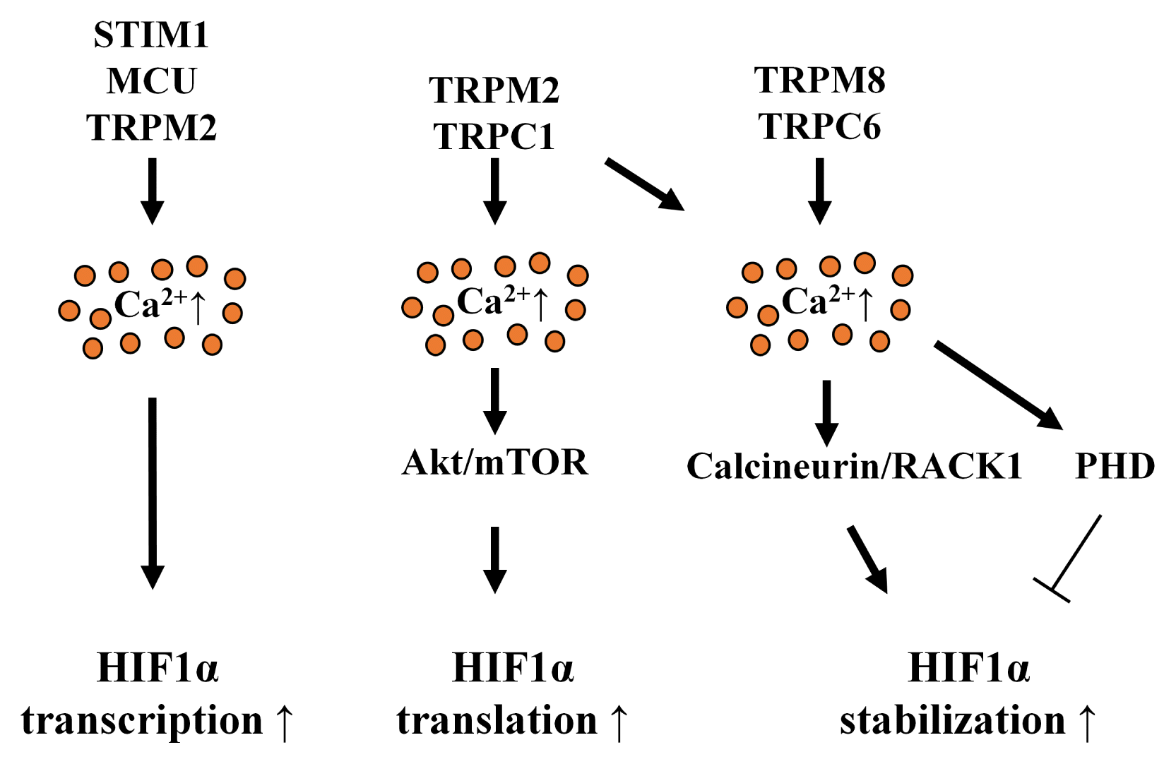
Hypoxia-induced extracellular calcium influx stimulates HIF1α translation, which contributes to the upregulation of approximately 50% of HIF1α protein levels (64). Calcium-stimulated protein kinase C (PKC) α is required to translate mTOR-dependent HIF1α under hypoxia-suppressing global protein synthesis (64). In addition, the translation of HIF1α is controlled by activating mTORC1-mediated S6K1 (64, 65). Under normoxia, silencing TRPC1 attenuates the translation of HIF1α by inhibiting receptor-operated calcium re-entry (66). TRPC1 expression is increased during incubation under hypoxic conditions, which induces HIF1α protein levels via the Akt pathway. However, the effect of TRPC1 on HIF1α regulation is independent of HIF1A gene transcription and HIF1α stability (60). These studies indicate that TRPC1-induced calcium influx stimulates HIF1α gene translation under both normoxia and hypoxia. Most research in this area has focused on the role of specific hyper-activated calcium channels in cancer biology. However, only a few studies have described the relationship between calcium channels and HIF1 in stem cells. However, many stem cell studies have reported that intracellular calcium signaling plays a critical role in the somatic differentiation and migration of stem cells (6769). These findings suggest that HIF1α can be induced by calcium signaling in stem cells under both normoxia and hypoxia.
In addition to clarifying the regulatory role of calcium in HIF1A gene transcription and HIF1α translation, previous studies have examined the relationship between calcium and HIF1α stability. Intracellular calcium chelation by BAPTA stops the interaction between VHL and HIF1 α through PHD inhibition, which further reduces the nuclear accumulation and binding of HIF1α and HIF1 to the gene promoter of carbonyl anhydrase 9 (70). Moreover, the overexpression of the thermo-sensitive calcium channel TRPM8 stabilizes HIF1α by inducing the oxygen-independent de-phosphorylation of RACK1 and inducing the binding of RACK1 to HIF1α and calcineurin, a calcium-dependent serine/threonine phosphatase (71). Calcineurin-induced RACK1 de-phosphorylation blocks the dimerization of RACK1, which further inhibits the oxygen-independent degradation of HIF1α by RACK1 (72). Silencing TRPC6 increases intracellular α-ketoglutarate levels, which promotes the prolyl hydroxylation of HIF1α under hypoxic conditions (73). However, the interplay between calcium and FIH remains poorly understood.

2OGDDs

PHD and FIH are oxygen-consuming enzymes belonging to the 2OGDD family of proteins. These proteins are capable of regulating the stability of HIF1α and epigenetics (74). In fact, 2-oxoglutarate (2OG), oxygen, and cofactors [such as a ferrous iron (Fe2+)] are necessary to activate the 2OGDD protein family (75). Succinate and fumarate (a succinate metabolite formed by succinate dehydrogenase) competes for the 2OG-binding site, which subsequently inactivates PHD and leads to the stabilization of HIF1 in embryonic stem cells under normoxic conditions (7678). The succinate concentration in the mitochondrial matrix is lower than 0.5 mM under normoxia, but it increases to approximately 6 mM under hypoxia (79). Previous research has indicated the crucial role of the intracellular 2OG-to-succinate ratio in maintaining the pluripotency of embryonic stem cells. Indeed, naive mESCs exhibit high activities of intracellular 2OG and 2OGDD enzymes (80). Specifically, the addition of 2OG increases the pluripotency of mESCs, whereas treatment with succinate promotes the differentiation of mESCs (80, 81). Additionally, naive human pluripotent stem cells show high concentrations of 2OG, which appear to be critical in maintaining stemness (82).
Recent advances have been made in our understanding of the mechanisms involved in the stabilization of HIF1α regulated by 2OGDDs (Fig. 3). ROS produced from mitochondrial complex III represents PHD-inhibiting factors for stabilizing HIF1α under hypoxia (83). Mitochondrial ROS inhibits HIF1α activity via PHD2 dimerization, which is induced by the oxidation of cysteine residues required for Fe2+ and ascorbate (74, 84). Additionally, ferritin expression (stimulated by LPS) lowers intracellular Fe2+ levels, which inhibits PHD-mediated hydroxylation, resulting in the stabilization of HIF1α under normoxia (85). Under cellular hypoxic conditions, FIH (a 2OGDD protein) is more sensitive to peroxide and oxygen than to PHDs (86, 87). HIF1α is the most susceptible isoform of HIF1 when FIH is modified (88, 89). Deletion of FIH in mouse embryonic fibroblast cells stimulates glucose and oxidative metabolism, which increases oxygen availability under normoxia (90). Furthermore, levels of FIH expression are higher in skeletal muscles than in other tissues. The loss of FIH in skeletal muscle accelerates adaptation to hypoxia, mediated by HIF1, which enhances glycolysis and cellular metabolic responses to hypoxia (90). Moreover, ascorbate decreases HIF1 activity by reducing iron at the hydroxylase activation site in FIH, even after HIF1α is stabilized by 1% O2, DMOG, and CoCl2 (91). In contrast, ascorbate and ascorbate-2-phosphate have been shown to inhibit HIF1α stability in both PHD-or FIH-independent manner (92). Collectively, these findings imply that 2OGDD regulators (including 2OG, oxygen, Fe2+, and ascorbate) are potential targets for modulating the HIF1-mediated hypoxic response in stem cells.
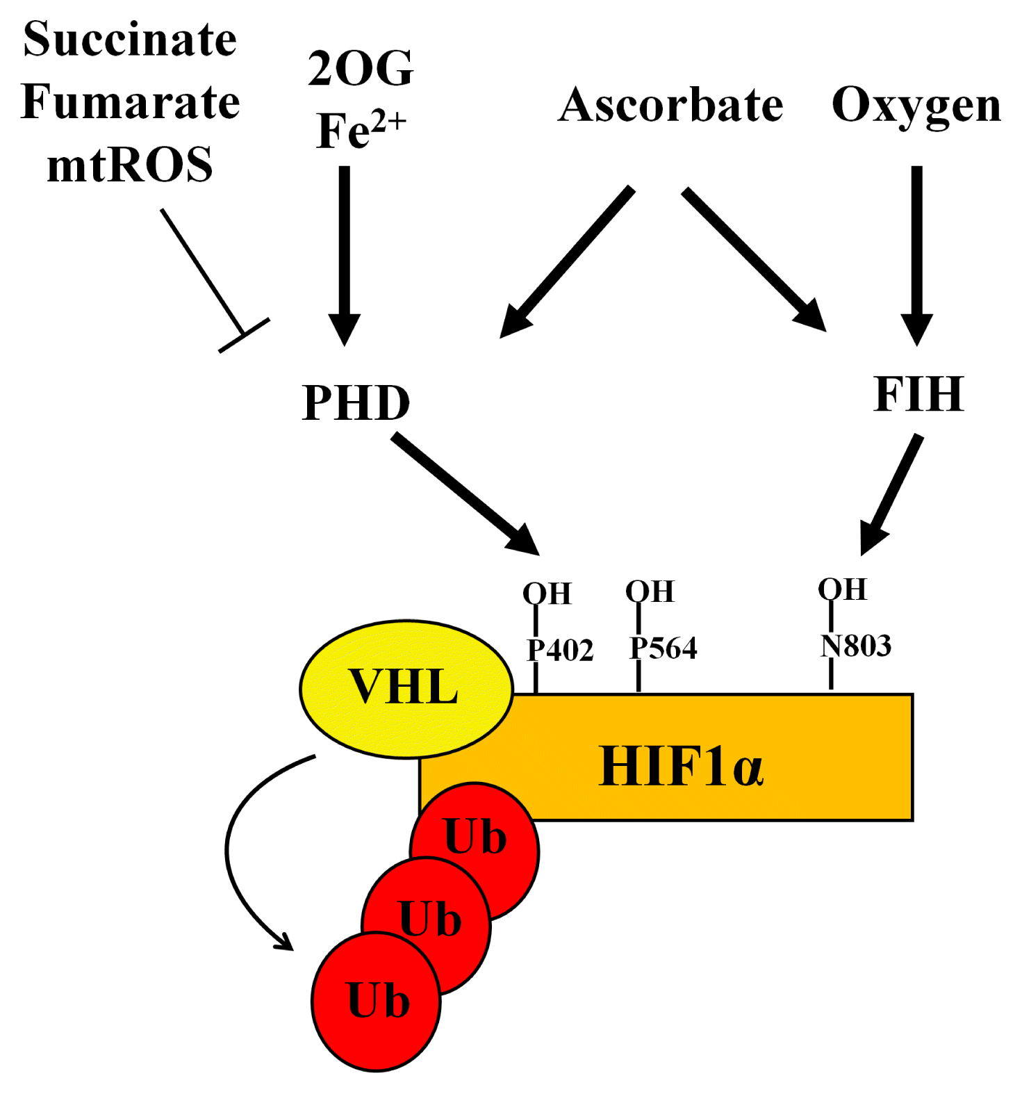
Microtubule network and importin

Considering that HIF1α stability is upregulated by VHL inhibition under hypoxia, HIF1α nuclear translocation may be a potential target for enhancing HIF1 activity in hypoxic stem cells. However, the mechanism of the HIF1α nuclear translocation process in stem cells under hypoxia is poorly understood. It has been reported that HIF1α nuclear translocation is tightly regulated by microtubule network stability, cytoplasmic dynein activity, dynein adaptor proteins, and importins (Fig. 4). Although many investigators have studied the regulation of microtubule network under hypoxia, the effect of hypoxia on microtubule network is still controversial (9396). Several studies have shown that hypoxia treatment stabilizes microtubule network, thus enhancing the chemoresistance of tumor cells (93, 94). Conversely, other studies have reported that hypoxia reduces microtubule stability and polymerization (95, 96). Despite these conflicting results, other studies have shown that stabilizing microtubules during hypoxia is critical for the nuclear translocation of HIF1α and for enhancing the chemoresistance of tumor cells (9799). Chronic hypoxia suppresses the nuclear translocation of HIF1α by disrupting the microtubule network, which can be recovered by taxol (a microtubule stabilizer) pretreatment (97). In addition, stabilized microtubule inhibits Ran-mediated VHL nuclear translocation, leading to the stabilization of HIF1α in the nucleus (100). HIF1α interacts indirectly with microtubule via cytoplasmic dynein, a minus end-directed motor protein (99). Thus, the interaction between HIF1α and dynein is critical for HIF1α nuclear translocation. Dynein-associated cargo transport is regulated by dynein-interacting adaptor proteins, such as dynactin, LIS1, NUDEL, NUDE, Hook, and BICD (101103). Additions of N-terminal fragment BICD2 and dynactin stabilize the formation of dynein–dynactin complexes, which enhance the processivity of dynein (104). In addition, recent our study demonstrated that BICD1 mediates interaction between dynein and HIF1α, critical for nuclear translocation of HIF1α in MSCs under hypoxia (105). Therefore, further investigations into the relationships between dynein adaptor proteins and HIF1α may provide new strategies to improve HIF1α activity, which could increase the efficiency of hypoxia adaptation in stem cells.
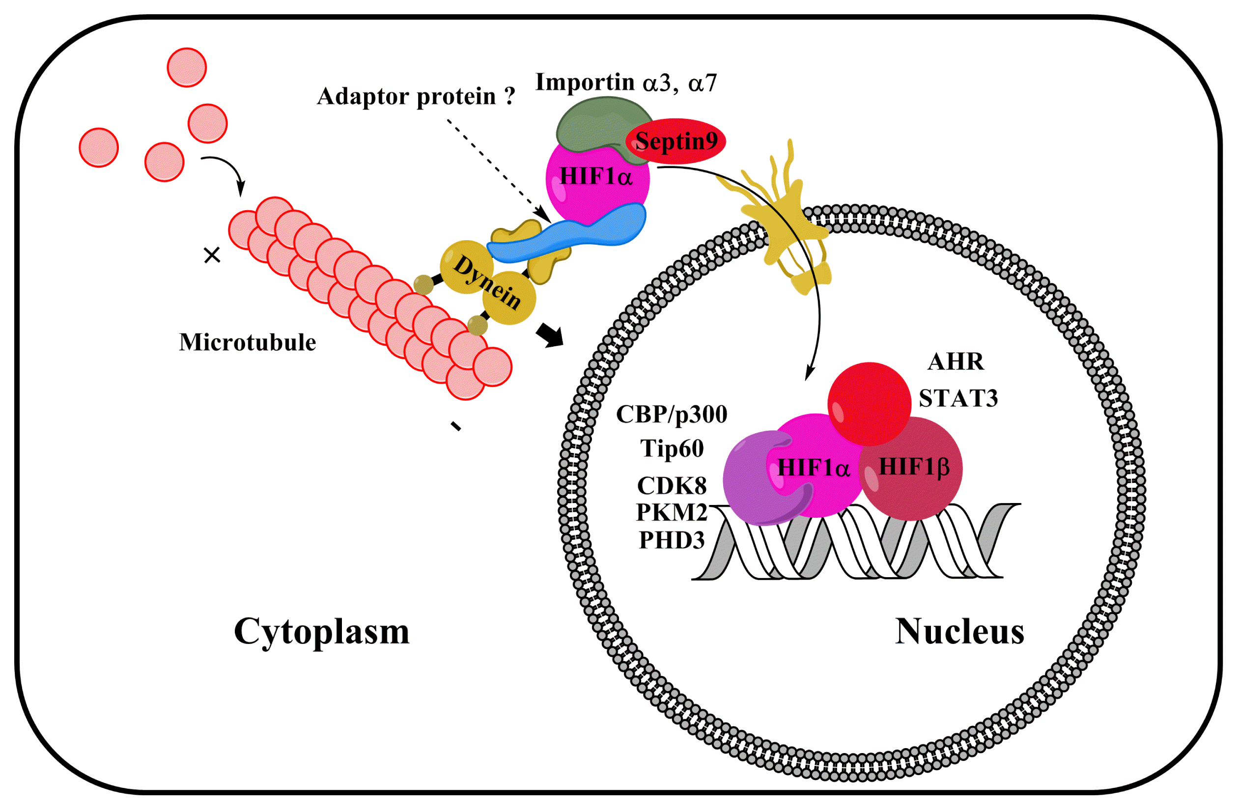
Under hypoxia, the nuclear translocation of HIF1α depends on a nuclear localization signal (NLS) (106). Importin α binds to the NLS region of HIF1α, forming a heterodimer complex with importin β, which in turn allows HIF1α to dock with RanBP2, a component of nuclear pore complexes (107109). It is well known that RanBP2, a SUMO E3 ligase, plays a key role in the nuclear importation of gene transcription factors (110). A major isotype of importin α that regulates the nuclear translocation of HIF1α appears to be cell type specific. One study reported that the NLS of HIF1α interacts with several types of importin α in U2OS osteosarcoma cells, including α1, α3, α5, and α7 (106). In addition, importin α3 is a major isotype regulating the nuclear translocation of HIF1α (106). Another study indicates that an importin α7 is required for the nuclear accumulation of HIF1α and the activity of HIF1 in HeLa cells (111). However, it is not yet known which isotypes of importin α drive the nuclear translocation of HIF1α in stem cells. In addition, a recent study of prostate cancer cells showed that isoform 1 of Septin 9, a GTP-binding protein, facilitates HIF1α nuclear translocation via its interaction with importin α7 (112). Likewise, calcium stimulates interactions between S100A6 and armadillo repeats of importin α, followed by the inhibition of NLS formation of cargo–importin α transport complexes that regulate the nuclear translocation of cargo proteins (113). These findings indicate that the interaction between importin α and HIF1 α improves the nuclear translocation of HIF1α.

HIF1 coactivators

Nuclear-translocated HIF1α interacts with various coactivators including CBP/p300, Tip60, CDK8, pyruvate kinase M2 (PKM2), PHD3, STAT3, and aryl hydrocarbon receptor (AHR) that are important in regulating HIF1 gene transcription activity (Fig. 4). In particular, lysine acetyl-transferase CBP/p300 is known to coactivate HIF1 gene transcription (114, 115). Mutational analysis has revealed that the CTAD of HIF1α is required for HIF1α to interact with CBP/p300, which is essential for the recruitment of SRC-1 to HIF1α under hypoxic conditions (116, 117). However, inhibiting the interaction between CBP/p300 and HIF1 via deletions of the first constant domain of CBP inhibits 35–50% of the HIF1-responsive genes, suggesting that CBP/p300 is not sufficient to activate HIF1 (118).
The Tip60 chromatin-modifying complex is regarded as a HIF1 coactivator and is required for HIF1α to activate RNA polymerase II and histone acetylation (119). Based on an analysis of transcriptome data, HIF1α employs TIP60, CDK8, or both as HIF1 coactivators. Their contributions to HIF1-responsive gene expression are >60% in colorectal cancer cells (119). Additionally, several studies have suggested that PKM2 and PHD3 are HIF1 coactivators. For example, Luo et al. reported that PKM2 mediates PHD3-dependent HIF1 transactivation and glycolytic reprogramming in HeLa cells (120). Consistent with this finding, Schoepflin et al. showed that PHD3 is a PKM2-independent HIF1 coactivator in pulposus cells of the cell nucleus (121). Collectively, these findings indicate that the major HIF1 coactivating partner differs according to the cell type. Therefore, further investigations are required to better understand the contribution of HIF1 coactivators in HIF1-target genes transcription in stem cells.
Recent studies have presented several gene transcription factors, such as STAT3 and AHR, as interacting partners with HIF1α (20, 122). In cancer stem cells, carboplatin-induced HIF1 activates signaling in calcium-dependent STAT3, thereby inducing pluripotency and enrichment in stem cells (123). Glioma stem-like cells in hypoxic conditions reveal high expression levels of vasorin (regulated by the HIF1α/STAT3 gene transcription complex), which is closely associated with tumorigenic capacity mediated by enhanced Notch1 signaling (20). Vasorin binds to Notch1 in the intracellular domain, which stimulates the production of Notch1 by γ-secretase (20). In addition, STAT3 and HIF1α synergistically stimulate the transcription levels of HIF1-target genes (124). Moreover, HIF1α has been reported to interact directly with the γ-secretase complex to regulate Notch signaling (125). These findings imply that HIF1α has both transcriptional and non-transcriptional roles in activating Notch signaling genes.
AHR is a transcription factor stimulated by various types of small molecules produced by gut flora during nutrient metabolism and by environmental stimuli (126). Similar to HIF1α, AHR also shares HIF1β as an interacting partner for dimerization and for activating gene transcription. Some studies have demonstrated that HIF1 β is essential for sustaining glycolysis in CD8+ effector T cells, suggesting a crosstalk between AHR and HIF1α (127, 128). HIF1α and AHR have also been reported to cooperate during the metabolic reprogramming of lymphocytes, including type 1 regulatory T cells and macrophages (129131). Furthermore, kynurenine (a tryptophan metabolite) acts as an endogenous ligand to activate AHR (132). Kynurenine activates AHR via the PKC pathway, which stimulates the interaction between AHR and HIF1 β, which in turn upregulates gene transcription activity of HIF1 (122).

Conclusions

Although several studies have advanced our understanding of HIF1α regulating processes, most research has focused only on the HIF1α regulating mechanism in cancer cells; however, additional studies on stem cells are needed to demonstrate the regulatory mechanism and physiological role of HIF1α. HIF1-mediated adaptation to hypoxia is essential in maintaining the biological functions and survival of transplanted stem cells. Therefore, HIF1α has the potential to improve the therapeutic efficacy of stem cell transplants. Recent studies have demonstrated that hypoxic preconditioning and HIF1α overexpression increases the transplant efficacy and immunomodulatory functions of stem cells (25, 41, 133). Moreover, oxygen-independent HIF1α induction via the regulation of calcium, 2OGDDs, microtubule network, and coactivators may provide a novel strategy to modulate and enhance the survival of transplanted stem cells. Therefore, a comprehensive understanding of the underlying mechanisms that affect HIF1α biology in stem cells will provide novel insights into stem cell biology and regenerative medicine.

Acknowledgments

This research was supported by National R&D Program through the National Research Foundation of Korea (NRF) funded by the Ministry of Science, ICT & Future Planning (NRF-2013M3A9B4076541, NRF-2017R1A2B200 8661) and BK21 PLUS Program for Creative Veterinary Science Research.

Notes

Potential Conflict of Interest

The authors have no conflicting financial interest.

References

1. Zhang CC, Sadek HA. Hypoxia and metabolic properties of hematopoietic stem cells. Antioxid Redox Signal. 2014; 20:1891–1901. DOI: 10.1089/ars.2012.5019. PMID: 23621582. PMCID: 3967354.
crossref
2. Chen J, Kang JG, Keyvanfar K, Young NS, Hwang PM. Long-term adaptation to hypoxia preserves hematopoietic stem cell function. Exp Hematol. 2016; 44:866–873.e4. DOI: 10.1016/j.exphem.2016.04.010. PMID: 27118043.
crossref
3. Choudhry H, Harris AL. Advances in hypoxia-inducible factor biology. Cell Metab. 2018; 27:281–298. DOI: 10.1016/j.cmet.2017.10.005. PMID: 29129785.
crossref
4. Gu W, Gaeta X, Sahakyan A, Chan AB, Hong CS, Kim R, Braas D, Plath K, Lowry WE, Christofk HR. Glycolytic metabolism plays a functional role in regulating human pluripotent stem cell state. Cell Stem Cell. 2016; 19:476–490. DOI: 10.1016/j.stem.2016.08.008. PMID: 27618217. PMCID: 5055460.
crossref
5. Seo BN, Ryu JM, Yun SP, Jeon JH, Park SS, Oh KB, Park JK, Han HJ. Delphinidin prevents hypoxia-induced mouse embryonic stem cell apoptosis through reduction of intracellular reactive oxygen species-mediated activation of JNK and NF-κB, and Akt inhibition. Apoptosis. 2013; 18:811–824. DOI: 10.1007/s10495-013-0838-2. PMID: 23584725.
crossref
6. Son TW, Yun SP, Yong MS, Seo BN, Ryu JM, Youn HY, Oh YM, Han HJ. Netrin-1 protects hypoxia-induced mitochondrial apoptosis through HSP27 expression via DCC-and integrin α6β4-dependent Akt, GSK-3β, and HSF-1 in mesenchymal stem cells. Cell Death Dis. 2013; 4:e563. DOI: 10.1038/cddis.2013.94. PMID: 23538444. PMCID: 3615739.
7. Bader AM, Klose K, Bieback K, Korinth D, Schneider M, Seifert M, Choi YH, Kurtz A, Falk V, Stamm C. Hypoxic preconditioning increases survival and pro-angiogenic capacity of human cord blood mesenchymal stromal cells in vitro. PLoS One. 2015; 10:e0138477. DOI: 10.1371/journal.pone.0138477. PMID: 26380983. PMCID: 4575058.
crossref
8. Liu YY, Chiang CH, Hung SC, Chian CF, Tsai CL, Chen WC, Zhang H. Hypoxia-preconditioned mesenchymal stem cells ameliorate ischemia/reperfusion-induced lung injury. PLoS One. 2017; 12:e0187637. DOI: 10.1371/journal.pone.0187637. PMID: 29117205. PMCID: 5678873.
crossref
9. Lee HJ, Ryu JM, Jung YH, Oh SY, Lee SJ, Han HJ. Novel pathway for hypoxia-induced proliferation and migration in human mesenchymal stem cells: involvement of HIF-1 α, FASN, and mTORC1. Stem Cells. 2015; 33:2182–2195. DOI: 10.1002/stem.2020. PMID: 25825864.
crossref
10. Lee HJ, Jung YH, Choi GE, Ko SH, Lee SJ, Lee SH, Han HJ. BNIP3 induction by hypoxia stimulates FASN-dependent free fatty acid production enhancing therapeutic potential of umbilical cord blood-derived human mesenchymal stem cells. Redox Biol. 2017; 13:426–443. DOI: 10.1016/j.redox.2017.07.004. PMID: 28704726. PMCID: 5508529.
crossref
11. Lee HJ, Ryu JM, Jung YH, Lee KH, Kim DI, Han HJ. Glycerol-3-phosphate acyltransferase-1 upregulation by O-GlcNAcylation of Sp1 protects against hypoxia-induced mouse embryonic stem cell apoptosis via mTOR activation. Cell Death Dis. 2016; 7:e2158. DOI: 10.1038/cddis.2015.410. PMID: 27010859. PMCID: 4823928.
crossref
12. Semenza GL, Nejfelt MK, Chi SM, Antonarakis SE. Hypoxia-inducible nuclear factors bind to an enhancer element located 3′ to the human erythropoietin gene. Proc Natl Acad Sci U S A. 1991; 88:5680–5684. DOI: 10.1073/pnas.88.13.5680. PMID: 2062846. PMCID: 51941.
crossref
13. Wang GL, Jiang BH, Rue EA, Semenza GL. Hypoxia-inducible factor 1 is a basic-helix-loop-helix-PAS heterodimer regulated by cellular O2 tension. Proc Natl Acad Sci U S A. 1995; 92:5510–5514. DOI: 10.1073/pnas.92.12.5510. PMID: 7539918. PMCID: 41725.
crossref
14. Reyes H, Reisz-Porszasz S, Hankinson O. Identification of the Ah receptor nuclear translocator protein (Arnt) as a component of the DNA binding form of the Ah receptor. Science. 1992; 256:1193–1195. DOI: 10.1126/science.256.5060.1193. PMID: 1317062.
crossref
15. Jiang BH, Zheng JZ, Leung SW, Roe R, Semenza GL. Transactivation and inhibitory domains of hypoxia-inducible factor 1α. Modulation of transcriptional activity by oxygen tension. J Biol Chem. 1997; 272:19253–19260. DOI: 10.1074/jbc.272.31.19253. PMID: 9235919.
16. Azimi I. The interplay between HIF-1 and calcium signalling in cancer. Int J Biochem Cell Biol. 2018; 97:73–77. DOI: 10.1016/j.biocel.2018.02.001. PMID: 29407528.
crossref
17. Mahon PC, Hirota K, Semenza GL. FIH-1: a novel protein that interacts with HIF-1α and VHL to mediate repression of HIF-1α transcriptional activity. Genes Dev. 2001; 15:2675–2686. DOI: 10.1101/gad.924501. PMID: 11641274. PMCID: 312814.
crossref
18. Lando D, Peet DJ, Gorman JJ, Whelan DA, Whitelaw ML, Bruick RK. FIH-1 is an asparaginyl hydroxylase enzyme that regulates the transcriptional activity of hypoxia-inducible factor. Genes Dev. 2002; 16:1466–1471. DOI: 10.1101/gad.991402. PMID: 12080085. PMCID: 186346.
crossref
19. Semenza GL. HIF-1 mediates metabolic responses to intra-tumoral hypoxia and oncogenic mutations. J Clin Invest. 2013; 123:3664–3671. DOI: 10.1172/JCI67230. PMID: 23999440. PMCID: 3754249.
crossref
20. Man J, Yu X, Huang H, Zhou W, Xiang C, Huang H, Miele L, Liu Z, Bebek G, Bao S, Yu JS. Hypoxic induction of vasorin regulates Notch1 turnover to maintain glioma stem-like cells. Cell Stem Cell. 2018; 22:104–118.e6. DOI: 10.1016/j.stem.2017.10.005. PMID: 29198941. PMCID: 5756127.
crossref
21. Semenza GL. HIF-1 and mechanisms of hypoxia sensing. Curr Opin Cell Biol. 2001; 13:167–171. DOI: 10.1016/S0955-0674(00)00194-0. PMID: 11248550.
crossref
22. Lee JH, Han YS, Yoon YM, Yun CW, Yun SP, Kim SM, Kwon HY, Jeong D, Baek MJ, Lee HJ, Lee SJ, Han HJ, Lee SH. Role of HSPA1L as a cellular prion protein stabilizer in tumor progression via HIF-1α/GP78 axis. Oncogene. 2017; 36:6555–6567. DOI: 10.1038/onc.2017.263. PMID: 28759037.
crossref
23. Lee HJ, Ryu JM, Jung YH, Lee SJ, Kim JY, Lee SH, Hwang IK, Seong JK, Han HJ. High glucose upregulates BACE1-mediated Aβ production through ROS-dependent HIF-1α and LXRα/ABCA1-regulated lipid raft reorganization in SK-N-MC cells. Sci Rep. 2016; 6:36746. DOI: 10.1038/srep36746. PMID: 27829662. PMCID: 5103190.
crossref
24. Menendez-Montes I, Escobar B, Palacios B, Gómez MJ, Izquierdo-Garcia JL, Flores L, Jiménez-Borreguero LJ, Aragones J, Ruiz-Cabello J, Torres M, Martin-Puig S. Myocardial VHL-HIF signaling controls an embryonic metabolic switch essential for cardiac maturation. Dev Cell. 2016; 39:724–739. DOI: 10.1016/j.devcel.2016.11.012. PMID: 27997827.
crossref
25. Lee JH, Yoon YM, Lee SH. Hypoxic preconditioning promotes the bioactivities of mesenchymal stem cells via the HIF-1α-GRP78-Akt axis. Int J Mol Sci. 2017; 18:DOI: 10.3390/ijms18061320. PMID: 28635661. PMCID: 5486141.
crossref
26. Yun SP, Lee MY, Ryu JM, Song CH, Han HJ. Role of HIF-1α and VEGF in human mesenchymal stem cell proliferation by 17β-estradiol: involvement of PKC, PI3K/Akt, and MAPKs. Am J Physiol Cell Physiol. 2009; 296:C317–326. DOI: 10.1152/ajpcell.00415.2008. PMID: 18987249.
crossref
27. Forristal CE, Nowlan B, Jacobsen RN, Barbier V, Walkinshaw G, Walkley CR, Winkler IG, Levesque JP. HIF-1α is required for hematopoietic stem cell mobilization and 4-prolyl hydroxylase inhibitors enhance mobilization by stabilizing HIF-1α. Leukemia. 2015; 29:1366–1378. DOI: 10.1038/leu.2015.8. PMID: 25578474. PMCID: 4498452.
crossref
28. Lee SH, Suh HN, Lee YJ, Seo BN, Ha JW, Han HJ. Midkine prevented hypoxic injury of mouse embryonic stem cells through activation of Akt and HIF-1α via low-density lipoprotein receptor-related protein-1. J Cell Physiol. 2012; 227:1731–1739. DOI: 10.1002/jcp.22897. PMID: 21688265.
crossref
29. Lee SH, Lee YJ, Han HJ. Effect of arachidonic acid on hypoxia-induced IL-6 production in mouse ES cells: involvement of MAPKs, NF-κB, and HIF-1α. J Cell Physiol. 2010; 222:574–585. PMID: 19950212. PMCID: 10.1002/jcp.21973.
crossref
30. Lee SH, Kim MH, Han HJ. Arachidonic acid potentiates hypoxia-induced VEGF expression in mouse embryonic stem cells: involvement of Notch, Wnt, and HIF-1α. Am J Physiol Cell Physiol. 2009; 297:C207–216. DOI: 10.1152/ajpcell.00579.2008. PMID: 19339510.
crossref
31. Semenza GL. Hypoxia-inducible factor 1: master regulator of O2 homeostasis. Curr Opin Genet Dev. 1998; 8:588–594. DOI: 10.1016/S0959-437X(98)80016-6. PMID: 9794818.
32. Semenza GL. Targeting HIF-1 for cancer therapy. Nat Rev Cancer. 2003; 3:721–732. DOI: 10.1038/nrc1187. PMID: 13130303.
crossref
33. Masoud GN, Li W. HIF-1α pathway: role, regulation and intervention for cancer therapy. Acta Pharm Sin B. 2015; 5:378–389. DOI: 10.1016/j.apsb.2015.05.007. PMID: 26579469. PMCID: 4629436.
crossref
34. Dengler VL, Galbraith M, Espinosa JM. Transcriptional regulation by hypoxia inducible factors. Crit Rev Biochem Mol Biol. 2014; 49:1–15. DOI: 10.3109/10409238.2013.838205. PMID: 24099156. PMCID: 4342852.
crossref
35. Ernens I, Goodfellow SJ, Innes F, Kenneth NS, Derblay LE, White RJ, Scott PH. Hypoxic stress suppresses RNA polymerase III recruitment and tRNA gene transcription in cardiomyocytes. Nucleic Acids Res. 2006; 34:286–294. DOI: 10.1093/nar/gkj402. PMID: 16407335. PMCID: 1326236.
crossref
36. Minet E, Ernest I, Michel G, Roland I, Remacle J, Raes M, Michiels C. HIF1A gene transcription is dependent on a core promoter sequence encompassing activating and inhibiting sequences located upstream from the transcription initiation site and cis elements located within the 5′UTR. Biochem Biophys Res Commun. 1999; 261:534–540. DOI: 10.1006/bbrc.1999.0995. PMID: 10425220.
crossref
37. Belaiba RS, Bonello S, Zähringer C, Schmidt S, Hess J, Kietzmann T, Görlach A. Hypoxia up-regulates hypoxia-inducible factor-1α transcription by involving phosphatidylinositol 3-kinase and nuclear factor κB in pulmonary artery smooth muscle cells. Mol Biol Cell. 2007; 18:4691–4697. DOI: 10.1091/mbc.e07-04-0391. PMID: 17898080. PMCID: 2096613.
crossref
38. Tosatto A, Sommaggio R, Kummerow C, Bentham RB, Blacker TS, Berecz T, Duchen MR, Rosato A, Bogeski I, Szabadkai G, Rizzuto R, Mammucari C. The mitochondrial calcium uniporter regulates breast cancer progression via HIF-1α. EMBO Mol Med. 2016; 8:569–585. DOI: 10.15252/emmm.201606255. PMID: 27138568. PMCID: 4864890.
crossref
39. Chamboredon S, Ciais D, Desroches-Castan A, Savi P, Bono F, Feige JJ, Cherradi N. Hypoxia-inducible factor-1 α mRNA: a new target for destabilization by tristetraprolin in endothelial cells. Mol Biol Cell. 2011; 22:3366–3378. DOI: 10.1091/mbc.e10-07-0617. PMID: 21775632. PMCID: 3172262.
crossref
40. Forristal CE, Wright KL, Hanley NA, Oreffo RO, Houghton FD. Hypoxia inducible factors regulate pluripotency and proliferation in human embryonic stem cells cultured at reduced oxygen tensions. Reproduction. 2010; 139:85–97. DOI: 10.1530/REP-09-0300. PMID: 19755485. PMCID: 2791494.
crossref
41. Lv B, Li F, Fang J, Xu L, Sun C, Han J, Hua T, Zhang Z, Feng Z, Jiang X. Hypoxia inducible factor 1α promotes survival of mesenchymal stem cells under hypoxia. Am J Transl Res. 2017; 9:1521–1529. PMID: 28386377. PMCID: 5376042.
42. Gingras AC, Raught B, Sonenberg N. Regulation of translation initiation by FRAP/mTOR. Genes Dev. 2001; 15:807–826. DOI: 10.1101/gad.887201. PMID: 11297505.
crossref
43. Harada H, Itasaka S, Kizaka-Kondoh S, Shibuya K, Morinibu A, Shinomiya K, Hiraoka M. The Akt/mTOR pathway assures the synthesis of HIF-1α protein in a glucose-and reoxygenation-dependent manner in irradiated tumors. J Biol Chem. 2009; 284:5332–5342. DOI: 10.1074/jbc.M806653200. PMID: 19098000.
crossref
44. Dodd KM, Yang J, Shen MH, Sampson JR, Tee AR. mTORC1 drives HIF-1α and VEGF-A signalling via multiple mechanisms involving 4E-BP1, S6K1 and STAT3. Oncogene. 2015; 34:2239–2250. DOI: 10.1038/onc.2014.164. PMID: 24931163. PMCID: 4172452.
crossref
45. Sakamoto T, Weng JS, Hara T, Yoshino S, Kozuka-Hata H, Oyama M, Seiki M. Hypoxia-inducible factor 1 regulation through cross talk between mTOR and MT1-MMP. Mol Cell Biol. 2014; 34:30–42. DOI: 10.1128/MCB.01169-13. PMID: 24164895. PMCID: 3911284.
crossref
46. Land SC, Tee AR. Hypoxia-inducible factor 1α is regulated by the mammalian target of rapamycin (mTOR) via an mTOR signaling motif. J Biol Chem. 2007; 282:20534–20543. DOI: 10.1074/jbc.M611782200. PMID: 17502379.
crossref
47. Brugarolas J, Lei K, Hurley RL, Manning BD, Reiling JH, Hafen E, Witters LA, Ellisen LW, Kaelin WG Jr. Regulation of mTOR function in response to hypoxia by REDD1 and the TSC1/TSC2 tumor suppressor complex. Genes Dev. 2004; 18:2893–2904. DOI: 10.1101/gad.1256804. PMID: 15545625. PMCID: 534650.
crossref
48. Vadysirisack DD, Ellisen LW. mTOR activity under hypoxia. Methods Mol Biol. 2012; 821:45–58. DOI: 10.1007/978-1-61779-430-8_4. PMID: 22125059. PMCID: 3960283.
crossref
49. Humar R, Kiefer FN, Berns H, Resink TJ, Battegay EJ. Hypoxia enhances vascular cell proliferation and angiogenesis in vitro via rapamycin (mTOR)-dependent signaling. FASEB J. 2002; 16:771–780. DOI: 10.1096/fj.01-0658com. PMID: 12039858.
crossref
50. Lv B, Hua T, Li F, Han J, Fang J, Xu L, Sun C, Zhang Z, Feng Z, Jiang X. Hypoxia-inducible factor 1 α protects mesenchymal stem cells against oxygen-glucose deprivation-induced injury via autophagy induction and PI3K/AKT/mTOR signaling pathway. Am J Transl Res. 2017; 9:2492–2499. PMID: 28559999. PMCID: 5446531.
51. Koyasu S, Kobayashi M, Goto Y, Hiraoka M, Harada H. Regulatory mechanisms of hypoxia-inducible factor 1 activity: Two decades of knowledge. Cancer Sci. 2018; 109:560–571. DOI: 10.1111/cas.13483. PMID: 29285833. PMCID: 5834787.
crossref
52. Moreno M, Fernández V, Monllau JM, Borrell V, Lerin C, de la Iglesia N. Transcriptional profiling of hypoxic neural stem cells identifies calcineurin-NFATc4 signaling as a major regulator of neural stem cell biology. Stem Cell Reports. 2015; 5:157–165. DOI: 10.1016/j.stemcr.2015.06.008. PMID: 26235896. PMCID: 4618660.
crossref
53. Seta KA, Yuan Y, Spicer Z, Lu G, Bedard J, Ferguson TK, Pathrose P, Cole-Strauss A, Kaufhold A, Millhorn DE. The role of calcium in hypoxia-induced signal transduction and gene expression. Cell Calcium. 2004; 36:331–340. DOI: 10.1016/j.ceca.2004.02.006. PMID: 15261489.
crossref
54. Wang Q, Gao S, Luo Y, Kang QY. Compound anisodine affects the proliferation and calcium overload of hypoxia-induced rat retinal progenitor cells and brain neural stem cells via the p-ERK1/2/HIF-1α/VEGF pathway. Exp Ther Med. 2017; 14:600–608. DOI: 10.3892/etm.2017.4528. PMID: 28672973. PMCID: 5488403.
crossref
55. Mottet D, Michel G, Renard P, Ninane N, Raes M, Michiels C. Role of ERK and calcium in the hypoxia-induced activation of HIF-1. J Cell Physiol. 2003; 194:30–44. DOI: 10.1002/jcp.10176. PMID: 12447987.
crossref
56. Corallino S, Malinverno C, Neumann B, Tischer C, Palamidessi A, Frittoli E, Panagiotakopoulou M, Disanza A, Malet-Engra G, Nastaly P, Galli C, Luise C, Bertalot G, Pece S, Di Fiore PP, Gauthier N, Ferrari A, Maiuri P, Scita G. A RAB35-p85/PI3K axis controls oscillatory apical protrusions required for efficient chemotactic migration. Nat Commun. 2018; 9(1):1475. DOI: 10.1038/s41467-018-03571-8. PMID: 29662076. PMCID: 5902610.
crossref
57. Valsecchi V, Pignataro G, Del Prete A, Sirabella R, Matrone C, Boscia F, Scorziello A, Sisalli MJ, Esposito E, Zambrano N, Di Renzo G, Annunziato L. NCX1 is a novel target gene for hypoxia-inducible factor-1 in ischemic brain preconditioning. Stroke. 2011; 42:754–763. DOI: 10.1161/STROKEAHA.110.597583. PMID: 21293012.
crossref
58. Li Y, Guo B, Xie Q, Ye D, Zhang D, Zhu Y, Chen H, Zhu B. STIM1 mediates hypoxia-driven hepatocarcinogenesis via interaction with HIF-1. Cell Rep. 2015; 12:388–395. DOI: 10.1016/j.celrep.2015.06.033. PMID: 26166565.
crossref
59. Wang J, Xu C, Zheng Q, Yang K, Lai N, Wang T, Tang H, Lu W. Orai1, 2, 3 and STIM1 promote store-operated calcium entry in pulmonary arterial smooth muscle cells. Cell Death Discov. 2017; 3:17074. DOI: 10.1038/cddiscovery.2017.74. PMID: 29188077. PMCID: 5702854.
crossref
60. Xiang L, Chen XJ, Wu KC, Zhang CJ, Zhou GH, Lv JN, Sun LF, Cheng FF, Cai XB, Jin ZB. miR-183/96 plays a pivotal regulatory role in mouse photoreceptor maturation and maintenance. Proc Natl Acad Sci U S A. 2017; 114:6376–6381. DOI: 10.1073/pnas.1618757114. PMID: 28559309. PMCID: 5474811.
crossref
61. Jenkins J, Papkovsky DB, Dmitriev RI. The Ca2+/Mn2+-transporting SPCA2 pump is regulated by oxygen and cell density in colon cancer cells. Biochem J. 2016; 473:2507–2518. DOI: 10.1042/BCJ20160477. PMID: 27316461.
crossref
62. Ronkainen VP, Skoumal R, Tavi P. Hypoxia and HIF-1 suppress SERCA2a expression in embryonic cardiac myocytes through two interdependent hypoxia response elements. J Mol Cell Cardiol. 2011; 50:1008–1016. DOI: 10.1016/j.yjmcc.2011.02.017. PMID: 21382378.
crossref
63. Chen SJ, Hoffman NE, Shanmughapriya S, Bao L, Keefer K, Conrad K, Merali S, Takahashi Y, Abraham T, Hirschler-Laszkiewicz I, Wang J, Zhang XQ, Song J, Barrero C, Shi Y, Kawasawa YI, Bayerl M, Sun T, Barbour M, Wang HG, Madesh M, Cheung JY, Miller BA. A splice variant of the human ion channel TRPM2 modulates neuroblastoma tumor growth through hypoxia-inducible factor (HIF)-1/2α. J Biol Chem. 2014; 289:36284–36302. DOI: 10.1074/jbc.M114.620922. PMID: 25391657. PMCID: 4276889.
crossref
64. Hui AS, Bauer AL, Striet JB, Schnell PO, Czyzyk-Krzeska MF. Calcium signaling stimulates translation of HIF-α during hypoxia. FASEB J. 2006; 20:466–475. DOI: 10.1096/fj.05-5086com. PMID: 16507764.
crossref
65. Yuan G, Nanduri J, Khan S, Semenza GL, Prabhakar NR. Induction of HIF-1α expression by intermittent hypoxia: involvement of NADPH oxidase, Ca2+ signaling, prolyl hydroxylases, and mTOR. J Cell Physiol. 2008; 217:674–85. DOI: 10.1002/jcp.21537. PMID: 18651560. PMCID: 2696817.
66. Asghar MY, Magnusson M, Kemppainen K, Sukumaran P, Löf C, Pulli I, Kalhori V, Törnquist K. Transient Receptor Potential Canonical 1 (TRPC1) channels as regulators of sphingolipid and VEGF receptor expression: implications for thyroid cancer cell migration and proliferation. J Biol Chem. 2015; 290:16116–16131. DOI: 10.1074/jbc.M115.643668. PMID: 25971967. PMCID: 4481213.
crossref
67. Pchelintseva E, Djamgoz MBA. Mesenchymal stem cell differentiation: control by calcium-activated potassium channels. J Cell Physiol. 2018; 233:3755–3768. DOI: 10.1002/jcp.26120. PMID: 28776687.
crossref
68. Sun S, Liu Y, Lipsky S, Cho M. Physical manipulation of calcium oscillations facilitates osteodifferentiation of human mesenchymal stem cells. FASEB J. 2007; 21:1472–1480. DOI: 10.1096/fj.06-7153com. PMID: 17264165.
crossref
69. Jiang LH, Mousawi F, Yang X, Roger S. ATP-induced Ca2+-signalling mechanisms in the regulation of mesenchymal stem cell migration. Cell Mol Life Sci. 2017; 74:3697–3710. DOI: 10.1007/s00018-017-2545-6. PMID: 28534085. PMCID: 5597679.
crossref
70. Berchner-Pfannschmidt U, Petrat F, Doege K, Trinidad B, Freitag P, Metzen E, de Groot H, Fandrey J. Chelation of cellular calcium modulates hypoxia-inducible gene expression through activation of hypoxia-inducible factor-1α. J Biol Chem. 2004; 279:44976–44986. DOI: 10.1074/jbc.M313995200. PMID: 15322093.
crossref
71. Yu S, Xu Z, Zou C, Wu D, Wang Y, Yao X, Ng CF, Chan FL. Ion channel TRPM8 promotes hypoxic growth of prostate cancer cells via an O2-independent and RACK1-mediated mechanism of HIF-1α stabilization. J Pathol. 2014; 234:514–525. DOI: 10.1002/path.4413. PMID: 25065497.
crossref
72. Liu YV, Hubbi ME, Pan F, McDonald KR, Mansharamani M, Cole RN, Liu JO, Semenza GL. Calcineurin promotes hypoxia-inducible factor 1α expression by dephosphorylating RACK1 and blocking RACK1 dimerization. J Biol Chem. 2007; 282:37064–37073. DOI: 10.1074/jbc.M705015200. PMID: 17965024. PMCID: 3754800.
crossref
73. Li S, Wang J, Wei Y, Liu Y, Ding X, Dong B, Xu Y, Wang Y. Crucial role of TRPC6 in maintaining the stability of HIF-1α in glioma cells under hypoxia. J Cell Sci. 2015; 128:3317–3329. DOI: 10.1242/jcs.173161. PMID: 26187851.
74. Bargiela D, Burr SP, Chinnery PF. Mitochondria and hypoxia: metabolic crosstalk in cell-fate decisions. Trends Endocrinol Metab. 2018; 29:249–259. DOI: 10.1016/j.tem.2018.02.002. PMID: 29501229.
crossref
75. Clifton IJ, McDonough MA, Ehrismann D, Kershaw NJ, Granatino N, Schofield CJ. Structural studies on 2-oxoglutarate oxygenases and related double-stranded β-helix fold proteins. J Inorg Biochem. 2006; 100:644–669. DOI: 10.1016/j.jinorgbio.2006.01.024. PMID: 16513174.
crossref
76. Hewitson KS, Liénard BM, McDonough MA, Clifton IJ, Butler D, Soares AS, Oldham NJ, McNeill LA, Schofield CJ. Structural and mechanistic studies on the inhibition of the hypoxia-inducible transcription factor hydroxylases by tricarboxylic acid cycle intermediates. J Biol Chem. 2007; 282:3293–3301. DOI: 10.1074/jbc.M608337200. PMID: 17135241.
crossref
77. Gimenez-Roqueplo AP, Favier J, Rustin P, Mourad JJ, Plouin PF, Corvol P, Rötig A, Jeunemaitre X. The R22X mutation of the SDHD gene in hereditary paraganglioma abolishes the enzymatic activity of complex II in the mitochondrial respiratory chain and activates the hypoxia pathway. Am J Hum Genet. 2001; 69:1186–1197. DOI: 10.1086/324413. PMID: 11605159. PMCID: 1235531.
crossref
78. Isaacs JS, Jung YJ, Mole DR, Lee S, Torres-Cabala C, Chung YL, Merino M, Trepel J, Zbar B, Toro J, Ratcliffe PJ, Linehan WM, Neckers L. HIF overexpression correlates with biallelic loss of fumarate hydratase in renal cancer: novel role of fumarate in regulation of HIF stability. Cancer Cell. 2005; 8:143–153. DOI: 10.1016/j.ccr.2005.06.017. PMID: 16098467.
crossref
79. Tretter L, Patocs A, Chinopoulos C. Succinate, an intermediate in metabolism, signal transduction, ROS, hypoxia, and tumorigenesis. Biochim Biophys Acta. 2016; 1857:1086–1101. DOI: 10.1016/j.bbabio.2016.03.012. PMID: 26971832.
crossref
80. Carey BW, Finley LW, Cross JR, Allis CD, Thompson CB. Intracellular α-ketoglutarate maintains the pluripotency of embryonic stem cells. Nature. 2015; 518:413–416. DOI: 10.1038/nature13981. PMID: 25487152. PMCID: 4336218.
crossref
81. Hwang IY, Kwak S, Lee S, Kim H, Lee SE, Kim JH, Kim YA, Jeon YK, Chung DH, Jin X, Park S, Jang H, Cho EJ, Youn HD. Psat1-dependent fluctuations in α-ketoglutarate affect the timing of ESC differentiation. Cell Metab. 2016; 24:494–501. DOI: 10.1016/j.cmet.2016.06.014. PMID: 27476977.
crossref
82. TeSlaa T, Chaikovsky AC, Lipchina I, Escobar SL, Hochedlinger K, Huang J, Graeber TG, Braas D, Teitell MA. α-Ketoglutarate accelerates the initial differentiation of primed human pluripotent stem cells. Cell Metab. 2016; 24:485–493. DOI: 10.1016/j.cmet.2016.07.002. PMID: 27476976. PMCID: 5023506.
crossref
83. Chandel NS, Maltepe E, Goldwasser E, Mathieu CE, Simon MC, Schumacker PT. Mitochondrial reactive oxygen species trigger hypoxia-induced transcription. Proc Natl Acad Sci U S A. 1998; 95:11715–11720. DOI: 10.1073/pnas.95.20.11715. PMID: 9751731. PMCID: 21706.
crossref
84. Lee G, Won HS, Lee YM, Choi JW, Oh TI, Jang JH, Choi DK, Lim BO, Kim YJ, Park JW, Puigserver P, Lim JH. Oxidative dimerization of PHD2 is responsible for its inactivation and contributes to metabolic reprogramming via HIF-1α activation. Sci Rep. 2016; 6:18928. DOI: 10.1038/srep18928. PMID: 26740011. PMCID: 4703963.
crossref
85. Siegert I, Schödel J, Nairz M, Schatz V, Dettmer K, Dick C, Kalucka J, Franke K, Ehrenschwender M, Schley G, Beneke A, Sutter J, Moll M, Hellerbrand C, Wielockx B, Katschinski DM, Lang R, Galy B, Hentze MW, Koivunen P, Oefner PJ, Bogdan C, Weiss G, Willam C, Jantsch J. Ferritin-mediated iron sequestration stabilizes hypoxia-inducible factor-1α upon LPS activation in the presence of ample oxygen. Cell Rep. 2015; 13:2048–2055. DOI: 10.1016/j.celrep.2015.11.005. PMID: 26628374.
crossref
86. Masson N, Singleton RS, Sekirnik R, Trudgian DC, Ambrose LJ, Miranda MX, Tian YM, Kessler BM, Schofield CJ, Ratcliffe PJ. The FIH hydroxylase is a cellular peroxide sensor that modulates HIF transcriptional activity. EMBO Rep. 2012; 13:251–257. DOI: 10.1038/embor.2012.9. PMID: 22310300. PMCID: 3323130.
crossref
87. Tarhonskaya H, Hardy AP, Howe EA, Loik ND, Kramer HB, McCullagh JS, Schofield CJ, Flashman E. Kinetic investigations of the role of factor inhibiting hypoxia-inducible factor (FIH) as an oxygen sensor. J Biol Chem. 2015; 290:19726–19742. DOI: 10.1074/jbc.M115.653014. PMID: 26112411. PMCID: 4528135.
crossref
88. Bracken CP, Fedele AO, Linke S, Balrak W, Lisy K, Whitelaw ML, Peet DJ. Cell-specific regulation of hypoxia-inducible factor (HIF)-1α and HIF-2α stabilization and transactivation in a graded oxygen environment. J Biol Chem. 2006; 281:22575–22585. DOI: 10.1074/jbc.M600288200. PMID: 16760477.
crossref
89. Koivunen P, Hirsilä M, Günzler V, Kivirikko KI, Myllyharju J. Catalytic properties of the asparaginyl hydroxylase (FIH) in the oxygen sensing pathway are distinct from those of its prolyl 4-hydroxylases. J Biol Chem. 2004; 279:9899–9904. DOI: 10.1074/jbc.M312254200. PMID: 14701857.
crossref
90. Sim J, Cowburn AS, Palazon A, Madhu B, Tyrakis PA, Macías D, Bargiela DM, Pietsch S, Gralla M, Evans CE, Kittipassorn T, Chey YCJ, Branco CM, Rundqvist H, Peet DJ, Johnson RS. The factor inhibiting HIF asparaginyl hydroxylase regulates oxidative metabolism and accelerates metabolic adaptation to hypoxia. Cell Metab. 2018; 27:898–913.e7. DOI: 10.1016/j.cmet.2018.02.020. PMID: 29617647. PMCID: 5887987.
crossref
91. Kuiper C, Dachs GU, Currie MJ, Vissers MC. Intracellular ascorbate enhances hypoxia-inducible factor (HIF)-hydroxylase activity and preferentially suppresses the HIF-1 transcriptional response. Free Radic Biol Med. 2014; 69:308–317. DOI: 10.1016/j.freeradbiomed.2014.01.033. PMID: 24495550.
crossref
92. Miles SL, Fischer AP, Joshi SJ, Niles RM. Ascorbic acid and ascorbate-2-phosphate decrease HIF activity and malignant properties of human melanoma cells. BMC Cancer. 2015; 15:867. DOI: 10.1186/s12885-015-1878-5. PMID: 26547841. PMCID: 4636772.
crossref
93. Peng WX, Pan FY, Liu XJ, Ning S, Xu N, Meng FL, Wang YQ, Li CJ. Hypoxia stabilizes microtubule networks and decreases tumor cell chemosensitivity to anticancer drugs through Egr-1. Anat Rec (Hoboken). 2010; 293:414–420. DOI: 10.1002/ar.21086. PMID: 20169563.
crossref
94. Yoon SO, Shin S, Mercurio AM. Hypoxia stimulates carcinoma invasion by stabilizing microtubules and promoting the Rab11 trafficking of the α6β4 integrin. Cancer Res. 2005; 65:2761–2769. DOI: 10.1158/0008-5472.CAN-04-4122. PMID: 15805276.
crossref
95. Fang YD, Xu X, Dang YM, Zhang YM, Zhang JP, Hu JY, Zhang Q, Dai X, Teng M, Zhang DX, Huang YS. MAP4 mechanism that stabilizes mitochondrial permeability transition in hypoxia: microtubule enhancement and DYNLT1 interaction with VDAC1. PLoS One. 2011; 6:e28052. DOI: 10.1371/journal.pone.0028052. PMID: 22164227. PMCID: 3229508.
crossref
96. Hu JY, Chu ZG, Han J, Dang YM, Yan H, Zhang Q, Liang GP, Huang YS. The p38/MAPK pathway regulates microtubule polymerization through phosphorylation of MAP4 and Op18 in hypoxic cells. Cell Mol Life Sci. 2010; 67:321–333. DOI: 10.1007/s00018-009-0187-z. PMID: 19915797.
crossref
97. Guo H, Zheng H, Wu J, Ma HP, Yu J, Yiliyaer M. The key role of microtubules in hypoxia preconditioning-induced nuclear translocation of HIF-1α in rat cardiomyocytes. PeerJ. 2017; 5:e3662. DOI: 10.7717/peerj.3662. PMID: 28828258. PMCID: 5560226.
crossref
98. McGrogan BT, Gilmartin B, Carney DN, McCann A. Taxanes, microtubules and chemoresistant breast cancer. Biochim Biophys Acta. 2008; 1785:96–132. DOI: 10.1016/j.bbcan.2007.10.004. PMID: 18068131.
crossref
99. Carbonaro M, Escuin D, O’Brate A, Thadani-Mulero M, Giannakakou P. Microtubules regulate hypoxia-inducible factor-1α protein trafficking and activity: implications for taxane therapy. J Biol Chem. 2012; 287:11859–11869. DOI: 10.1074/jbc.M112.345587. PMID: 22367210. PMCID: 3320934.
crossref
100. Jiang X, Zhang D, Zhang H, Huang Y, Teng M. Role of Ran-regulated nuclear-cytoplasmic trafficking of pVHL in the regulation of microtubular stability-mediated HIF-1α in hypoxic cardiomyocytes. Sci Rep. 2015; 5:9193. DOI: 10.1038/srep09193. PMID: 25779090. PMCID: 4361876.
crossref
101. Kardon JR, Vale RD. Regulators of the cytoplasmic dynein motor. Nat Rev Mol Cell Biol. 2009; 10:854–865. DOI: 10.1038/nrm2804. PMID: 19935668. PMCID: 3394690.
crossref
102. Schroeder CM, Vale RD. Assembly and activation of dynein-dynactin by the cargo adaptor protein Hook3. J Cell Biol. 2016; 214:309–318. DOI: 10.1083/jcb.201604002. PMID: 27482052. PMCID: 4970328.
crossref
103. Olenick MA, Tokito M, Boczkowska M, Dominguez R, Holzbaur EL. Hook adaptors induce unidirectional processive motility by enhancing the dynein-dynactin interaction. J Biol Chem. 2016; 291:18239–18251. DOI: 10.1074/jbc.M116.738211. PMID: 27365401. PMCID: 5000072.
crossref
104. Splinter D, Razafsky DS, Schlager MA, Serra-Marques A, Grigoriev I, Demmers J, Keijzer N, Jiang K, Poser I, Hyman AA, Hoogenraad CC, King SJ, Akhmanova A. BICD2, dynactin, and LIS1 cooperate in regulating dynein recruitment to cellular structures. Mol Biol Cell. 2012; 23:4226–4241. DOI: 10.1091/mbc.e12-03-0210. PMID: 22956769. PMCID: 3484101.
crossref
105. Lee HJ, Jung YH, Oh JY, Choi GE, Chae CW, Kim JS, Lim JR, Kim SY, Lee SJ, Seong JK, Han HJ. BICD1 mediates HIF1α nuclear translocation in mesenchymal stem cells during hypoxia adaptation. Cell Death Differ. 2018; DOI: 10.1038/s41418-018-0241-1. PMID: 30464225. [Epub ahead of print].
crossref
106. Depping R, Steinhoff A, Schindler SG, Friedrich B, Fagerlund R, Metzen E, Hartmann E, Köhler M. Nuclear translocation of hypoxia-inducible factors (HIFs): involvement of the classical importin α/β pathway. Biochim Biophys Acta. 2008; 1783:394–404. DOI: 10.1016/j.bbamcr.2007.12.006. PMID: 18187047.
crossref
107. Miyamoto Y, Yamada K, Yoneda Y. Importin α: a key molecule in nuclear transport and non-transport functions. J Biochem. 2016; 160:69–75. DOI: 10.1093/jb/mvw036. PMID: 27289017.
crossref
108. Hutten S, Flotho A, Melchior F, Kehlenbach RH. The Nup358-RanGAP complex is required for efficient importin α/β-dependent nuclear import. Mol Biol Cell. 2008; 19:2300–2310. DOI: 10.1091/mbc.e07-12-1279. PMID: 18305100. PMCID: 2366868.
crossref
109. Hamada M, Haeger A, Jeganathan KB, van Ree JH, Malureanu L, Wälde S, Joseph J, Kehlenbach RH, van Deursen JM. Ran-dependent docking of importin-β to RanBP2/Nup358 filaments is essential for protein import and cell viability. J Cell Biol. 2011; 194:597–612. DOI: 10.1083/jcb.201102018. PMID: 21859863. PMCID: 3160583.
crossref
110. Wälde S, Thakar K, Hutten S, Spillner C, Nath A, Rothbauer U, Wiemann S, Kehlenbach RH. The nucleoporin Nup358/RanBP2 promotes nuclear import in a cargo-and transport receptor-specific manner. Traffic. 2012; 13:218–233. DOI: 10.1111/j.1600-0854.2011.01302.x. PMID: 21995724.
crossref
111. Chachami G, Paraskeva E, Mingot JM, Braliou GG, Görlich D, Simos G. Transport of hypoxia-inducible factor HIF-1α into the nucleus involves importins 4 and 7. Biochem Biophys Res Commun. 2009; 390:235–240. DOI: 10.1016/j.bbrc.2009.09.093. PMID: 19788888.
crossref
112. Tazat K, Schindler S, Depping R, Mabjeesh NJ. Septin 9 isoform 1 (SEPT9_i1) specifically interacts with importin-α 7 to drive hypoxia-inducible factor (HIF)-1α nuclear translocation. Cytoskeleton (Hoboken). 2018; DOI: 10.1002/cm.21450. PMID: 29742803. [Epub ahead of print].
113. Takata M, Shimamoto S, Yamaguchi F, Tokuda M, Tokumitsu H, Kobayashi R. Regulation of nuclear localization signal-importin α interaction by Ca2+/S100A6. FEBS Lett. 2010; 584:4517–4523. DOI: 10.1016/j.febslet.2010.09.052. PMID: 20965181.
crossref
114. Arany Z, Huang LE, Eckner R, Bhattacharya S, Jiang C, Goldberg MA, Bunn HF, Livingston DM. An essential role for p300/CBP in the cellular response to hypoxia. Proc Natl Acad Sci U S A. 1996; 93:12969–12973. DOI: 10.1073/pnas.93.23.12969. PMID: 8917528. PMCID: 24030.
115. Ebert BL, Bunn HF. Regulation of transcription by hypoxia requires a multiprotein complex that includes hypoxia-inducible factor 1, an adjacent transcription factor, and p300/CREB binding protein. Mol Cell Biol. 1998; 18:4089–4096. DOI: 10.1128/MCB.18.7.4089. PMID: 9632793. PMCID: 108993.
crossref
116. Ruas JL, Poellinger L, Pereira T. Functional analysis of hypoxia-inducible factor-1 α-mediated transactivation. Identification of amino acid residues critical for transcriptional activation and/or interaction with CREB-binding protein. J Biol Chem. 2002; 277:38723–38730. DOI: 10.1074/jbc.M205051200. PMID: 12133832.
crossref
117. Ruas JL, Poellinger L, Pereira T. Role of CBP in regulating HIF-1-mediated activation of transcription. J Cell Sci. 2005; 118:301–311. DOI: 10.1242/jcs.01617. PMID: 15615775.
crossref
118. Kasper LH, Boussouar F, Boyd K, Xu W, Biesen M, Rehg J, Baudino TA, Cleveland JL, Brindle PK. Two transactivation mechanisms cooperate for the bulk of HIF-1-responsive gene expression. EMBO J. 2005; 24:3846–3858. DOI: 10.1038/sj.emboj.7600846. PMID: 16237459. PMCID: 1283945.
crossref
119. Perez-Perri JI, Dengler VL, Audetat KA, Pandey A, Bonner EA, Urh M, Mendez J, Daniels DL, Wappner P, Galbraith MD, Espinosa JM. The TIP60 complex is a conserved coactivator of HIF1A. Cell Rep. 2016; 16:37–47. DOI: 10.1016/j.celrep.2016.05.082. PMID: 27320910. PMCID: 4957981.
crossref
120. Luo W, Hu H, Chang R, Zhong J, Knabel M, O’Meally R, Cole RN, Pandey A, Semenza GL. Pyruvate kinase M2 is a PHD3-stimulated coactivator for hypoxia-inducible factor 1. Cell. 2011; 145:732–744. DOI: 10.1016/j.cell.2011.03.054. PMID: 21620138. PMCID: 3130564.
crossref
121. Schoepflin ZR, Silagi ES, Shapiro IM, Risbud MV. PHD3 is a transcriptional coactivator of HIF-1α in nucleus pulposus cells independent of the PKM2-JMJD5 axis. FASEB J. 2017; 31:3831–3847. DOI: 10.1096/fj.201601291R. PMID: 28495754. PMCID: 5572688.
crossref
122. Gabriely G, Wheeler MA, Takenaka MC, Quintana FJ. Role of AHR and HIF-1α in glioblastoma metabolism. Trends Endocrinol Metab. 2017; 28:428–436. DOI: 10.1016/j.tem.2017.02.009. PMID: 28318896. PMCID: 5438779.
crossref
123. Lu H, Chen I, Shimoda LA, Park Y, Zhang C, Tran L, Zhang H, Semenza GL. Chemotherapy-induced Ca2+ release stimulates breast cancer stem cell enrichment. Cell Rep. 2017; 18:1946–1957. DOI: 10.1016/j.celrep.2017.02.001. PMID: 28228260.
crossref
124. Pawlus MR, Wang L, Hu CJ. STAT3 and HIF1α cooperatively activate HIF1 target genes in MDA-MB-231 and RCC4 cells. Oncogene. 2014; 33:1670–1679. DOI: 10.1038/onc.2013.115. PMID: 23604114. PMCID: 3868635.
crossref
125. Villa JC, Chiu D, Brandes AH, Escorcia FE, Villa CH, Maguire WF, Hu CJ, de Stanchina E, Simon MC, Sisodia SS, Scheinberg DA, Li YM. Nontranscriptional role of Hif-1α in activation of γ-secretase and notch signaling in breast cancer. Cell Rep. 2014; 8:1077–1092. DOI: 10.1016/j.celrep.2014.07.028. PMID: 25131208. PMCID: 4346175.
crossref
126. Murray IA, Patterson AD, Perdew GH. Aryl hydrocarbon receptor ligands in cancer: friend and foe. Nat Rev Cancer. 2014; 14:801–814. DOI: 10.1038/nrc3846. PMID: 25568920. PMCID: 4401080.
crossref
127. Quintana FJ, Sherr DH. Aryl hydrocarbon receptor control of adaptive immunity. Pharmacol Rev. 2013; 65:1148–1161. DOI: 10.1124/pr.113.007823. PMID: 23908379. PMCID: 3799235.
crossref
128. Finlay DK, Rosenzweig E, Sinclair LV, Feijoo-Carnero C, Hukelmann JL, Rolf J, Panteleyev AA, Okkenhaug K, Cantrell DA. PDK1 regulation of mTOR and hypoxia-inducible factor 1 integrate metabolism and migration of CD8+ T cells. J Exp Med. 2012; 209:2441–2453. DOI: 10.1084/jem.20112607. PMID: 23183047. PMCID: 3526360.
crossref
129. Mascanfroni ID, Takenaka MC, Yeste A, Patel B, Wu Y, Kenison JE, Siddiqui S, Basso AS, Otterbein LE, Pardoll DM, Pan F, Priel A, Clish CB, Robson SC, Quintana FJ. Metabolic control of type 1 regulatory T cell differentiation by AHR and HIF1-α. Nat Med. 2015; 21:638–646. DOI: 10.1038/nm.3868. PMID: 26005855. PMCID: 4476246.
crossref
130. Blouin CC, Pagé EL, Soucy GM, Richard DE. Hypoxic gene activation by lipopolysaccharide in macrophages: implication of hypoxia-inducible factor 1α. Blood. 2004; 103:1124–1130. DOI: 10.1182/blood-2003-07-2427. PMID: 14525767.
crossref
131. Sekine H, Mimura J, Oshima M, Okawa H, Kanno J, Igarashi K, Gonzalez FJ, Ikuta T, Kawajiri K, Fujii-Kuriyama Y. Hypersensitivity of aryl hydrocarbon receptor-deficient mice to lipopolysaccharide-induced septic shock. Mol Cell Biol. 2009; 29:6391–6400. DOI: 10.1128/MCB.00337-09. PMID: 19822660. PMCID: 2786870.
crossref
132. Opitz CA, Litzenburger UM, Sahm F, Ott M, Tritschler I, Trump S, Schumacher T, Jestaedt L, Schrenk D, Weller M, Jugold M, Guillemin GJ, Miller CL, Lutz C, Radlwimmer B, Lehmann I, von Deimling A, Wick W, Platten M. An endogenous tumour-promoting ligand of the human aryl hydrocarbon receptor. Nature. 2011; 478:197–203. DOI: 10.1038/nature10491. PMID: 21976023.
crossref
133. Martinez VG, Ontoria-Oviedo I, Ricardo CP, Harding SE, Sacedon R, Varas A, Zapata A, Sepulveda P, Vicente A. Overexpression of hypoxia-inducible factor 1 α improves immunomodulation by dental mesenchymal stem cells. Stem Cell Res Ther. 2017; 8:208. DOI: 10.1186/s13287-017-0659-2. PMID: 28962641. PMCID: 5622468.

Fig. 1
Schematic structures of HIF1α and HIF1β domains. Both HIF1α and HIF1β possess bHLH and PAS domains for the formation of heterodimeric complexes and for DNA binding. HIF1α has two transactivation domains (NTAD and CTAD) and an inhibitory domain (ID), whereas HIF1β possesses only the CTAD domain. PHD hydroxylases possess two proline residues (P402 and P564) in the NTAD domain, whereas FIH hydroxylases possess an asparagine residue (N803) in the CTAD domain in HIF1α. These hydroxylated residues are ubiquitinated by VHL.
ijsc-12-008f1.gif
Fig. 2
Regulatory mechanism of calcium on HIF1α induction. Calcium channels regulating intracellular calcium levels induce HIF1α expression by inducing gene transcription, translation, and stabilization. STIM1, MCU, and TRPM2-activated intracellular calcium signaling increase HIF1A mRNA expression. TRPM2 and TRPC1 activate the Akt/mTORC1 pathway, which increases HIF1α translation. TRPM8 and TRPC6 stabilize HIF1α via the calcineurin/RACK pathway and by PHD-mediated prolyl hydroxylation, respectively.
ijsc-12-008f2.gif
Fig. 3
Regulatory factors of 2OGDDs for HIF1α stabilization. Succinate, fumarate, and mitochondrial ROS inhibit PHD activation, leading to HIF1α stability. Conversely, 2OG, Fe2+, and ascorbate are required for PHD activation, followed by VHL-induced HIF1α ubiquitination. FIH is more sensitive to oxygen than VHL. Like PHD, ascorbate also activates FIH, which leads to asparaginyl hydroxylation, leading in turn to the destabilization of HIF1α.
ijsc-12-008f3.gif
Fig. 4
Roles of microtubule network, importin, and coactivators in the nuclear translocation and activation of gene transcription in HIF1α. HIF1α nuclear translocation is regulated by microtubule stability and dynein activation. Interaction between HIF1α and importins α3 and α7 is important for the import of HIF1α into the cell nucleus. Dynein adaptor proteins (including BICD) may regulate the dynein-associated nuclear translocation of HIF1α. CBP/p300, Tip60, CDK8, PKM2, and PHD3 bind to HIF1α to coactivate gene transcription. Other gene transcription factors, such as STAT3 and AHR, also interact with HIF1α by synergistically activating HIF1-target genes expression.
ijsc-12-008f4.gif
TOOLS
Similar articles