

 PDF
PDF Citation
 Print
Print


Introduction
The parotid gland is a purely serous gland that secretes about 25 to 35 percent of the whole daily salivary secretions (1). Cisplatin (cis-diammine dichloro platinum II, CDDP) is an effective, important antineoplastic drug which is applied for the managing of many types of tumors (2). It has general cytotoxic as well as many oral side effects like xerostomia, mucositis, bleeding tendency and dental infections (3). Cisplatin was also shown to affect submandibular as well as parotid salivary glands (4, 5).
Stem-cell therapy is a growing promising approach to treat many diseases and as a source of stem cells, adult bone marrow is considered to be a reliable, functional source of stem cells that can self-renew, proliferate and differentiate into many cell types (6). Bone marrow mesenchymal stem cells (BMSCs) were proven to retain a wide range of differentiation capabilities in vitro that is not restricted to mesodermal tissues but includes tissues normally derived from other embryonic germ layers like endoderm-derived hepatocytes (7) and ectoderm-derived neurons (8).
BMSCs were proven to enhance tissue regeneration after cisplatin induced cytotoxicity in many organs like kidneys (9) and ovaries (10). They also showed the ability to increase the proliferation capacity of injured cells in different tissues (9, 11–13). This may be mediated by the paracrine release of cytokines and growth factors like IL-6 (Interleukin-6), HGF (Hepatocyte growth factor) and VEGF (Vascular endothelial growth factor), and anti-apoptotic factors like caspase-3 (11).
Almost all studies regarding bone marrow stem cell therapy for salivary glands were concerned only with the damaging effect of radiotherapy on the submandibular glands (12, 14, 15) while none studied its effect on chemotherapy induced salivary gland injury and no available studies on parotid glands could be found so far, neither by systemic nor local routes. Also no studies detected the effect of combined treatment with both local and systemic routes together on salivary glands or any other organs. So, this work will be conducted to evaluate the effect of different routes of injection of bone marrow stem cells on parotid glands of rats receiving cisplatin histologically, ultra-ctructurally and immunohistochemically.
Go to : 
Materials and Methods
All experimental procedures were taken under a protocol approved by the Ethical Committee of Faculty of Dentistry, Mansoura University and according to the ARRIVE guidlines (Animal Research: Reporting In Vivo Experiments) for reporting animal research.
Rat bone marrow mesenchymal stem cells harvesting
Bone marrow mesenchymal stem cells were obtained from dissected femur bones of adult Sprague-Dawley rats as described by Sumita et al. (12) where Dulbecco’s Modified Eagle’s Medium (DMEM) was used containing 10% fetal bovine serum (FBS) obtained from Lonza (Verviers, Belgium), with antibiotic-antimycotic reagent, L-glutamine and D-glucose at 37°C in a humidified atmosphere that contained 5% CO2. Cells were used for the experiment after the third passage. Mesenchymal stem cells features were demonstrated by typical spindle-shaped morphology and adherence to plastic walls. Cells were also characterized by fluorescence-activated cell sorting (FACS) analysis as described by Lotfy et al. (16). BMSCs were negative for the hematopoietic lineage marker CD45 and positive for CD29 and CD90. Flourescent labeled antibodies were obtained from eBioscience (San Diego, CA, USA).
Stem cells labeling
Cells were labeled by Sacrofere (Iron sucrose) from Amoun Pharmaceutical Co. (El Obour city, Cairo, Egypt). For this, 20 μl/ml iron was added to the cell medium two hours before transplantation. Some Iron oxide-labeled cells were tested for positive reaction with prussian blue (PB) on histological slides before transplantation into rats. Almost all of the labeled cells showed positive staining.
Animals and study design
One hundred and forty male Sprague-Dawley rats of 200~250 g weight (5~6 months of age) were kept under standard housing conditions in cages of 6 rats each with normal access to food and water in the animal house in Mansoura Experimental Research Center (MERC). Animals were acclimatized for at least 2 weeks before starting the study and then they were randomly divided into 3 groups as follows:
Group I (PBS group)
Sixty rats received Phosphate buffered saline (PBS) and considered as negative control. They were divided into 3 subgroups of twenty rats each as follows:
Group II (Cisplatin group)
Twenty rats received single intraperitoneal injection of Cisplatin drug (Hospira, UK Limited, Warwickshire) (10 mg/kg) each (17) and considered as positive control.
Group III (Experimental group)
The rest of the animals (60 rats) received the same dose of Cisplatin and were divided into three subgroups each of twenty rats and received stem cell therapy as follows:
Subgroup A (Local transplantation group)
Received intraparotid transplantation of BMSCs (5×10⁵ cells) (18) suspended in 0.5 ml of PBS at day 1 after cisplatin administration.
Subgroup B (Systemic administration group)
Received intravenous injection of BMSCs (2×10⁶ cells) (19) suspended in 0.5 ml of PBS via the tail vein at day 1 after cisplatin administration.
Histological evaluation
Animals were euthanized with overdose of halothane at the specified periods then the parotid glands were excised and processed for:
1) Haematoxylin and Eosin staining as a routine stain.
2) Immunohistochemical staining: with anti PCNA (Proliferating cell nuclear antigen) antibody to detect cell proliferation.
3) Prussian blue staining: to detect the iron oxide labeled BMSCs in the tissues of the parotid gland.
4) Transmission electron microscopic study.
Tissue processing
For heamatoxylin and eosin, immunohistochemical and Prussian blue staining the parotid gland specimens were fixed in 10% formaldehyde diluted with phosphate buffered saline and processed with an automated tissue processor. Specimens were embedded in paraffin blocks and cut into sections of 4 μm. For each histological block, several sections were prepared for examination.
Heamatoxylin and eosin staining
Tissue sections were deparaffinized with xylol, rehydrated in descending grades of alcohol and stained with the basophilic heamatoxylin stain. Sections were washed under tap water and then stained with the acidophilic eosin stain.
Immunohistochemical staining
Sections were mounted on silane-coated glass slides, deparaffinized with xylol and then rehydrated. Endogenous peroxidase was blocked using H2O2 at 3% and antigen retrieval was done at high temperature with citrate buffer 0.01M (pH 6.0) according to the manufacturer’s instructions. Slides were then incubated with the primary monoclonal PCNA antibody (Abcam, Cambridge, UK) and the secondary biotinylated antibody followed by Streptoavidin-Biotin Peroxidase complex. Development with chromogene substrate diaminobenzidine (DAB) was done and finally the slides were counterstained with Harris hematoxylin. Negative controls were obtained using non-immune serum instead of the primary antibody. Positive reaction was seen as brown staining of the nuclei.
Digital image analysis
Five different fields of each slide were photographed using Olympus digital camera installed on Olympus microscope with 1/2 X photo-adaptor, using 40 X objective. The images were analyzed on Intel Core I3 based computer using Video Test Morphology software with a specific built-in automated object. The positive immuno-stained nuclei were counted regardless of intensity and then the percentage of positive nuclei of the total number of nuclei was calculated.
Prussian blue (PB) staining
Slides were kept for 20 minutes in a mixed solution of 20% HCL and 10% potassium ferrocyanide of equal proportions. They were then washed for 3 times then fast red was used as a counterstain for 5 minutes. On examination, the cytoplasm showed a pink color, the nuclei red and the areas containing the ferric iron were bright blue.
Transmission electron microscopic study
Specimens were fixed for 1 hour in buffered glutaraldehyde (2.5%) at 4°C temperature followed by 2 hours in osmium tetroxide (1%). Dehydration was then done by immersion in ethanol of increasing concentrations. Specimens were then placed in propylene oxide, and then resin Epon 812 was used for embedding. Samples were cut into ultrathin sections (60 nm), contrasted with 4% uranyl acetate and Reynold’s lead citrate and finally examined by a transmission electron microscope.
Statistical analysis
PCNA (Proliferating cell nuclear antigen) labeling index data was tabulated, coded then analyzed using the computer program Statistical Package for Social Science (SPSS, Version 17.0, Chicago, IL, USA) to obtain descriptive data which was calculated in the form of Mean±Standard deviation (SD). Two way Analysis of Variance (ANOVA) test was used to detect the effect of different groups and different time periods on the PCNA proliferation index. Statistical significance (S) was considered when p-value was ≤0.05.
Go to :
Results
In the present study, some animals died during the study course and were replaced to keep the number of each group.
Histological and ultrastructural results (Fig. 1, 2)
Group I
The salivary glands of the rats receiving PBS with different routes showed normal structure with rounded serous acini, pyramidal cells bordering narrow lumens, basal spherical nuclei and regular duct systems. Nuclei were euchromatic with regular outlines. Densely packed homogenous secretory granules and rough endoplasmic reticulum (rER) with parallel condensed cisternae were observed.
Group II
Histological sections in parotid glands treated with cisplatin showed different signs of degeneration at all periods. The secretory acini showed ill-defined outlines and multiple intracytoplasmic vacuoles. The connective tissue septa appeared widened with fibrosis and inflammatory infiltrate and eosinophilic material condensation was observed between the acini. Vacuolation was also noted in the lining of some excretory ducts. Moreover, complete degeneration of whole acini leaving empty spaces could be observed.
By electron microscopy, some acinar nuclei appeared irregular and fragmented with condensed heterochromatin. Secretory granules were also non-homogenous with increased electrolucency. In addition, dilated rER cisternae and some degenerated mitochondria could be noticed.
Group III
Local transplantation group
Showed some improvement in the gland tissues if compared to the cisplatin group. At days 3 and 6 the salivary acini and nuclei still had irregular outlines with intracytoplasmic vacuolation and non-homogenous secretory granules. However, the nuclei showed some mitotic figures and the ducts appeared normal unlike the positive control group. At days 10 and 15 the histological appearance of the gland was more improved showing more regular secretory acini and nuclear outlines.
Systemic administration group
At day 3, results similar to the local group at the same period were found with no much obvious improvement, but starting from day 6 better acinar, nuclear morphology and secretory granules appearance was noticed with increased number of nuclei showing signs of mitosis and less degenerative vacuolations. At day 15 much improvement was observed where the parotid sections appeared nearly normal with well-arranged acini and nuclei, regularly arranged saccules of rER and electrodense homogenous secretory granules which are similar in shape and size.
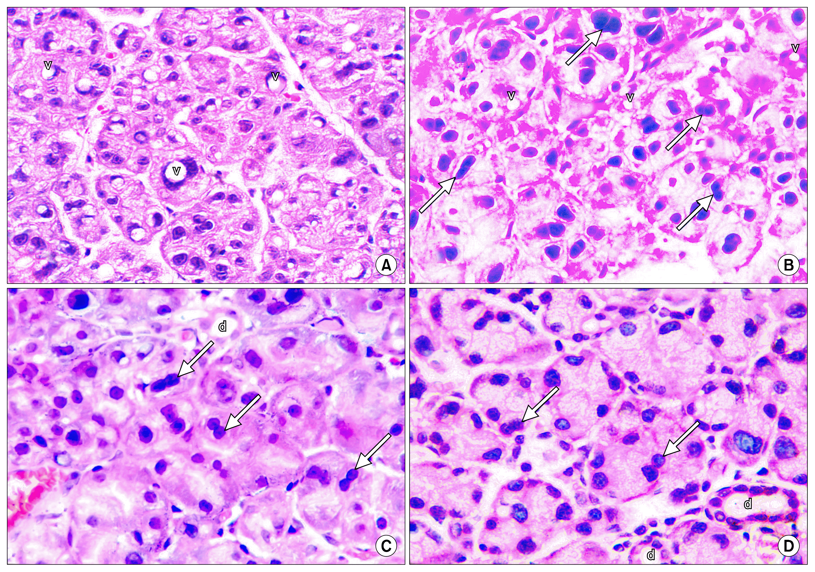 | Fig. 1Photomicrographs of parotid glands at day 10. (A) Cisplatin group showing severe intracytoplasmic vacuolation with irregular nuclear and acinar outlines. (B) Local BMSCs administration group showing some vacuoles and some mitotic figures. (C) Systemic BMSCs administration group showing regular nuclei with some mitotic figures and normal ducts. (D) Combination BMSCs administration group showing normal ducts, regular acinar and nuclear outlines and some mitotic figures. (v) vacuoles, (arrows) mitotic figures, (d) ducts (H&E, 400×).
|
 | Fig. 2Electron micrographs of parotid glands at day 10. (A) Cisplatin group showing irregular and atrophied nuclei, dilated rER and cytoplasmic vacuoles. (B) Local BMSCs administration group showing more regular nuclei but with condensed heterochromatin, dilated rER and many lysosomes. (C) Systemic BMSCs administration group showing regular euochromatic nuclei with homogenous secretory granules bordering a narrow lumen. (D) Combination BMSCs administration group showing euochromatic nucleus, regular rER and normal mitochondria. (N) nucleus, (rER) rough endoplasmic reticulum, (v) vacuoles, (L) lysosomes, (SG) secretory granules, (Lu) lumen, (M) mitochondria.
|
Immunohistochemical results (Fig. 3)

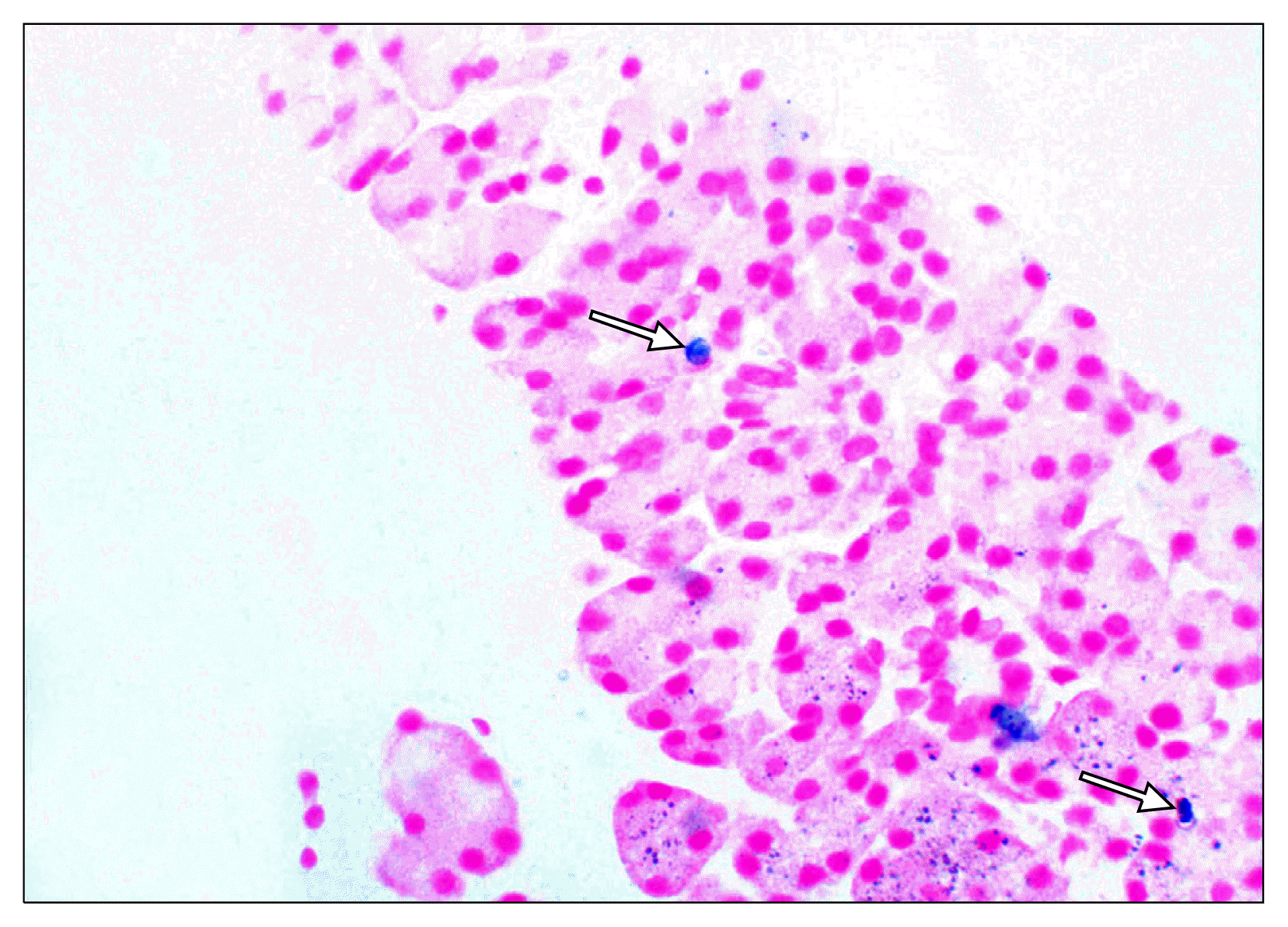
Prussian blue staining results (Fig. 4)
For the experimental groups only, some positive cells appeared in the parotid glands at all periods as granular blue or purple deposits.
Statistical results
Two way ANOVA (Analysis of Variance) test was used to detect the effect of different groups and different time periods on the PCNA proliferation index. Statistical significance (S) was considered when p value was ≤0.05.
The summary table (Table 1) shows a statistically significant interaction between the effects of groups and time on the proliferation index. The analysis also showed significance of the main effect of groups and the main effect of time.
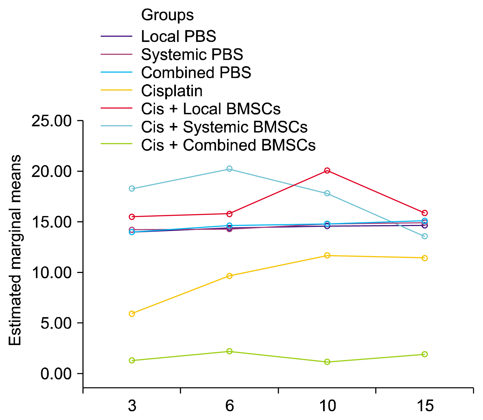
The marginal means of different groups, as presented in Table 2, show that the cisplatin receiving group had much depressed proliferation index than the negative control PBS groups indicating the suppressive effect of cisplatin on the cell cycle. The local group showed higher proliferation than the cisplatin group but not reaching the normal control levels. Systemic and combination groups had elevated proliferation indices than PBS groups may be to compensate the damage that occurred to the gland but their values were insignificantly different from each other. This shows that systemic and combination stem cell therapy had better effects in enhancing proliferation of the affected glands more than local therapy.
Table 2
Marginal means of different groups over time and statistical relation between them.
| Local PBS | Systemic PBS | Combined PBS | Cisplatin | Cisplatin+Local BMSCs | Cisplatin+Systemic BMSCs | Cisplatin+Combined BMSCs | |
|---|---|---|---|---|---|---|---|
| Mean | 14.41 | 14.51 | 14.64 | 1.64 | 9.67 | 16.80 | 17.47 |
| ±SD | 1.75 | 1.87 | 1.68 | 0.52 | 2.80 | 3.37 | 4.34 |
| P1 | 1.00 | 1.00 | <0.001* | <0.001* | <0.001* | <0.001* | |
| P2 | 1.00 | <0.001* | <0.001* | <0.001* | <0.001* | ||
| P3 | <0.001* | <0.001* | <0.001* | <0.001* | |||
| P4 | <0.001* | <0.001* | <0.001* | ||||
| P5 | <0.001* | <0.001* | |||||
| P6 | 1.00 |
![]()
The marginal means of different time periods over groups as presented in Table 3 show that cellular proliferation in the glands increased through the experiment starting from day 3 to compensate for the cisplatin induced cellular damage where proliferation at day 6 and 10 was significantly higher than day 3 reaching its peak at day 10 then dropped back at day 15 to be insignificant from day 3.
The line graph in Fig. 5 shows the course of each group through the experiment at each time period. The PBS groups appeared to be stable at all periods. Cisplatin group had a very much lowered index that was almost static through different periods. Local administration group was below the normal PBS line but showed increased proliferation. Systemic and combination groups showed higher proliferation indices than the control PBS group from the beginning, reaching a peak and then coming down to approach its level.
Go to :
Discussion
Cisplatin generates its antitumour effect by inducing cytotoxic changes leading to apoptosis or programmed cell death (20) by binding to cellular DNA (21), or mitochondrial DNA (22). It also induces reactive oxygen species production leading to increased lipid peroxidation and Ca2+ influx which can mediate apoptosis (23).
A known complication of cisplatin drug is acute kidney injury where administration of 5 ml/kg cisplatin in the abdominal cavity is associated with development of acute renal failure in rats within 72 hours (24). This may count for the death of some animals during the study course.
In the cisplatin control group, the histological and ultrastructural examinations showed many changes in the gland structure. These results agreed with the study by Kitashima who detected irregularity and swelling of the acinar cells with abnormal nuclei, large fused secretory granules with light and dark areas, enlarged fragmented rER, and swollen mitochondria in submandibular glands of rats receiving cisplatin (4).
Changes in the nuclear outlines and chromatin distribution as seen in the current study may be related to the mechanism of action of cisplatin where it affects the nucleus causing DNA platination and cytotoxic changes that may end with apoptosis. Typical morphological changes are characteristic for apoptosis as cell shrinkage, DNA fragmentation and chromatin condensation (25) similar to the changes observed in the present study. So, these changes might be signs of degeneration or a pre-apoptosis stage of acinar cells.
Intracytoplasmic vacuoles may be attributed to the damage of some intracellular structures and according to Kitashima (4), membranes of adjacent secretory granules can fuse forming intracytoplasmic vacuoles. Electrolucent secretory granules as those seen in the parotid gland sections in the present study are usually encountered in cases of sialedenosis and are thought to be signs of changes in the salivary protein component (26).
According to McInnes (27), eosinophilic materials, as those seen in the cisplatin control group, can be seen as an abnormal change in mice mucosal epithelium, sub-mucosal glands and ducts, and are thought to be accumulation of proteinaceous materials in the tissues.
The proposed mechanisms by which bone marrow cells improve organ functions have been investigated over a long period. Initial reports proposed the ability of BM cells to (trans) differentiate into cells of a non-marrow/non-hematopoietic lineage (28). Then reports on fusion of BM cells with cells of other tissues were documented (29). The third proposed mechanism was vasculogenesis from endothelial progenitor cells contained within BM (30). Lastly, the most recent mechanism is that BM cells provide a local paracrine effect on endogenous cells that may be achieved by secreting anti-apoptotic, anti-inflammatory and proliferation promoting factors (31).
Previous studies on the effect of local BMSCs transplantation into the submandibular glands of irradiated mice showed preserved acinar cells and gland morphologies along with improved gland functions as salivary flow rate and glandular weight (13, 14).
Similar studies were conducted by Tran et al. (15) and Sumita et al. (12) who injected BMSCs in tail veins of mice and rats with irradiated submandibular glands respectively. These studies detected better submandibular gland functions, increased proliferation, decreased apoptosis and increased vascularity as well.
In this study, it was noticed that parotid gland tissues started to show signs of recovery in the experimental local group at day 10 whereas in the systemic group starting from day 6. Moreover, at the end of the study; at day 15, the final histological picture of the gland sections treated with systemic BMSCs was more improved than the local intraglandular transplantation group. This may point to a better effect of systemic BMSCs administration than local transplantation route.
In consistency with our results, a study on the effect of different routes of BMSCs administration on a rat model of liver fibrosis compared between intraperitoneal, intra-hepatic and intravenous routes and the results of the study showed the intravenous route to be the most effective in regeneration of the fibrosed liver functions and histology. The authors justified their results by detecting elevated levels of IL-10 in the intravenous group than the other 2 groups where they suggested that the presence of BMSCs in the venous blood stimulates IL-10 release which can modulate the host immune response and enhances the healing process (32). Also in accordance with our results, a study on the effect of BMSCs on persistent orofacial pain rat model detected improvement in both intravenous and local injection routes but the results of the systemic group lasted for about 5 months while the local lasted only for 1 month (33) which gives an advantage to the systemic over the local route.
Some studies detected equal results clinically and histologically as well as equal incorporation of BMSCs into the target tissue by both local and systemic ways (34, 35). On the other hand, some studies reported better results with local transplantation than systemic BMSCs administration in oral mucosal ulcers as well as bone defect injuries (19, 36). Also, the regenerative effect of intravenous administration of BMSCs or BM soup on irradiated mice submandibular glands was found to be comparable to the local intraglandular route but with four times the required dose (15).
Local administration of BMSCs has shown efficiency in healing different injuries in many tissues as fore-mentioned. However, local infusion is likely not clinically feasible in many cases due to its potentially high degree of invasiveness (e.g., into the heart or brain), and locally administered cells may die before significantly contributing to the healing response due to diffusion limitations of nutrients and oxygen (37). Moreover, according to many studies it was reported that the main problem facing the efficacy of local direct stem cell injections is the ability of the injected cells to reside in the injury site and that is why different types of carriers like injectable hydrogel carriers, tissue grafts and cell-sheets are used to enable stem cell localization (38).
In addition to that, it was noted that in previous studies of local BMSCs administration on submandibular glands, the glands were surgically exposed first then injected directly (13, 15) but in this study for the parotid glands, the authors managed to assess the ability of the externally injected BMSCs to reach the gland and contribute to its regeneration. Also, the number of injected stem cells in the local administration group was less than some of the previous studies. These findings may justify the less histological improvement noted in the local transplantation group in comparison to the other experimental groups.
As far as we know, no available studies using a combination protocol for stem cell therapy could be found but the results of the combination group are close to those of the systemic intravenous group which gives no advantage for using both delivery routes together, besides the risk of increasing the stem cells dose and increasing manipulation procedures for the recipient.
In the experimental local intraparotid BMSCs transplantation group, the PCNA proliferation index was increased compared to the control group. This is in accordance with the results by Lim et al. (13) who found increased proliferation index as marked by PCNA and decreased apoptosis index as marked by TUNEL in the irradiated submandibular gland tissues treated with intraglandular BMSCs.
In the systemic intravenous BMSCs administration group, the average number of positive immunolabeled PCNA cells was significantly increased indicating the better effect of intravenous than the intraglandular BMSCs delivery routes. Similarly, in the study by Sumita et al. (12), intravenous BMSCs administration led to increased PCNA proliferation marker in irradiated submandibular glands.
No previous studies detecting the delivery of stem cells to the parotid glands were found but other studies confirmed the ability of BMSCs to home to submandibular glands after local as well as systemic administration with different labeling methods (14, 39).
Iron oxide labeled stem cells were detected in all experimental groups at all periods. As far as we know, only one in vivo study was found using iron sucrose for labeling of MSCs which were injected into lapine intravertebral discs and were detected after sacrifice by prussian blue staining technique (40).
Lastly it was concluded that BMSCs can reach the parotid glands by local as well as systemic routes and can enhance the healing potential and the proliferative capacity of the parotid gland cells after cisplatin administration. These results can be subjected for further experimental trials before clinical application on patients receiving chemotherapy to ameliorate their salivary gland functions during treatment.
Go to :
 XML Download