Journal List > Int J Stem Cells > v.8(2) > 1158932

Hosseini, Farahmandnia, Kazemi, Shakibajahromi, Sarvestani, and Khodabande: A Novel Cell Therapy Method for Recovering after Brain Stroke in Rats

Abstract

Background

Nowadays, stroke leads to a significant part of the adult mortality and morbidity and also it could result in some neurological deficits in the patients’ lives. Cell therapy has opened a new approach to treat the brain ischemia and reduce its terrible effects on the patients’ lives. There are several articles which show that the cell therapy could be beneficial for treating brain stroke. In this study, we have planned to present a new cell therapy method for stroke by administration of Mesenchymal stem cells and differentiated neural stem cells without astrocytes.

Method and Materials

The Mesenchymal stem cells were isolated from tibia and femur of a 250~300 g rat and they were cultured in DMEM/F12, 10% fetal bovine serum, 1% Pen/Strep. Neural stem cells were isolated from 14 days rat embryo ganglion eminence and were cultured in NSA media containing Neurobasal, 2% B27, bFGF 10 ng/ml and EGF 20 ng/ml after 5 days they formed some neurospheres. The isolated neural stem cells were differentiated to neural lineages by adding 5% fetal bovine serum to their culture media. After 48 hours the astrocytes were depleted by using MACS kit.

Results

The group that received Mesenchymal stem cells systemically and differentiated neural stem cells without astrocytes had the best neurological outcomes and the least infarct volume and apoptosis. It could be understood that this cell therapy method might cause almost full recovery after brain stoke.

Conclusion

Using combination cell therapy with Mesenchymal stem cells and differentiated neural stem cells with removed astrocyte could provide a novel method for curing brain stroke.

Introduction

Stroke is the second most important cause of death in industrial countries and the most significant cause of adult disability around the world (1, 2). Neurological deficits contain paralysis, memory impairment, parestheisa, slurred speech; etc (3). stroke is a result of an artery occlusion that leads to lack of oxygen and glucose, this nutrient shortage could cause some irreversible damage in brain tissue (4). Inflammation caused by loss of blood perfusion plays an important role in stroke pathophysiology, secreting of some cytokines such as interleukin1 (IL-1), Tumor necrosis factor-α (TNF-α), interleukin-6 (IL-6), Transforming growth factor-β (TGB-β) is resulting the post stoke inflammation (5, 6). Neuronal loss and brain damage is the secondary brain damage which the first neural loss is because of diminishing blood perfusion (7). the inflammatory cytokines could be secreted from microglia or astrocytes (8). Matrix metalloproteinases are a type of proteolytic enzyme that could make remodeling after stroke and is secreted by microglia, astrocyte, etc (9).
These days, there is only one approved drug for stroke which is rt-PA, it could be efficient according to thrombolytic effects; however, it has some side effects such as increasing the risk of bleeding and golden time limitation (10). Several hypotheses around mesenchymal stem cell therapy for stroke to account for therapeutic effects of this method have been suggested containing reducing inflammation, increasing angiogenesis, etc (11). This type of stem cells could be beneficial for reducing the inflammation and preparing the microenvironment by secreting some nutrients cytokines such as basic fibroblast growth factor (bFGF) and vascular endothelial growth factor (VEGF) (12). Neural stem cells is another type of stem cells which has been shown a promising approach to treat stroke and they are capable of differentiating to three neural lineage including neurons, astrocytes and oligodendrocytes (13).
In this study we have planned to transplant Mesenchymal stem cells systemically in acute phase (12 hours after ischemia) for modulating the immune system and differentiated neural stem cells with astrocyte depletion transplantation 3 days after brain ischemia for promoting neural regeneration.

Materials and Methods

Animal preparation

Seventy five Sprague Dawley male rats weighting 250~300 g were selected randomly and they were divided in 7 groups (n=15) as below:
  • Control group: Control group which received no intervention and treatment

  • Sham group: Sham group which were underwent middle cerebral Artery occlusion (MCAO) operation without any treatment

  • Third Group: The group which received systemic Mesenchymal stem cells injection 12 hours after inducing MCAO

  • Forth Group: The group which received differentiated neural stem cells without astrocytes intraventicular injection 3 days after inducing MCAO

  • Fifth Group: The group which received differentiation neural stem cells (Neurons, Oligodendrocytes and astrocytes) intraventricular injection 3 days after inducing MCAO

  • Sixth Group: The group which received systemic Mesenchymal stem cells 12 hours and differentiated neural stem cells (including astrocytes) after 3 days of inducing MCAO

  • Seventh Group: The group which received systemic Mesenchymal stem cells 12 hours and differentiated neural stem cells without astrocytes after 3 days of inducing MCAO

The animals were kept with free access to food and water and all procedures were approved by Shiraz University of Medical Sciences ethical committee.

Mesenchymal Stem Cell Isolation, Expansion and characterization

The Mesenchymal stem cells were obtained from adult male rat femur and tibia. Briefly, the femur and tibia were isolated and the connective tissue was removed, after that the bones were flashed by Mesenchymal stem cells culture media (DMEM/F12, 10% fetal bovine serum, 1% Pen/Strep) and the culture media containing rat bone marrow was transferred to T-25 cm2 and incubated in 5% CO2 and 37°C. The media was changed every 2 days and after 7 days the flask was 80~90% confluent. The cultured cells were passaged by adding trypsin 0.05% and complete culture media.
Some Mesenchymal stem cells markers containing CD44 (CBL1508FMillipore), CD45 (05-1410 Millipore), CD90 (CBL1500F Millipore) and CD34 (ab192547Abcam) were assessed by immunocytochemistry method for the isolated cells.
The Mesenchymal stem cells (1000000 Cells/kg) were injected via tail vein 12 hours after inducing MCAO.

Mesenchymal stem cells Differentiation

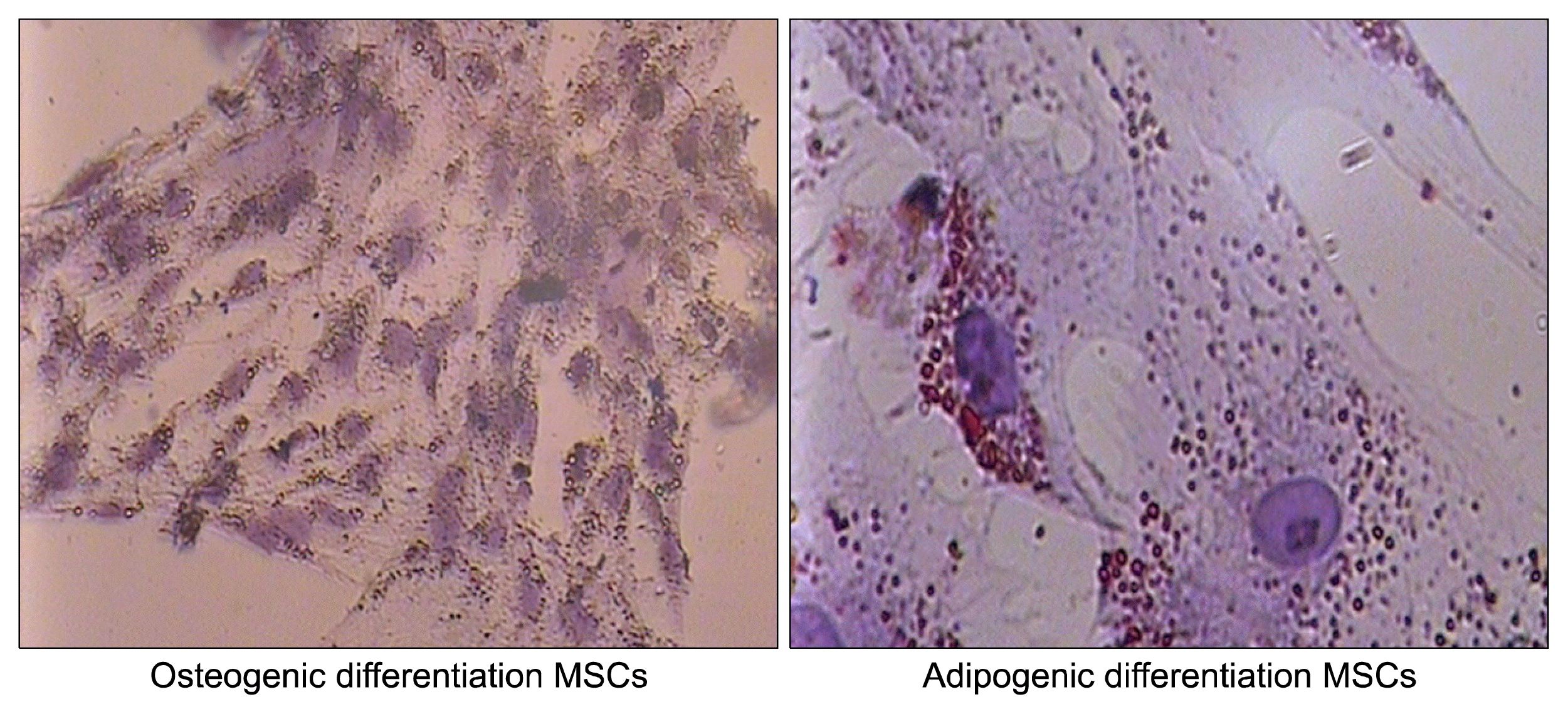
To evaluate the multipotent features of the isolated cells before laser treatment, BMSCs were cultured inadipogenic and osteogenic differentiation medium (Miltenyi biotech) for approximately 21 days. The cells stained with Alizarin Red for osteogenic differentiation and Oil red for adipogenic differentiation.

Neural Stem Cell Isolation, expansion and characterization

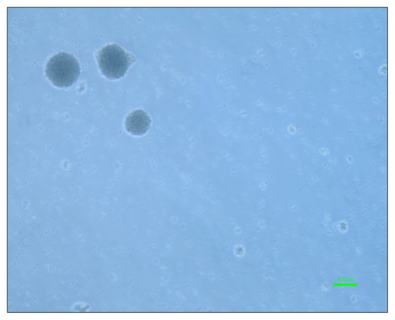
The neural stem cells were isolated by dissecting rat embryo 14 ganglion eminences and adding neural stem cell culture media (Neurobasal, 2% B27, bFGF 10 ng/ml, EGF 20 ng/ml and 1% pen/strep). After 5 days some spheres had formed in the culture flask which called neurospheres.
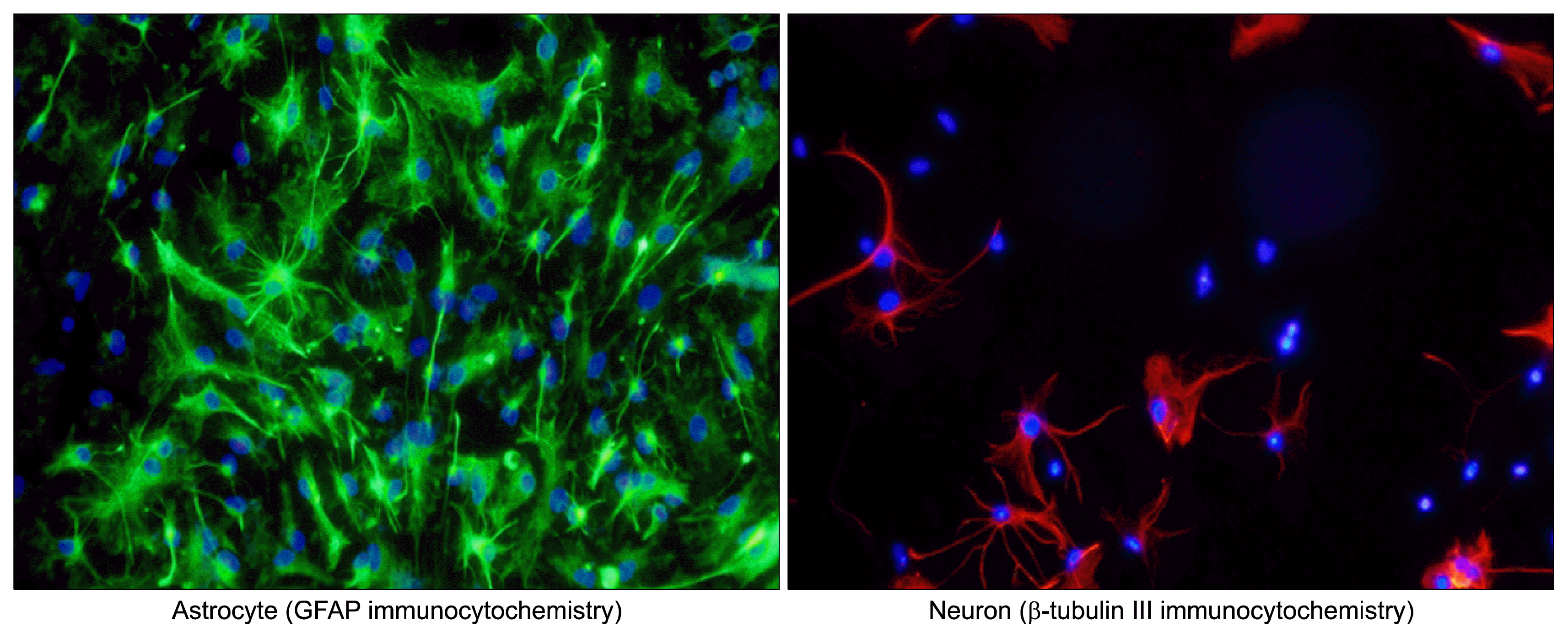
The spheres were trypsinized and the single cells were cultured in a Poly-l-lysin coated plate and 5% fetal bovine serum was added to the neural stem cells culture media to differentiate the NSCs to three neural lineages. The neural stem cells were differentiated to neurons, oligodendrocytes and astrocytes 3 days later. The neurons and astrocytes were stained with β-tubulin and GFAP by immunocytochemistry method.
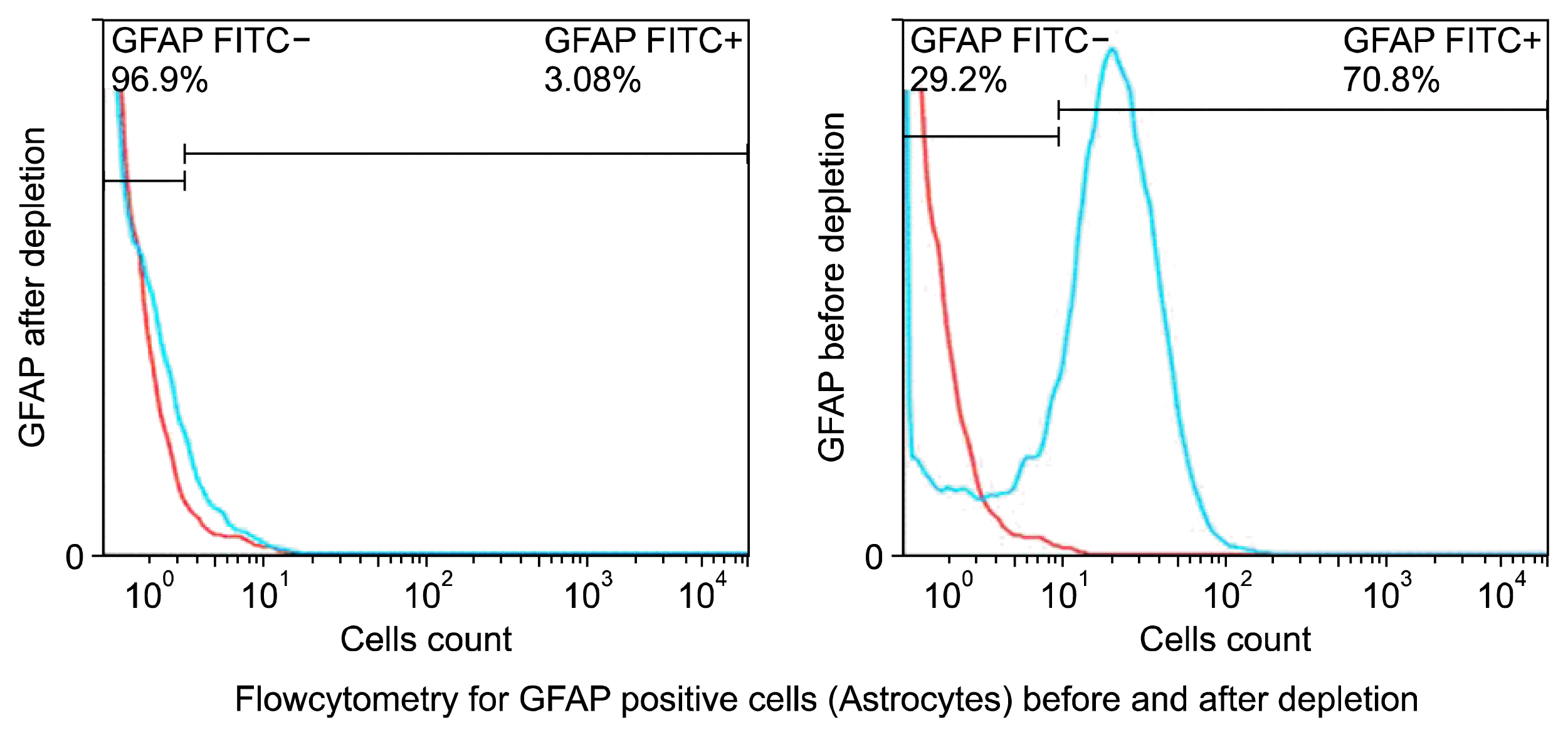
For preparing the differentiated neural stem cells for injecting the astrocyte were depleted with Astrocyte sorting kit (Miltenyi biotech, cat number: 130-096-052) and their depletion was assessed with flow cytometry method.
The differentiated neural stem cells minus astrocytes were transplanted stereotactically into lateral ventricle 3 days after MCAO.

Stereotactic Injection of Neural Stem Cells

The animals were anesthetized with halothane (induction 5% and maintenance 1%) and fixed to the stereotactical frame, the differentiated neural stem cells minus astrocytes were injected into right lateral ventricle at: Anterioposterior (AP)=−0.12 mm, mediolateral (ML)=1.6 mm, dorsoventricular (DV)=4.3 mm. Each rat received 200,000 cells in its right lateral ventricle.

Middle Cerebral Artery Occlusion (MCAO)

The rats were anesthetized by halothane (5% induction and 1.5% for maintenance) in the mixture of NO2 and O2 (50:50). MCAO induction was performed regarding to Koizumi’s method. Briefly, an incision was excised in the midline of the neck and the connective tissues, muscles and salivary glands were dissected till the carotid sheath was exposed, the sheath was removed and exposed carotid artery was separated from vagus nerve, then 2 loose sutures were prepared in common carotid artery and the external carotid artery was clamped, after that a small incision was excised between the sutures on common carotid artery and a silicon coated 4.0 nylon with round tip was passed through the common carotid artery to induce the occlusion of middle cerebral artery, after 45 minutes the 4.0 Nylon was removed and the sutures were tighten up and also the clamp on external carotid artery was opened to maintain the brain blood perfusion after the operation (14, 15).

Neurological Function Assessment

Neurological function was assessed every two days for all rats during 28 days of experiment. The neurological examination was scored on six-score scale. The scores are following as below (16):
  • Score of 0: No neurological deficit

  • Score of 1: Failure to extend left forepaw completely. It shows mild focal neurological deficit

  • Score of 2: Circling to the left. It means a moderate focal neurological deficit

  • Score of 3: Falling to the left. It indicates a sever focal neurological deficit

  • Score of 4: Not walking spontaneously and decreasing level of consciousness.

  • Score of 5: Death due to brain ischemia

Histology

After 28 days, the rats were anesthetized with Halothane and were fixed with normal saline followed Paraformaldehyde 4%, cry sections (20 μm) were mounted on silicon coated slides and stained with Hematoxin & Eosin.

Apoptosis evaluation with measurement of Caspase 3 activity

Activation of ICE family proteases/caspases begins apoptosis in mammalian cells. This assay is based on spectrophtorometric evaluation of chromophore p-nitroaniline (p-NA) after cleavage from labeled substrate DEVD-p-NA. The p-NA light emission could be measured by using spectrophotometer at 400~405 nm. For this assay Caspase 3 assay kit from abcam company used (ab39401).

Statistical Analysis

All the data were analyzed with One-way ANOVA method with Prism 6.00 software.

Results

Mesenchymal stem cells morphology and marker assessment

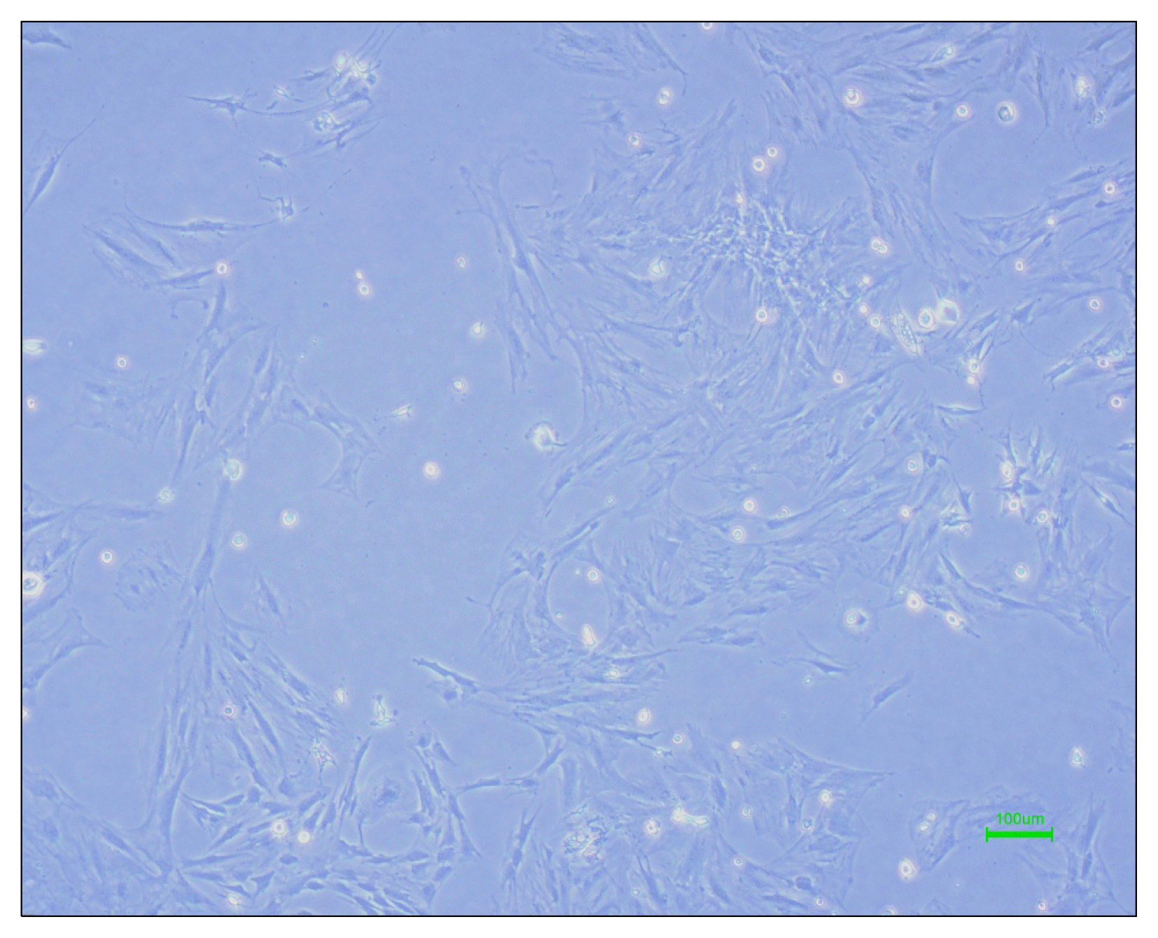
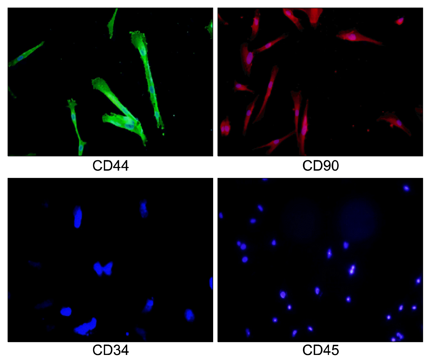
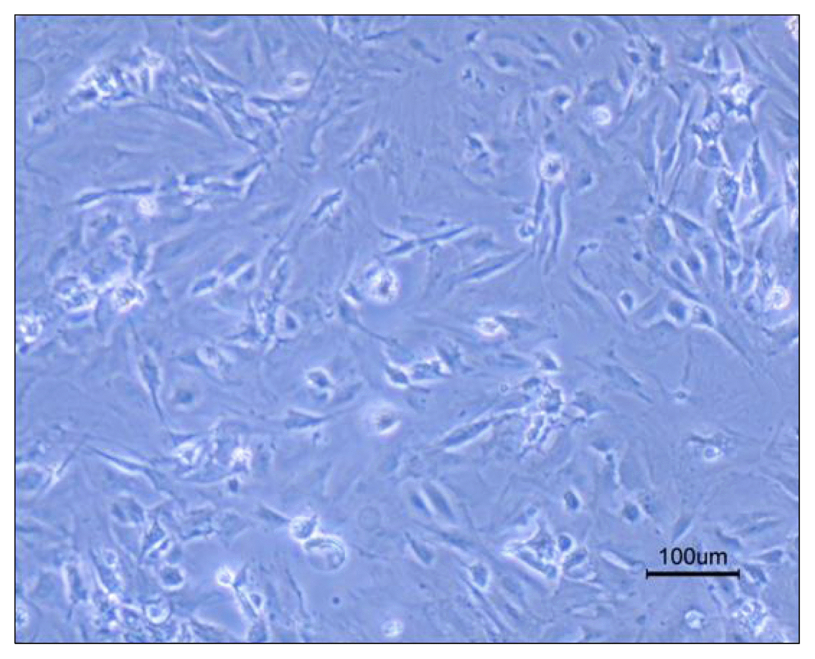
The isolated rat bone marrow Mesenchymal stem cells formed few thin and long cell processes and a large nucleus in their cell body could be observed (Fig. 1). These cells did not express hematopoietic stem cells markers (CD34 and CD45) and also they expressed the mesenchymal stem cells markers (CD44 and CD90) (Fig. 2, Table 1).

Differentiation Assay

Differentiation to adipocyte was confirmed by morphological changes after 7 days such as vacuole production and Oil red staining after 21 days seeding in adipogenic media.
Osteogenic differentiation was started with matrix deposition after 5~7 days and was confirmed with Alizarin Red staining after 14 days (Fig. 3).

Neural Stem Cells Characterization and Differentiation

Five days after culturing neural stem cells, some spheres formed in culture flasks which were called neurospheres (Fig. 4). 48 hours after adding fetal bovine serum to the neural stem cells media they have differentiated to three neural lineages cell (Neurons, Oligodendrocytes and Astrocytes) as it could be seen in Fig. 5 the differentiated neural stem cells have different morphology due to different provided cell types (Fig. 5).
For evaluation of neural and astrocytes differentiation they were stained with anti β-tubulin III and anti-GFAP by immunocytochemistry method (Fig. 6, Table 2).
The astrocyte depletion of differentiated neural stem cells was assessed by flowcytometry method (Fig. 7).

Neurological Examination

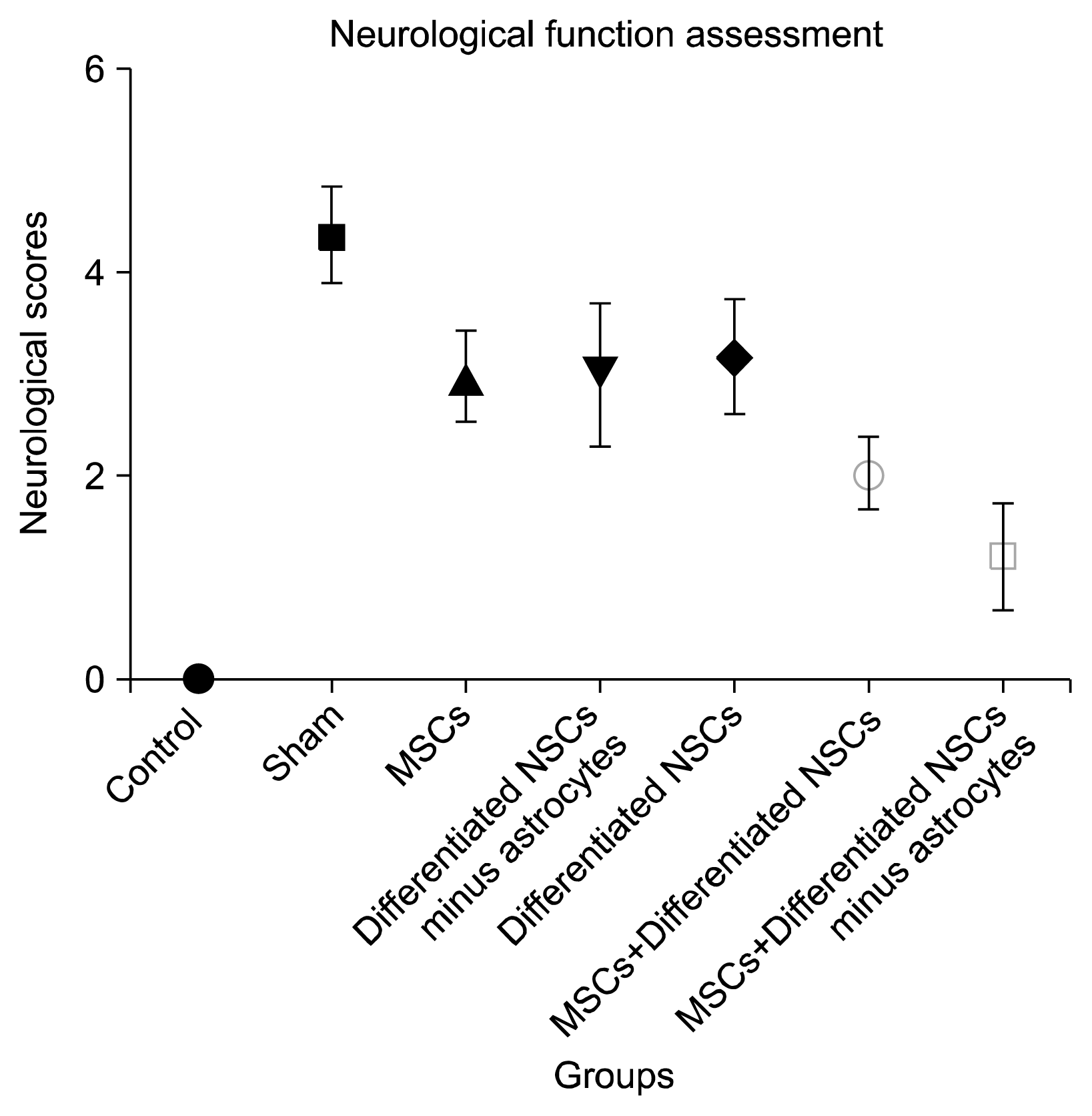
All seventy five rats were examined according to six-score scale by double blind examiner every two days during 28 days of experiment. The neurological scores were analyzed with SPSS 16.00 one-way ANOVA test and there is a significant difference between the groups which received cell therapy and the sham group. In addition, the group which received Mesenchymal stem cells and differentiated neural stem cells without astrocytes had the best neurological outcome between the groups received cell therapy with p value less than 0.05 (p value<0.05) (Fig. 8).

Histology

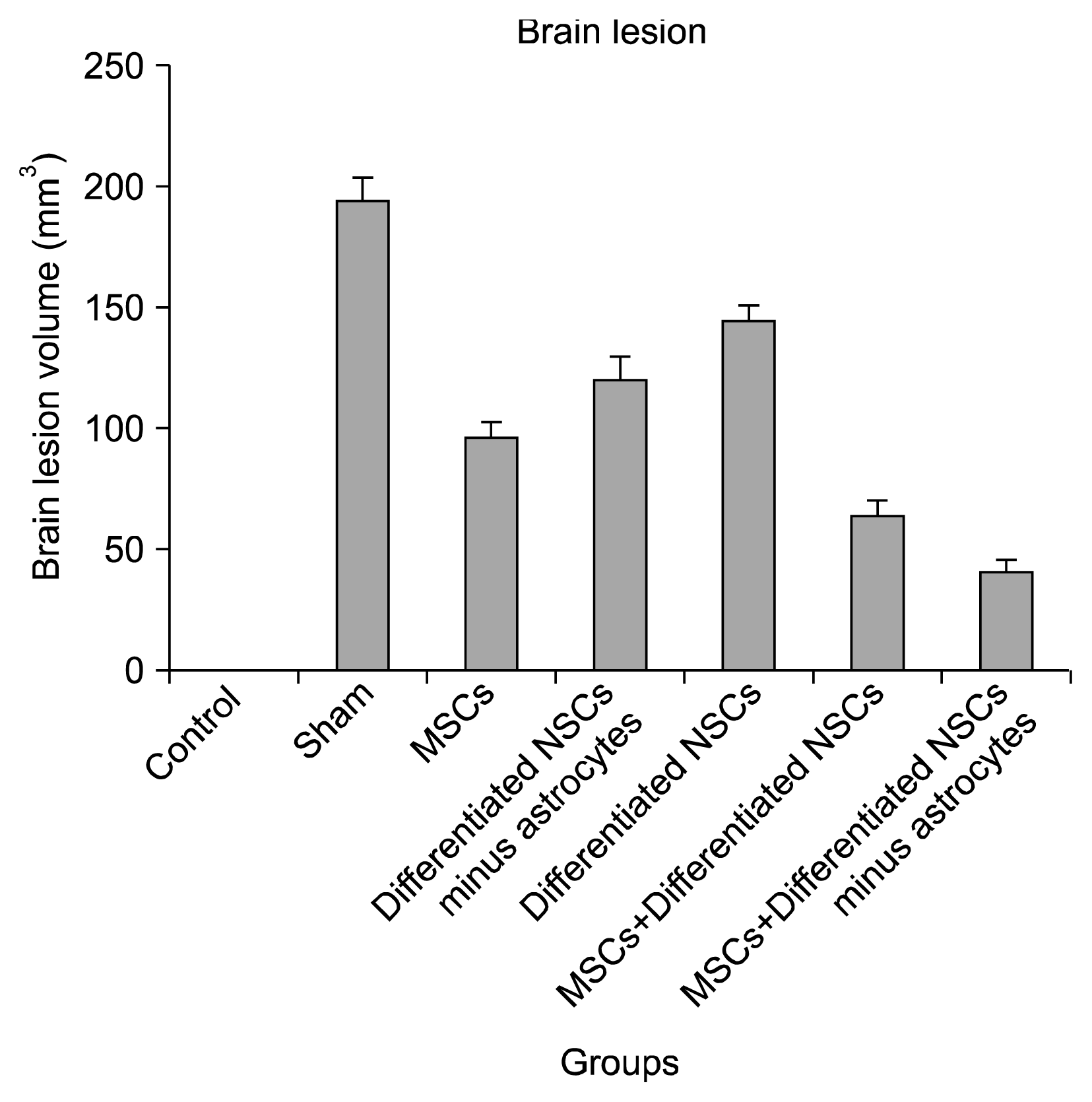
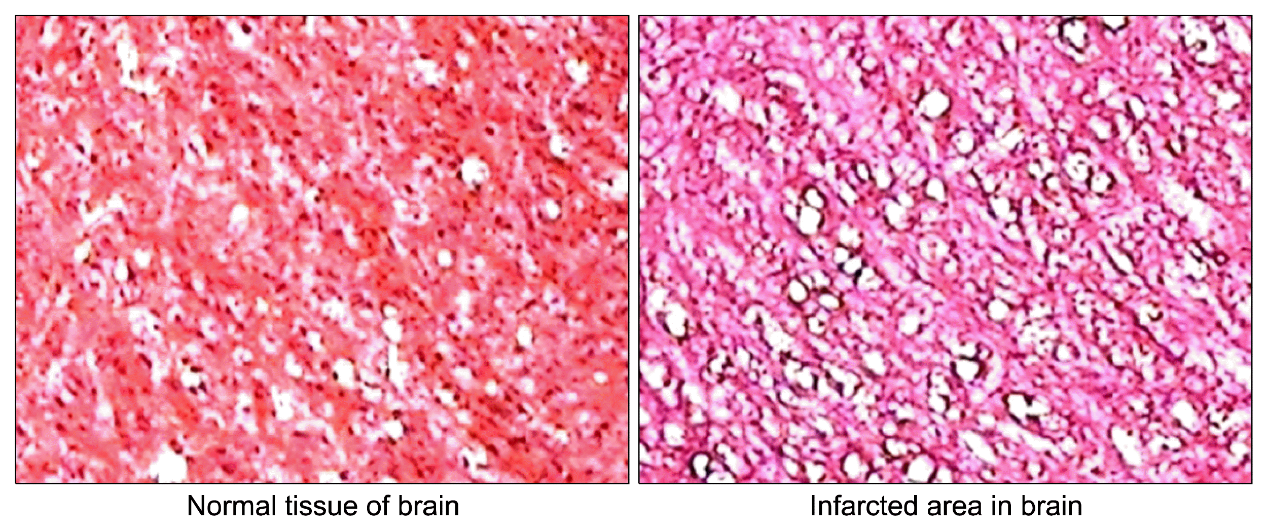
The coronal sections were selected with 1.2 mm interval and from the first one was 1 mm posterior to frontal pole. Damaged area was defined by some ischemic signs such as eosinophilic cytoplasm and pyknotic nuclei. The ischemic zones in the groups which were taken cell therapy were significantly decreased in comparison with the sham group. Also, the group received Mesenchymal stem cells and differentiated neural stem cells without astrocytes had the least ischemic area among the cell therapy groups (40.264 mm3±6.12) that shows it could be more effective than the other methods (Fig. 9 and 10).

Caspase 3 Activity Assay

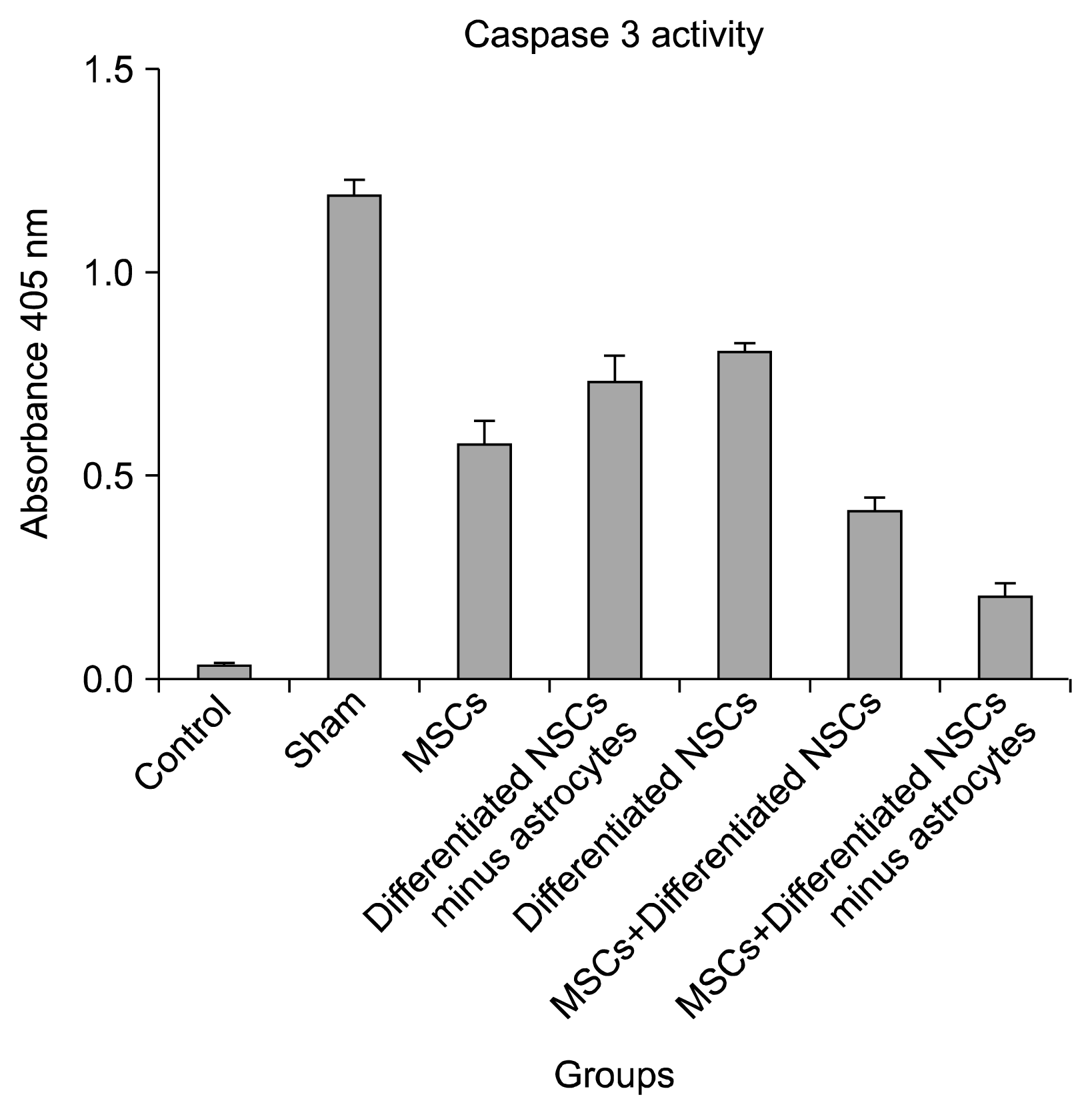
Some stem cells are capable of reducing apoptosis by down regulating some apoptotic genes such as caspase3 (14). Caspase 3 activity was assessed by spectrophotometer at 405 nm. The higher absorbance value revealed the higher Caspase 3activity and as a result the higher incidence of apoptosis. The result indicates that the group which received Mesenchymal stem cells and differentiated neural stem cells without astrocytes had the least amount of Caspase 3 activity between the rests of the groups and as a result the lowest amount of apoptosis (0.215±0.04) (Fig. 11).

Discussion

In this study we have presented a novel cell therapy method for almost treating stroke with Mesenchymal stem cells and differentiated neural stem cells with astrocytes depletion.
We have designed different cell therapy method according to different characteristics of Mesenchymal stem cells and differentiated neural stem cells and according the results the combination cell therapy with MSCs and differentiated NSCs without astrocytes could improve neurological function and also reduce the brain lesion volume and apoptosis after brain stroke.
Mesenchymal stem cells have been shown suppress T-cell activation and proliferation and also reduce T-cells response indirectly by suppression of CD 34+ progenitor cell differentiation (14, 1719). Some studies demonstrated that Mesenchymal stem cells are capable to have anti-inflammatory effects and secret some cytokines such as interleukin 10 (IL-10) and IL-6 according to their micro-environment and some pro-inflammatory cytokines including TNF-α, IFN-λ and IL-1β are potential to induce MSC anti-inflammatory effects (2022). Cheng et al. in 2015 have shown that Mesenchymal stem cells transplantation could improve neurological function after stroke by their ability of attenuating inflammatory factors and also modulating TGF-β (23). Calio et al. in 2014 Mesenchymal stem cells transplantation could protect the brain tissue against stress oxidative and diminish apoptosis after stroke (24). As previous studies and our results indicate, the Mesenchymal stem cells could make less volume infarct, better neurological outcomes and less apoptosis especially in acute phase of stroke. In all groups which treated with Mesenchymal stem cells alone or with other cells, neurological recovery and decreasing of apoptosis could be observed.
Neural stem cells are a type of stem cells exists mostly in subventricular zone (SVZ) and dentate gyrus (DG), they are able to migrate to degenerating area and regenerate the damaged zone by anti-inflammatory effects and differentiation to three neural lineages containing neurons, oligodendrocytes and astrocytes (2529). Despite of mentioned positive points of neural stem cells, they promote astrocytosis (30) and it might be an obstacle for reaching an optimum effect of neural stem cells transplantation. Stroke induces a series of changes in molecular and cellular aspects in brain tissue such as increasing morphology changes, cellular proliferation and genes expressions (3134). The expression of GFAP would be increased in stroke which leads to astrocytosis and formation of glial scares that could surround the ischemic zone and inhibit regeneration and endogenous neural stem cells migration (3539). Our results illustrates that the groups which received differentiated neural stem cells had better outcome and less apoptosis; however by omitting the astrocyte the outcome would be promoted and apoptosis would be diminished.
According to all above, we designed an experiment to define the effects of depleting astrocyte and a new method by combining differentiated neural stem cells and Mesenchymal stem cells for treating brain stroke. As our result shows depleting the astrocytes might have positive effects on recovering after stroke and it could be developed when the Mesenchymal stem cells were injected systemically.

Notes

Potential conflict of interest

The authors have no conflicting financial interest.

References

1. Wu D. Neuroprotection in experimental stroke with targeted neurotrophins. NeuroRx. 2005; 2:120–128. DOI: 10.1602/neurorx.2.1.120. PMID: 15717063. PMCID: 539332.
crossref
2. Broderick JP, William M. Feinberg Lecture: stroke therapy in the year 2025: burden, breakthroughs, and barriers to progress. Stroke. 2004; 35:205–211. DOI: 10.1161/01.STR.0000106160.34316.19.
3. Muir KW, Tyrrell P, Sattar N, Warburton E. Inflammation and ischaemic stroke. Curr Opin Neurol. 2007; 20:334–342. DOI: 10.1097/WCO.0b013e32813ba151. PMID: 17495630.
crossref
4. Dirnagl U, Iadecola C, Moskowitz MA. Pathobiology of ischaemic stroke: an integrated view. Trends Neurosci. 1999; 22:391–397. DOI: 10.1016/S0166-2236(99)01401-0. PMID: 10441299.
crossref
5. Zhu Y, Yang GY, Ahlemeyer B, Pang L, Che XM, Culmsee C, Klumpp S, Krieglstein J. Transforming growth factor-beta 1 increases bad phosphorylation and protects neurons against damage. J Neurosci. 2002; 22:3898–3909. PMID: 12019309.
crossref
6. Spera PA, Ellison JA, Feuerstein GZ, Barone FC. IL-10 reduces rat brain injury following focal stroke. Neurosci Lett. 1998; 251:189–192. DOI: 10.1016/S0304-3940(98)00537-0. PMID: 9726375.
crossref
7. Lakhan SE, Kirchgessner A, Hofer M. Inflammatory mechanisms in ischemic stroke: therapeutic approaches. J Transl Med. 2009; 7:97. DOI: 10.1186/1479-5876-7-97. PMID: 19919699. PMCID: 2780998.
crossref
8. Swanson RA, Ying W, Kauppinen TM. Astrocyte influences on ischemic neuronal death. Curr Mol Med. 2004; 4:193–205. DOI: 10.2174/1566524043479185. PMID: 15032713.
crossref
9. Ginsberg MD. Cerebrovascular disease: pathophysiology, diagnosis, and management. Blackwell Science, Bogousslavsky J. Malden, UK: Blackwell Science;1998. p. 193–204.
10. Furlan AJ, Katzan IL, Caplan LR. Thrombolytic therapy in acute ischemic stroke. Curr Treat Options Cardiovasc Med. 2003; 5:171–180. DOI: 10.1007/s11936-003-0001-4. PMID: 12777195.
crossref
11. Hirouchi M, Ukai Y. Current state on development of neuroprotective agents for cerebral ischemia. Nihon Yakurigaku Zasshi. 2002; 120:107–113. DOI: 10.1254/fpj.120.107. PMID: 12187623.
crossref
12. Rehman J, Traktuev D, Li J, Merfeld-Clauss S, Temm-Grove CJ, Bovenkerk JE, Pell CL, Johnstone BH, Considine RV, March KL. Secretion of angiogenic and antiapoptotic factors by human adipose stromal cells. Circulation. 2004; 109:1292–1298. DOI: 10.1161/01.CIR.0000121425.42966.F1. PMID: 14993122.
crossref
13. Chang DJ, Oh SH, Lee N, Choi C, Jeon I, Kim HS, Shin DA, Lee SE, Kim D, Song J. Contralaterally transplanted human embryonic stem cell-derived neural precursor cells (ENStem-A) migrate and improve brain functions in stroke-damaged rats. Exp Mol Med. 2013; 45:e53. DOI: 10.1038/emm.2013.93. PMID: 24232252. PMCID: 3849578.
crossref
14. Jiang XX, Zhang Y, Liu B, Zhang SX, Wu Y, Yu XD, Mao N. Human mesenchymal stem cells inhibit differentiation and function of monocyte-derived dendritic cells. Blood. 2005; 105:4120–4126. DOI: 10.1182/blood-2004-02-0586. PMID: 15692068.
crossref
15. Koizumi J, Yoshida Y, Nakazawa T, Ooneda G. Experimental studies of ischemic brain edema: 1. A new experimental model of cerebral embolism in rats in which recirculation can be introduced in the ischemic area. Jpn J Stroke. 1986; 8:1–8. DOI: 10.3995/jstroke.8.1.
16. Longa EZ, Weinstein PR, Carlson S, Cummins R. Reversible middle cerebral artery occlusion without craniectomy in rats. Stroke. 1989; 20:84–91. DOI: 10.1161/01.STR.20.1.84. PMID: 2643202.
crossref
17. Di Nicola M, Carlo-Stella C, Magni M, Milanesi M, Longoni PD, Matteucci P, Grisanti S, Gianni AM. Human bone marrow stromal cells suppress T-lymphocyte proliferation induced by cellular or nonspecific mitogenic stimuli. Blood. 2002; 99:3838–3843. DOI: 10.1182/blood.V99.10.3838. PMID: 11986244.
crossref
18. Krampera M, Glennie S, Dyson J, Scott D, Laylor R, Simpson E, Dazzi F. Bone marrow mesenchymal stem cells inhibit the response of naive and memory antigen-specific T cells to their cognate peptide. Blood. 2003; 101:3722–3729. DOI: 10.1182/blood-2002-07-2104.
crossref
19. Ivanova-Todorova E, Bochev I, Mourdjeva M, Dimitrov R, Bukarev D, Kyurkchiev S, Tivchev P, Altunkova I, Kyurkchiev DS. Adipose tissue-derived mesenchymal stem cells are more potent suppressors of dendritic cells differentiation compared to bone marrow-derived mesenchymal stem cells. Immunol Lett. 2009; 126:37–42. DOI: 10.1016/j.imlet.2009.07.010. PMID: 19647021.
crossref
20. DelaRosa O, Dalemans W, Lombardo E. Mesenchymal stem cells as therapeutic agents of inflammatory and auto-immune diseases. Curr Opin Biotechnol. 2012; 23:978–983. DOI: 10.1016/j.copbio.2012.05.005. PMID: 22682584.
crossref
21. Ma S, Xie N, Li W, Yuan B, Shi Y, Wang Y. Immunobiology of mesenchymal stem cells. Cell Death Differ. 2014; 21:216–225. DOI: 10.1038/cdd.2013.158. PMCID: 3890955.
crossref
22. Shi Y, Hu G, Su J, Li W, Chen Q, Shou P, Xu C, Chen X, Huang Y, Zhu Z, Huang X, Han X, Xie N, Ren G. Mesenchymal stem cells: a new strategy for immunosuppression and tissue repair. Cell Res. 2010; 20:510–518. DOI: 10.1038/cr.2010.44. PMID: 20368733.
crossref
23. Cheng Q, Zhang Z, Zhang S, Yang H, Zhang X, Pan J, Weng L, Sha D, Zhu M, Hu X, Xu Y. Human umbilical cord mesenchymal stem cells protect against ischemic brain injury in mouse by regulating peripheral immunoinflammation. Brain Res. 2015; 1594:293–304. DOI: 10.1016/j.brainres.2014.10.065.
crossref
24. Calió ML, Marinho DS, Ko GM, Ribeiro RR, Carbonel AF, Oyama LM, Ormanji M, Guirao TP, Calió PL, Reis LA, Simões Mde J, Lisbôa-Nascimento T, Ferreira AT, Bertoncini CR. Transplantation of bone marrow mesenchymal stem cells decreases oxidative stress, apoptosis, and hippocampal damage in brain of a spontaneous stroke model. Free Radic Biol Med. 2014; 70:141–154. DOI: 10.1016/j.freeradbiomed.2014.01.024. PMID: 24525001.
crossref
25. Eriksson PS, Perfilieva E, Björk-Eriksson T, Alborn AM, Nordborg C, Peterson DA, Gage FH. Neurogenesis in the adult human hippocampus. Nat Med. 1998; 4:1313–1317. DOI: 10.1038/3305. PMID: 9809557.
crossref
26. Curtis MA, Kam M, Nannmark U, Anderson MF, Axell MZ, Wikkelso C, Holtås S, van Roon-Mom WM, Björk-Eriksson T, Nordborg C, Frisén J, Dragunow M, Faull RL, Eriksson PS. Human neuroblasts migrate to the olfactory bulb via a lateral ventricular extension. Science. 2007; 315:1243–1249. DOI: 10.1126/science.1136281. PMID: 17303719.
crossref
27. Kornblum HI, Geschwind DH. Molecular markers in CNS stem cell research: hitting a moving target. Nat Rev Neurosci. 2001; 2:843–846. DOI: 10.1038/35097597. PMID: 11715062.
crossref
28. Bull ND, Bartlett PF. The adult mouse hippocampal progenitor is neurogenic but not a stem cell. J Neurosci. 2005; 25:10815–10821. DOI: 10.1523/JNEUROSCI.3249-05.2005. PMID: 16306394.
crossref
29. Taupin P. The therapeutic potential of adult neural stem cells. Curr Opin Mol Ther. 2006; 8:225–231. PMID: 16774042.
30. Batrakova EV, Gendelman HE, Kabanov AV. Cell-mediated drug delivery. Expert Opin Drug Deliv. 2011; 8:415–433. DOI: 10.1517/17425247.2011.559457. PMID: 21348773. PMCID: 3062753.
crossref
31. Ding S, Wang T, Cui W, Haydon PG. Photothrombosis ischemia stimulates a sustained astrocytic Ca2+ signaling in vivo. Glia. 2009; 57:767–776. DOI: 10.1002/glia.20804. PMCID: 2697167.
crossref
32. Li H, Zhang N, Sun G, Ding S. Inhibition of the group I mGluRs reduces acute brain damage and improves long-term histological outcomes after photothrombosis-induced ischaemia. ASN Neuro. 2013; 5:195–207. DOI: 10.1042/AN20130002. PMID: 23772679. PMCID: 3786425.
crossref
33. Panickar KS, Norenberg MD. Astrocytes in cerebral ischemic injury: morphological and general considerations. Glia. 2005; 50:287–298. DOI: 10.1002/glia.20181. PMID: 15846806.
crossref
34. Zamanian JL, Xu L, Foo LC, Nouri N, Zhou L, Giffard RG, Barres BA. Genomic analysis of reactive astrogliosis. J Neurosci. 2012; 32:6391–6410. DOI: 10.1523/JNEUROSCI.6221-11.2012. PMID: 22553043. PMCID: 3480225.
crossref
35. Haupt C, Witte OW, Frahm C. Up-regulation of Connexin43 in the glial scar following photothrombotic ischemic injury. Mol Cell Neurosci. 2007; 35:89–99. DOI: 10.1016/j.mcn.2007.02.005. PMID: 17350281.
crossref
36. Hayakawa K, Nakano T, Irie K, Higuchi S, Fujioka M, Orito K, Iwasaki K, Jin G, Lo EH, Mishima K, Fujiwara M. Inhibition of reactive astrocytes with fluorocitrate retards neurovascular remodeling and recovery after focal cerebral ischemia in mice. J Cereb Blood Flow Metab. 2010; 30:871–882. DOI: 10.1038/jcbfm.2009.257.
crossref
37. Barreto GE, Sun X, Xu L, Giffard RG. Astrocyte proliferation following stroke in the mouse depends on distance from the infarct. PLoS One. 2011; 6:e27881. DOI: 10.1371/journal.pone.0027881. PMID: 22132159. PMCID: 3221692.
crossref
38. Bao Y, Qin L, Kim E, Bhosle S, Guo H, Febbraio M, Haskew-Layton RE, Ratan R, Cho S. CD36 is involved in astrocyte activation and astroglial scar formation. J Cereb Blood Flow Metab. 2012; 32:1567–1577. DOI: 10.1038/jcbfm.2012.52. PMID: 22510603. PMCID: 3421096.
crossref
39. Shimada IS, Borders A, Aronshtam A, Spees JL. Proliferating reactive astrocytes are regulated by Notch-1 in the peri-infarct area after stroke. Stroke. 2011; 42:3231–3237. DOI: 10.1161/STROKEAHA.111.623280. PMID: 21836083. PMCID: 4469355.
crossref

Fig. 1
Rat Mesenchymal stem cells.
ijsc-08-191f1.tif
Fig. 2
Rat Mesenchymal stem cell immunocytochemistry for CD44, CD90, CD45 and CD34.
ijsc-08-191f2.tif
Fig. 3
Mesenchymal stem cells differentiation to adipocyte and osteocyte.
ijsc-08-191f3.tif
Fig. 4
Neurospheres 5 days after neural stem cell culture.
ijsc-08-191f4.tif
Fig. 5
Differentiated neural stem cells.
ijsc-08-191f5.tif
Fig. 6
Neural Stem Cell Differentiation and immunocytochemistry with GFAP and β-tubulin III antibody.
ijsc-08-191f6.tif
Fig. 7
Flow cytometry for assessing depletion of astrocytes. The blue line is the GFAP positive cells.
ijsc-08-191f7.tif
Fig. 8
Neurological scores in 28 days. The data was analyzed with One-way ANOVA and the p value is less than 0.05. The data between groups was analyzed with Tukey’s multiple comparison tests.
ijsc-08-191f8.tif
Fig. 9
H&E staining and ischemic area volume. The data was analyzed with One-way ANOVA and the p value is less than 0.05. The data between groups was analyzed with Tukey’s multiple comparison tests.
ijsc-08-191f9.tif
Fig. 10
H&E staining for brain tissue.
ijsc-08-191f10.tif
Fig. 11
Caspase 3 activity. The data was analyzed with One-way ANOVA and the p value is less than 0.05. The data between groups was analyzed with Tukey’s multiple comparison tests.
ijsc-08-191f11.tif
Table 1
Mesenchymal stem cells markers expression
CD44 CD90 CD 34 CD45
94.44%±2.91 84.25%±7.10 1.70%±0.84 0.94%±0.64
Table 2
Neural Stem Cells Differentiation to neurons and astrocytes
β-tubulin III GFAP
11.84%±3.98 84.25%± 5.62
TOOLS
Similar articles