Journal List > Int J Stem Cells > v.3(2) > 1158912

Abdel Aziz, EL Asmar, Mostafa, Salama, Atta, Mahfouz, Roshdy, Rashed, Sabry, Hasan, Mahmoud, and Elderwy: Reversal of Hepatic Fibrosis by Human CD34+ Stem/Progenitor Cell Transplantation in Rats

Abstract

Human umbilical cord blood (UCB) cells have many advantages as grafts for cell transplantation. Here, we transplant UCB cells into injured liver fibrosis, investigated the hepatic potential of UCB cells both in vitro and in vivo. a CCl4 rat model with liver fibrosis was prepared. Human (UCB) CD34+ stem cell was separated with MACS (magnetic cell sorting). Cells were cultured with and without hepatic differentiation medium. Rats were divided into 3 groups; group (1): control healthy, group (2): CCl4 injected rats and group 3: CCl4/CD34+injected rats with human differentiated and undifferentiated cells through intrahepatic (IH) and intravenous (IV) routes. A significant elevation was detected in serum albumin in CCl4/CD34+ compared to the CCl4 group (p<0.001). Serum ALT, had a significant decrease of its level after administration of stem cells compared to the CCl4 group (p<0.001). However, it was still significantly higher than control (p<0.001) with no significant difference between the groups that received stem cells. Histopathological examination of liver tissue showed that stem cells have a significant antifibrotic effect. Concerning gene expression, the collagen gene (rat) was highly expressed in the CCl4 group whereas its expression was significantly decreased after administration of stem cells. Human albumin and matrix metalloproteinase (MMP2) genes were expressed in liver tissues in the groups that received stem cells. Highest expression was in the group that received un-differentiated cells I.V. human UCB CD34+ stem cells can ameliorate liver fibrosis in rats.

Introduction

Liver transplantation is the gold standard treatment for end-stage liver failure and for numerous liver based in-born errors of metabolism. However, organ shortage remains a major limiting factor and alternative solutions are being examined in the liver therapy field. Liver cell transplantation is emerging with heartening success (1, 2), but is still limited by cell viability, modest engraftment and limited tissue availability. Increasing interest is carried to stem cells regarding the recent demonstration of their plasticity (3).
Several sources of stem cells have been proposed as sources for cell therapy. Embryonic stem cells are the most potent in terms of their differentiation potential but may be tumorigenic when transplanted in vivo, and their use is beset by ethical issues (4, 5). Adult stem cells may be found in any tissue (6), but hematopoietic tissue is most accessible. Hematopoietic tissue contains two types of stem cells, the mesenchymal and hematopoietic stem cells. Abdel Aziz et al. (7) showed that bone marrow -derived mesenchymal stem cells can ameliorate liver fibrosis in rats. Stem cells in hematopoietic tissue have been used for hematological reconstitution for many years (8). These cells are CD34+ and CD133+ and give rise to all lineages of blood cell differentiation. Thus, they have the advantage that they can be prospectively isolated from hematopoietic tissue in known numbers.
In humans, Alison et al. (9) and Theise et al. (10) showed that the adult human hematopoietic stem cell population can yield hepatocytes upon instruction by the appropriate environment. Korbling et al. (11) showed that hepatocytes are generated from the bone marrow of recipients of sex-mismatched bone marrow transplants at a high frequency that ranges from 4% to 7%. Moreover, Ng et al. (12) found that in human liver allografts, although most of the recipient-derived cells showed macrophage/Kupffer cell differentiation, recipient-derived hepatocytes were also present and constituted 0.62% of all the hepatocytes in the recipient. To examine the mechanisms by which human hematopoietic cells contribute to liver regeneration, the human-to-mouse xenogeneic transplantation model was used.
Several reports have shown that when human umbilical cord blood (UCB) cells (all cells, CD34+ cells, or CD45+ cells) are injected into mice through either the portal vein or the systemic circulation, they can form human hepatocyte-like cells in the murine liver environment (1320). However, even when there is massive liver damage, the frequency with which this hepatocytic differentiation occurs is low compared to that reported in human-to-human transplantation studies. This low level of efficiency makes it hard to clarify whether transdifferentiation or cell fusion is the primary mechanism that generates hepatocytes from human hematopoietic cells.
In the present study, we aimed to clarify the role played by human CD34+ stem cells to ameliorate liver fibrosis in rats. We also aimed to investigate whether there is an effect of the route of administration as well as the degree of differentiation of the stem cells.

Materials and Methods

Cell source

Human UCB was used for separation of mononuclear cells (MNCs) after obtaining an informed consent and research ethics committee approval.

Cell isolation

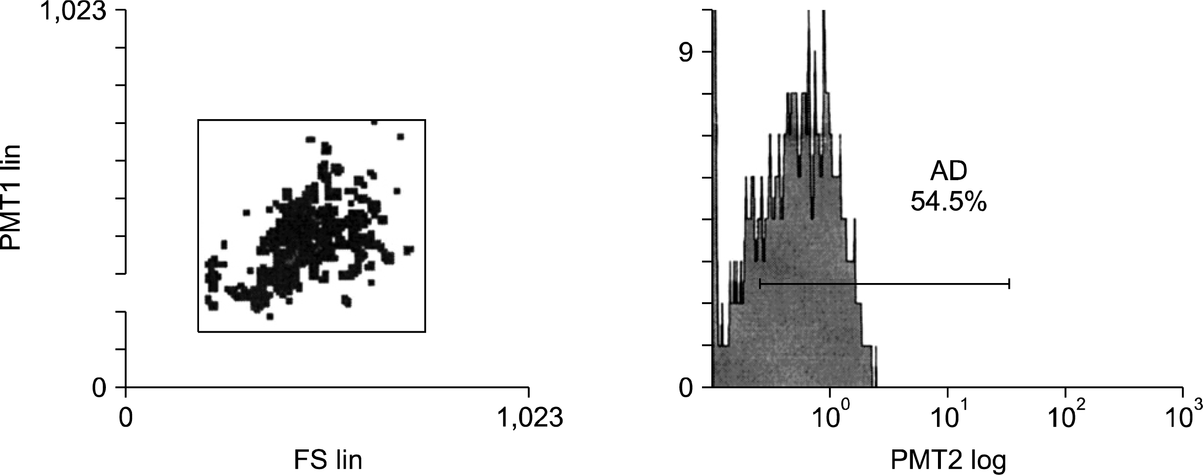
Anticoagulated cord blood was diluted 1:4 with phosphate-buffered saline (PBS) containing 2 mM EDTA (Gibco-Invitrogen, Grand Island, NY) The mononuclear cells (MNCs) were separated by centrifugation over a Ficoll-Paque (Gibco-Invitrogen, Grand Island, NY) density gradient at 400xg rpm for 35 minutes at 20°C. The MNC fraction was collected and washed first in PBS, then with MACS (magnetic cell sorting) buffer (PBS supplemented with 0.5% bovine serum albumin and 2 mM EDTA, pH 7.2). CD34+ cells were isolated from MNCs, using the CD34+ positive cell selection kit (MiniMacs; Miltenyi Biotec, Bergisch Gladbach, Germany). Isolation of CD34+ cells was confirmed by flow cytometry (Fig. 1).

In vitro cell culture and differentiation

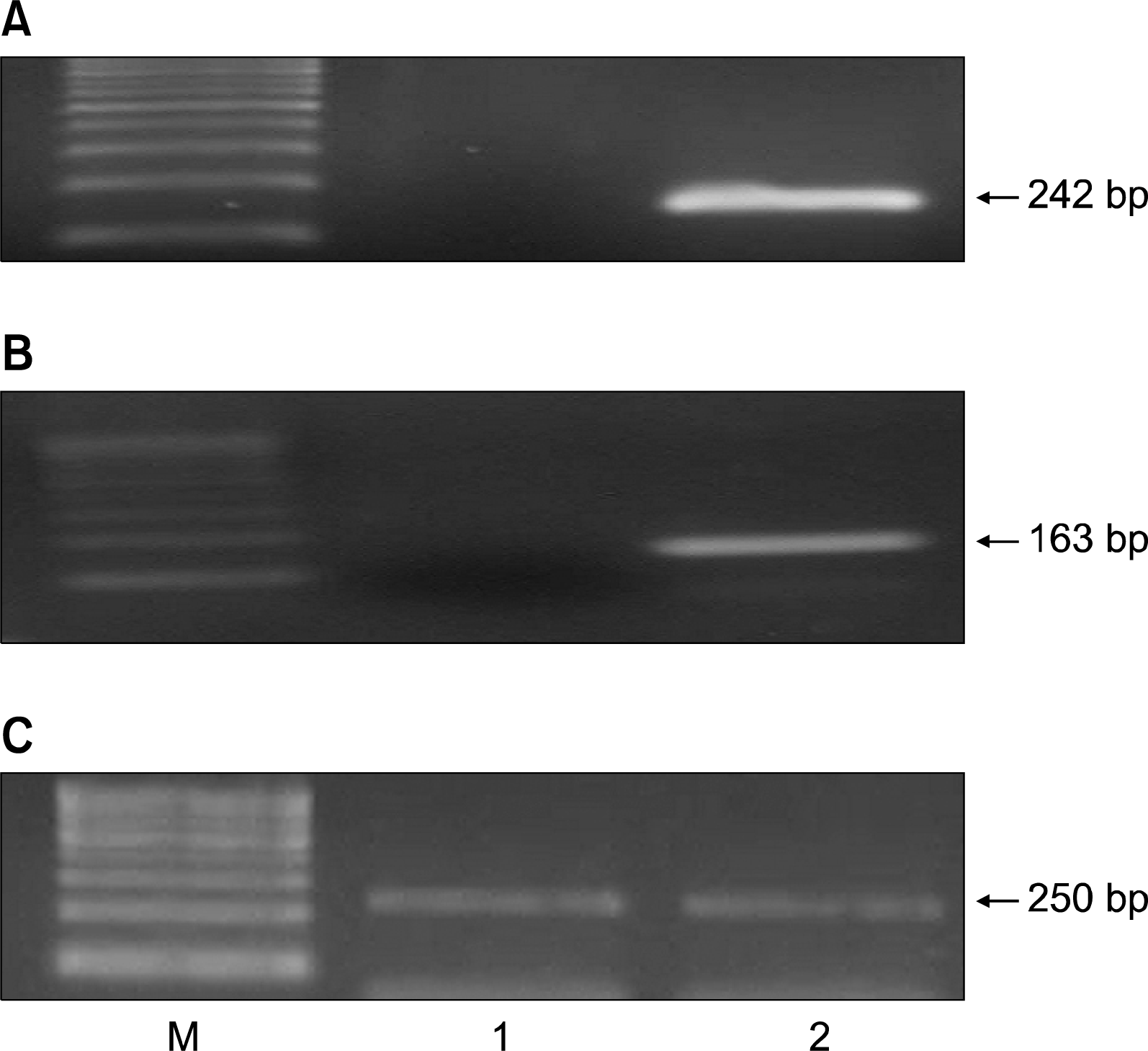
Isolated CD34+ cells were plated on 35-mm2 Petri dishes in minimal essential medium (MEM) supplemented with 15% fetal bovine serum (FBS) and incubated for 2 hours at 37°C and 5% CO2. After 2 hours, the non-adherent cell fraction was removed by washing the plates three times. Adherent CD34+ cells were cultured in -MEM supplemented with 30% FBS and cytokines (20 ng/ml stem cell factor [SCF], 1 ng/ml GM-SCF, 5 ng/ml IL-3, 100 ng/ml G-CSF and 20 ng/ml hepatocyte growth factor (HGF) at 37°C in 5% CO2 in air. (Modified from 21). Differentiation was confirmed by morphology (Fig. 2) and by detection of human albumin and α-fetoprotein gene expression in cells at 2 weeks from addition of hepatic differentiated medium.

In vitro labeling stem cells with PKH26

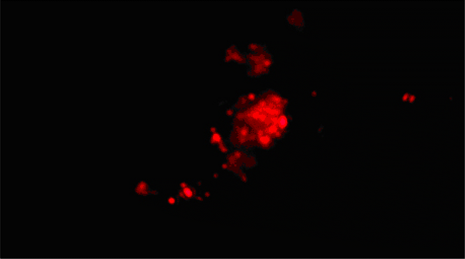
PKH26 is a red fluorochrome. It has excitation (551 nm) and emission (567 nm) characteristics compatible with rhodamine or phycoerythrin detection systems. The linkers are physiologically stable and show little to no toxic side-effects on cell systems. Labeled cells retain both biological and proliferating activity, and are ideal for in vitro cell labeling, in vitro proliferation studies and long term, in vivo cell tracking. In the current work, CD34+ cells and differentiated cells were labeled with PKH26 purchased from Sigma Company (Saint Louis, Missouri USA). Cells were centrifuged and washed twice in serum free medium. Cells were pelleted and suspended in dye solution. Cells were injected intravenously into rat tail vain. After one month, liver tissue was examined with a fluorescence microscope to detect and trace the cells stained with PKH26.

In vivo CCl4-induced liver fibrosis model and stem cell administration

Female white Albino rats (inbred strain (Cux1: HEL1)) were 6 weeks old, weighing between 150 and 200 g. Rats were bred and maintained in an air-conditioned animal house with specific pathogen-free conditions, and were subjected to a 12:12-h daylight/darkness and allowed unlimited access to chow and water. The morphological and behavioral changes of rats were monitored every day. Liver fibrosis was induced by CCl4 injected by subcutaneous route at a dose of 0.2 ml/100 g body weight of 40 ml/l CCl4 (Sigma, St Louis, USA) dissolved in equal volume of castor oil (Sigma, St. Louis, USA). The injection was given twice a week for 6 weeks (22). The same volume of castor oil alone was used as a control. The delay in administration of stem cells until 6 weeks of injection of CCl4 was suggested by histopathological examination of liver samples and also supported by the work of Zhao et al. (22). Stem cells were given at a dose of 107 cells per rat. All animal experiments received approval from the institutional animal care committee. On day 0, rats were divided into the following groups:
Control: 10 rats received 0.2 ml/100 g body weight of castor oil twice a week for 6 weeks; CCl4: 10 rats received 0.2 ml/100 g body weight of CCl4 by the schedule mentioned above. Liver fibrosis was determined by histopathological examination.
CCl4/cells: 40 rats received 0.2 ml/100 g body weight of CCl4 by the schedule mentioned above. The 40 rats were then randomly divided into four groups. On day 42: CCl4/I.V. CD34+, 10 rats were infused with a dose of 107 undifferentiated cells per rat intravenously (through tail vain); CCl4/I.H. CD34+, 10 rats were infused with 107 un-differentiated cells per rat intrahepatically. CCl4/I.V. differentiated CD34+, 10 rats were infused with a dose of 107 differentiated cells (at 2 weeks of differentiation) per rat intravenously; CCl4/I.H. differentiated CD34+, 10 rats were infused with 107 differentiated cells (at 2 weeks of differentiation) per rat intrahepatically.
After 4 weeks from stopping CCL4 and administration of stem cells, venous blood was collected from the retro-orbital vein. All rats were sacrificed with CO2 narcosis, and liver tissue was harvested for analysis.

Analysis of liver histopathology

Liver samples were collected into PBS and fixed overnight in 40 g/l paraformaldehyde in PBS at 4°C. Serial 5-μm sections of the right lobes of the livers were stained with hematoxylin and eosin (HE) and were examined histopathologically.

Morphometric study

The mean optical density of collagen in liver sections stained with Sirius red was measured. The image analyzer was first calibrated automatically to convert the measurement units (pixels) produced by the image analyzer program into actual micrometer units. These measurements were done using an objective lens of magnification 10, i.e. of total magnification 100. Ten readings were obtained in each specimen &the mean values were obtained.
The data obtained were subjected to statistical analysis using t-Student’s test and ANOVA.

PCR detection of rat collagen gene expression

Total RNA was extracted from liver tissue homogenate using RNeasy purification reagent (Qiagen, Valencia, CA). cDNA was generated from 5 μg of total RNA extracted with 1 μl (20 pmol) antisense primer and 0.8 μl superscript AMV reverse transcriptase for 60 min at 37°C. For PCR, 4 μl cDNAwas incubated with 30.5 μl water, 4 μl 25 mMMgCl2, 1 μl dNTPs (10 mM), 5 μl 10×PCR buffer, 0.5 μl (2.5 U) Taq polymerase and 2.5 μl of each primer containing 10 pmol. Primer sequences were as follows: forward 5′-GAACTTGGGGCAAGACAGTCA-3′, reverse 5′-GTCACGTTCAGTTGGTCAA-3′ (UniGene Rn.2953). The reaction mixture was subjected to 40 cycles of PCR amplification as follows: denaturation at 95°C for 1 min, annealing at 67°C for 1 min and extension at 72°C for 2 min. The PCR product yielded a 333 bp fragment on 1.5% agarose gel electrophoreses (23).

PCR detection of human albumin and α-fetoprotein gene expression

Total RNA was extracted from cultured cells (to confirm differentiation) and from liver tissue homogenate using RNeasy purification reagent (Qiagen, Valencia, CA). cDNA was generated from 5 μg of total RNA extracted with 1 μl (20 pmol) antisense primer and 0.8 μl superscript AMV reverse transcriptase for 60 min at 37°C. For PCR, 4 μl cDNA was incubated with 30.5 μl water, 4 μl 25 mM MgCl2, 1 μl dNTPs (10 mM), 5 μl 10× PCR buffer, 0.5 μl (2.5 U) Taq polymerase and 2.5 μl of each primer containing 10 pmol. The following oligonucleotide primers were used: for albumin (ALB) (Forward, 5′-GGCAGGGCTCAGTCAGTAATGA-3′, Reverse, 5′-AGGCC TACCCCAGCCAGTAG-3′), and for α-fetoprotein (AFP) (sense, 5′-TCCTGAATGGGAGAGGTCC-3′; antisense, 5′- TCTTGGCCAAAGGAGACG-3′), Amplification reactions were performed at 94°C for 30 seconds, 55°C for 30 seconds, and 72°C for 60 seconds for 30 cycles. The PCR product yielded a 242 bp and 163 bp fragments on 1.5% agarose gel electrophoreses for AFP and ALB respectively (24).

PCR detection of human MMP-2 gene expression

Total RNA was extracted from liver tissue homogenate using RNeasy purification reagent (Qiagen, Valencia, CA). cDNA was generated from 5 μg of total RNA extracted with 1 μl (20 pmol) antisense primer and 0.8 μl superscript AMV reverse transcriptase for 60 min at 37°C. For PCR, 4 μl cDNAwas incubated with 30.5 μl water, 4 μl 25 mM MgCl2, 1 μl dNTPs (10 mM), 5 μl 10× PCR buffer, 0.5 μl (2.5 U) Taq polymerase and 2.5 μl of each primer containing 10 pmol. Primer sequences were as follows: MMP-2 (Forward primer: 5′-CTGTGAGCCACAGA AGGTTG-3′ Reverse primer:5′-TGACTGTACTCCTCCC AGGC -3′ (GenBank Accession: G62045). The PCR product yielded a 614 bp fragment. The conditions for PCR reactions were: 50°C for 2 min and 95°C for 10 min, followed by 40 cycles of amplification at 95°C for 15 s and 60°C for 1 min.

PCR detection of β-actin

The presence of RNA in all tissues was assessed by analysis of the “house-keeping” gene β-actin. cDNA was generated from 1 μg of total RNA extracted with AMV reverse transcriptase for 60 min at 37°C. For PCR, 4 μl cDNA was incubated with 30.5 μl water, 4 μl 25 mM MgCl2, 1 μl dNTPs (10 mM), 5 μl 10× PCR buffer, 0.5 μl (2.5 U) Taq polymerase and 2.5 μl of each primer containing 10 pmol. Rat β-actin primers (forward 5′-TGTTGTCCCTGTATGCCTCT-3′, reverse 5′-TAATGTC ACGCACGATTTCC-3′) were designed from GenBank (accession no.J00691). The PCR product yielded 206 bp fragments.
Human β-actin primers were as follows: (forward 5′-TCCTGGGACCTAACGATTTTG-3′, reverse 5′-CATTTA TCCGTGTGCCGAC-3′, STS-W68401) to give a PCR product size of 250 bp. The reaction mixture was subjected to 40 cycles of PCR amplification as follows: denaturation at 95°C for 1 min, annealing at 57°C for 1 min and extension at 72°C for 2 min.

Semiquantitation of PCR products

DNA concentration was assessed using the gel documentation system (Bio Doc Analyze) provided by Biometra, to obtain a semiquantitative measurement of PCR products.

Statistical analysis

Data are expressed as mean±SD. Significant differences were determined by using ANOVA and post hoc tests for multiple comparisons using SPSS 15.0 computer Software. Results were considered significant at p<0.05.

Results

Isolated human UCB undifferentiated CD34+cells were identified by fluorescence-activated cell sorting (FACs) and revealed 54.5% positive for CD34 (Fig. 1).
In vitro differentiation of CD34+ cells into hepatocyte like cells was detected by changing in cell morphology (Fig. 2) and expression of human albumin and α fetoprotein genes in cultured cells (Fig. 3).
Cells labeled with the PKH26 showed strong red autofluorescence after transplantation in rats, confirming that these cells were actually seeded into the liver tissue (Fig. 4).
The results of the present study show a significant elevation in serum albumin after administration of stem cells compared to the CCl4 group (p<0.001). Highest level was found in the group that received undifferentiated cells I.V. The level was significantly higher than the group that received I.V. differentiated stem cells (p<0.001) but there was non-significant difference with other groups that received stem cells (p>0.05). As regards liver enzyme, ALT, there was a significant decrease of its level compared to the CCl4 group (p<0.001). However, it was still significantly higher than control (p<0.001) and there was non-significant difference between the groups that received stem cells (p>0.05) (Table 1).
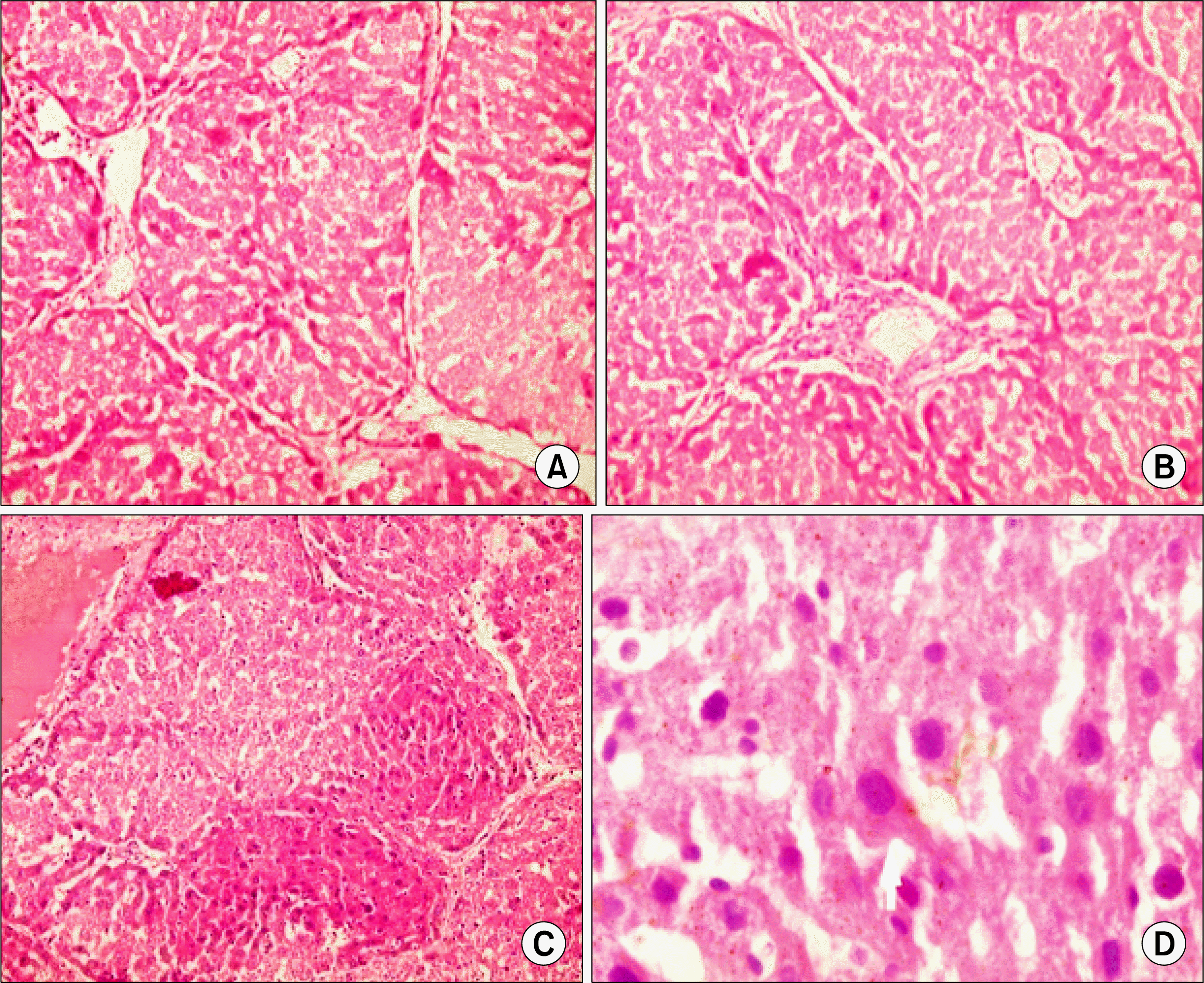
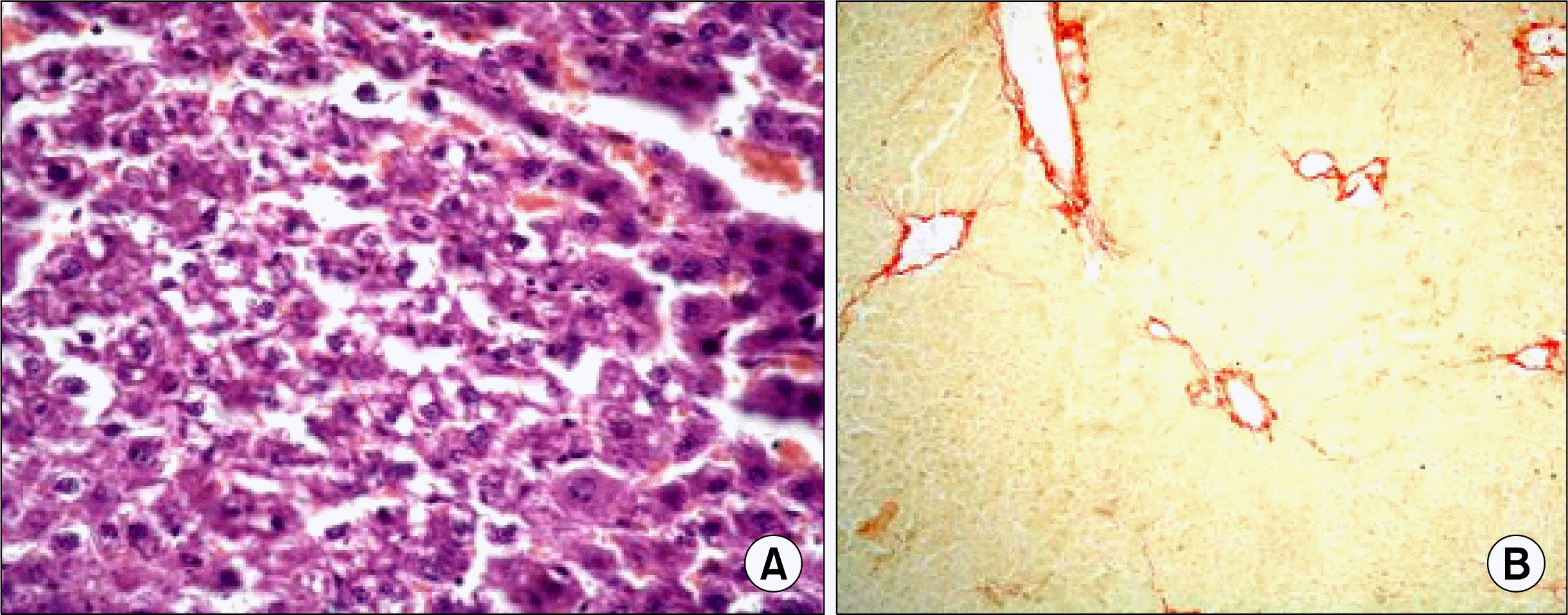
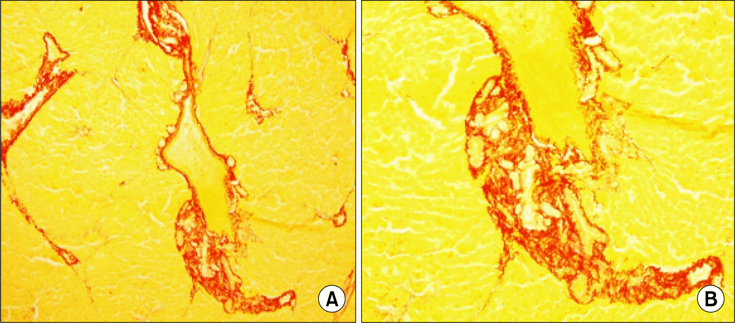
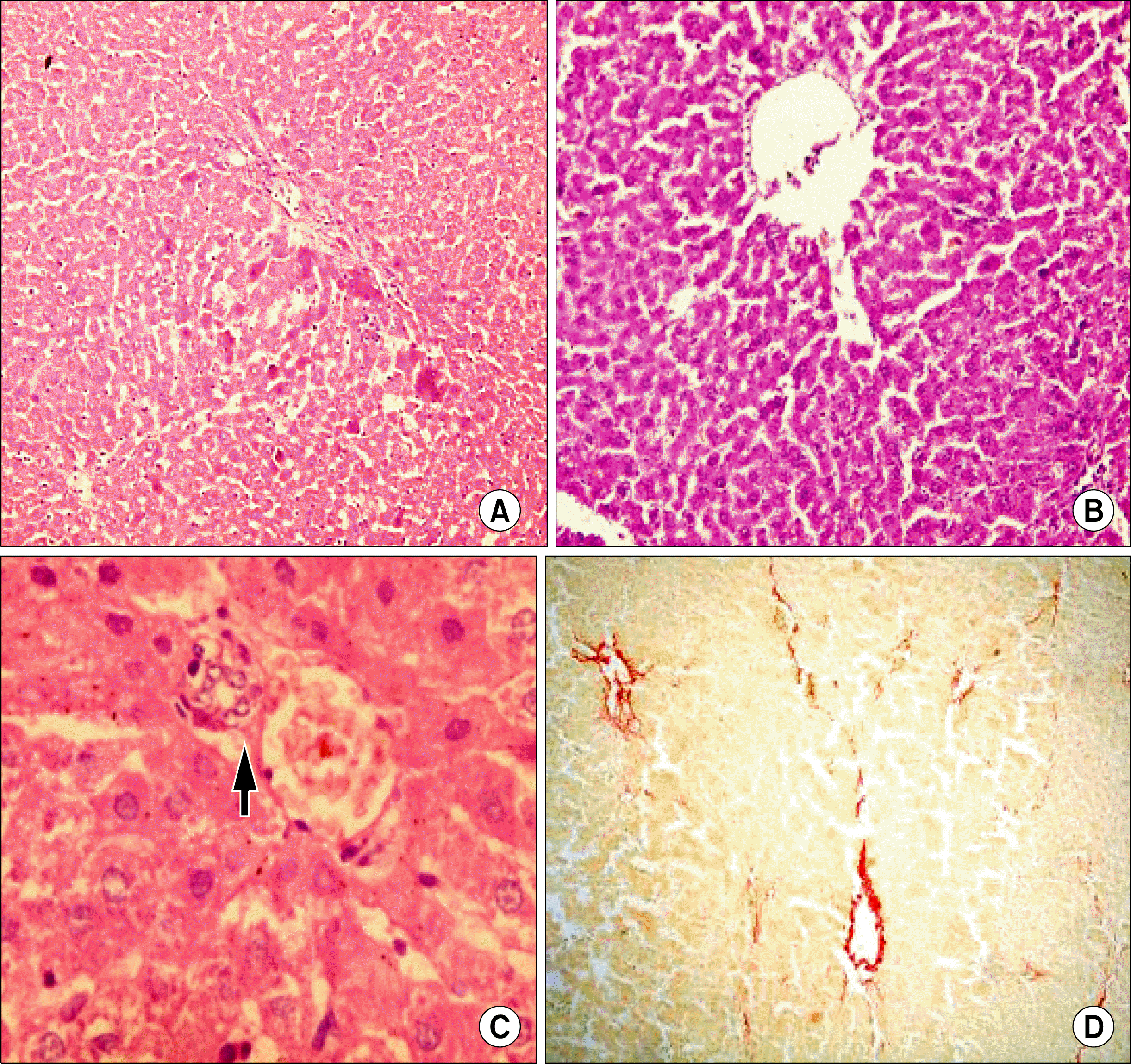
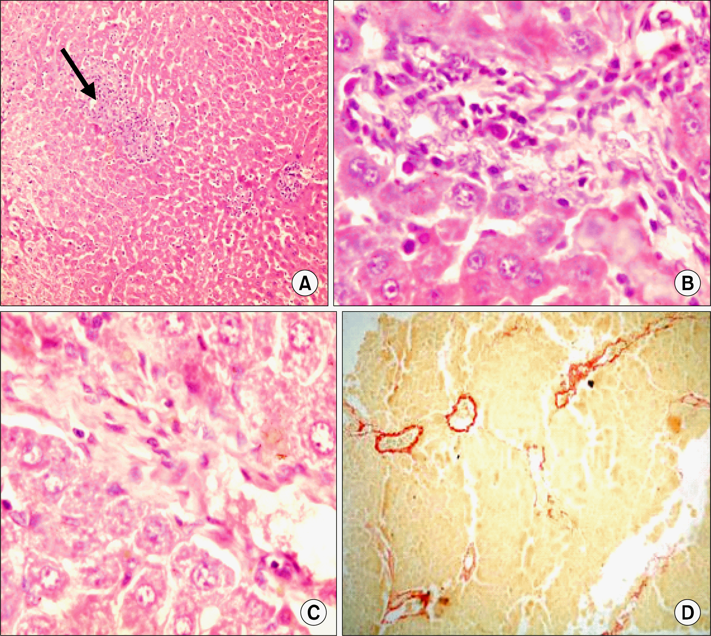
Histopathological examination of liver tissue showed that stem cells have a significant antifibrotic effect as evidenced by the decrease in liver collagen stained with Sirius red (morphometric study) compared to the CCl4 group together with improvement of liver histopathological picture as detected by Hematoxylin and Eosin (Fig. 510) (Table 2). Best results were obtained with I.V. administration of undifferentiated CD34+ cells (p<0.001 compared to CCL4 group). However, there was no significant difference between the groups that received stem cells.
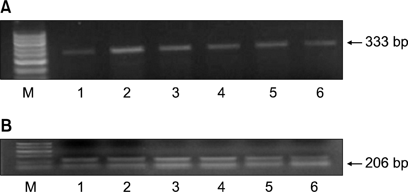
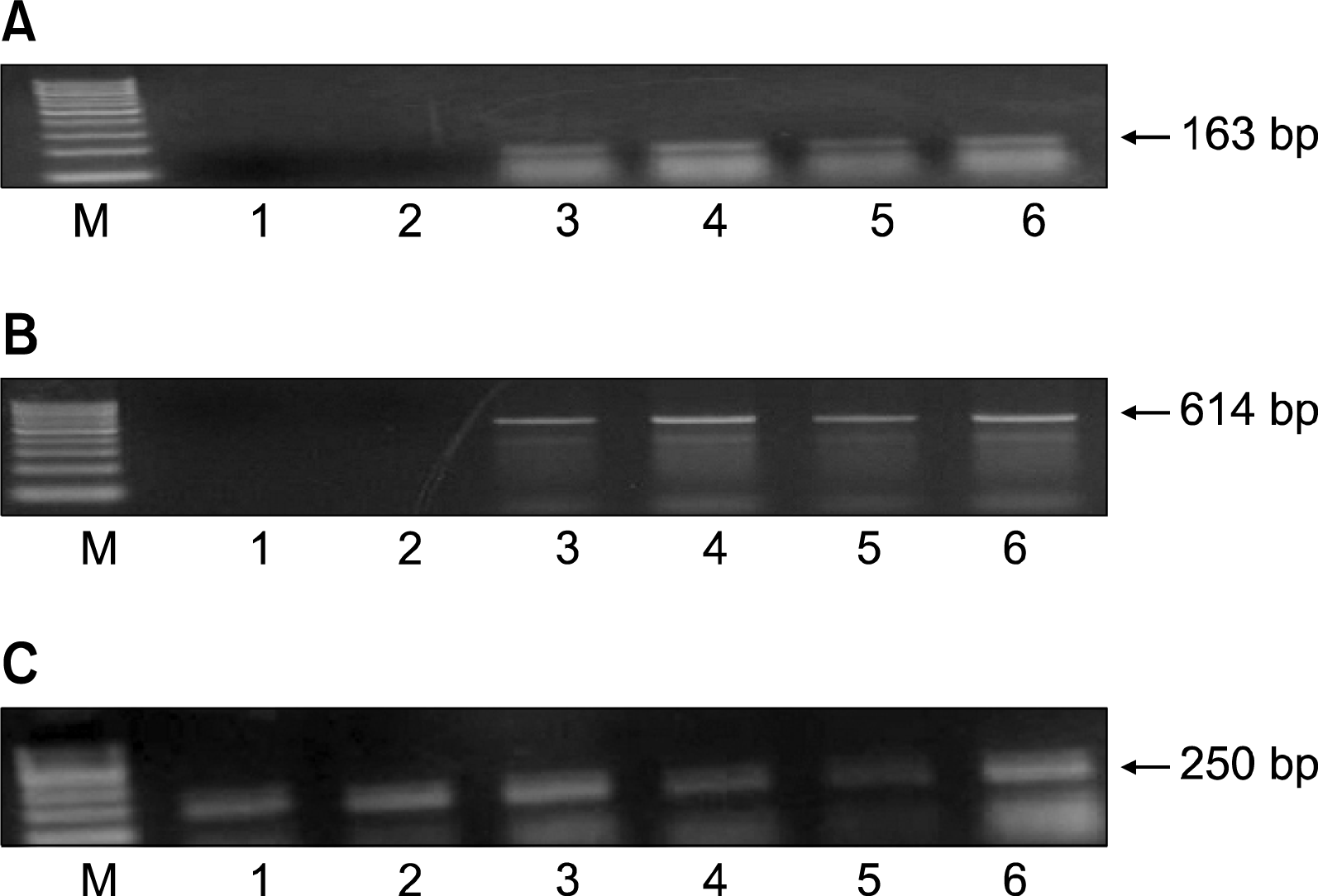
Concerning gene expression, the collagen gene (rat) was highly expressed in the CCl4 group whereas its band density was significantly decreased after administration of stem cells. Least expression was in the group that received undifferentiated cells I.V. However, there was non-significant difference between the different groups that received stem cells (p>0.05) (Fig. 11) (Table 3).
The human albumin gene was expressed in the differentiated cells. It was also detected in liver tissues in the groups that received stem cells. Highest expression was in the group that received undifferentiated cells I.V. The level was significantly higher than other groups that received stem cells (p<0.001) (Fig. 12A) (Table 3).
The human MMP2 gene was expressed in the groups that received stem cells. Highest expression was in the group that received undifferentiated cells I.V. The level was significantly higher than that in the group that received I.H. undifferentiated cells and I.V. differentiated cells (p<0.001). The difference was non-significant when compared to the group that received I.H. differentiated cells (p>0.05) (Fig. 12B) (Table 3).

Discussion

Cell based therapies are increasingly studied in various types of human diseases (2527). However, safety issues should be carefully considered in these novel treatment approaches. The essential requirements for stem cell therapy are (a) an easily procurable source of the stem cells themselves, (b) identification and characterization of the stem cell properties, (c) ability to increase (“expand”) cell numbers in culture reliably and reproducibly, (d) potential for differentiation of stem cell progeny into the desired tissue type, and (e) demonstration that the transplanted cells improve the function of damaged tissue.
Adult human bone marrow and peripheral blood are easily available sources of stem cells. They contain two major types of stem cells, the hematopoietic stem cells and the MSCs. Classically; the hematopoietic stem cells are the source of all of the circulating mature blood cells, whereas the MSCs provide the stromal cells constituting the micro-environment within the marrow cavities. More recent data indicate broader potential for MSCs (28) in particular. Gaia et al (29) mobilized CD34+ cells and observed bone marrow-derived cells may represent an easy immature cell source potentially useful for novel approaches for liver regeneration. Gordon et al. reported that HSCs infusion through the portal vein or hepatic artery was safe and may be effective in decompensated cirrhosis (21).
Human UCB, a rich source of hematopoietic stem cells, offers practical and ethical advantages. It has been reported that various adult stem cells transplanted into a damaged liver show characteristics of a hepatic lineage (30). UCB-derived progenitor cells were capable of differentiating into hepatocyte-like cells in vitro. UCB is thought to be an attractive cell source for cell therapy because of their young age and low infection rate compared with adult tissue MSC (31).
Owing to lack of knowledge on the intrinsic mechanisms regulating stem cell behaviors (i.e. self-renewal, maintenance, proliferation, differentiation, apoptosis and migration/homing), tissue formation and tissue homeostatic maintenance in humans, currently human beings still face the great difficulties in applying stem cells in regenerative medicine and treatments for human diseases. At present, much of this kind of knowledge was emerged from the in vitro and in vivo murine models. A preclinically and clinically relevant in vivo humanized animal model can and should more realistically imitate as closely as possible the in vivo situations in human (32).
Results of the present work demonstrated that UCB cells can proliferate into hepatocyte lineage cells in the original primary culture system in vitro and that UCB cells differentiate into functionally mature hepatocytes in vivo. To demonstrate that UCB cells have hepatic competence in vitro, we determined sufficient conditions for the expression of albumin in UCB cells in vitro. UCB cells were induced to express albumin and α-fetoprotein genes in media containing the combination of FGF-1, IL-6, GCSF, SCF, and HGF. Moreover, some of the albumin expressing cells showed the same characteristics as hepatic progenitor cells, such as hepatic oval cells, in the culture system. In the present study, the isolated CD34+ UCB cells did not neither express albumin mRNA nor α-fetoprotein mRNA. In vitro culture of the adherent CD34+ cell fraction derived from UCB with exposure to the combined effects of FGF-1, IL-6, GCSF, SCF, and HGF were necessary for differentiation into hepatocyte cell-like. It is also noted that albumin-producing cells could continue to proliferate after transplantation of UCB cells.
Chinzei et al. (33) reported that, after transplantation, there was no formation of tumors, such as teratomas, which can be observed in the transplantation of embryonic stem cells. For these reasons, cultured UCB cells could be a suitable source of cells for transplantation.
In the present work, it has been shown that human CD34+ cells derived from human UCB have the ability to improve liver function in rats with liver fibrosis as indicated by increased serum albumin and decreased ALT level. Histopathological examination of liver tissue confirmed this result. Administration of stem cells decreased collagen and increased MMP2 gene expression. Gene expression of MMP2 was assessed as MMPs are the major enzymes that degrade the various types of collagen (34). Their increased expression after administration of stem cells may account for the amelioration of liver fibrosis and improvement of liver function observed.
Results also showed that 1) the livers of recipient rats expressed human albumin and MMP2, and that 2) the livers of recipient rats expressed rat collagen. These findings can’t tell if the main mechanism of this phenomenon is cell fusion or transdifferentiation. Expression of human gene (albumin and MMP2) and rat gene (collagen) was detected in liver homogenate not in individual cell. Further study on the presence or absence of both human and mouse genomic DNAs in the cell would be recommended to clarify the mechanism by which hematopoietic cells regenerate hepatocytes.
The fusion process implies that a cell inserts its genetic content into another cell to form a resulting unit that acquires the ‘host’ phenotype. The resultant product creates a heterokaryon in which the nuclei do not always fuse. The concept of fusion has emerged after experiments on co-culture of BM cells with ESCs (35, 36). Fusion between hematopoietic cells and hepatocytes has been demonstrated (3742) and invalidated (4345). This is explained by the conceptual diversity of these studies and perhaps the complexity of the process itself. For example, fusion events can be acted by HSCs (46) or require homing of these cells and implication of progeny, as highlighted by studies showing fusion between myelomonocytic lineages and hepatocytes (3942). Interestingly, studies providing strong data about fusion used the FAH−/− model which produce mitotic and chromosomal abnormalities (47) that could strengthen the fusion process. It appears that selective pressure is necessary to induce relevant fusion processes which happen rarely in a non-injury model. How the cell fusion and plasticity phenomenon are parts of the same process or vary in importance according to the population used (stem cells versus committed cells (48), has to be explored by tracing the donor cells in their route and studying signalling pathways. Interestingly, fusion events have, to date, never been described with MSCs (49, 50).
Lagasse et al. (51) were the first to demonstrate that purified hematopoietic stem cells can differentiate into hepatocytes in vivo. Subsequently, congenic mouse-to-mouse transplantation of bone marrow populations rich in hematopoietic stem cells was used to show hepatic differentiation of hematopoietic cells. However, it is controversial how hematopoietic stem cells generate hepatocytes. Many researchers have used the human-to-mouse xenogeneic transplantation model, but with extremely low efficiency (<0.5%), making it difficult to analyze the mechanism by which hematopoietic stem cell transplantation induces liver regeneration.
Fujino et al. (52) transplanted small numbers of CD34+ cells (which represent hematopoietic stem cells) to exclude the effects of other cells types (e.g., the CD34 MSCs and other unknown stem cells). They demonstrated that the livers of the recipient mice expressed human albumin, HepPar1, and α-1-antitrypsin protein. In addition, they detected considerable numbers of human albumin-positive cells (an average of 2.73% of the total number of hepatocytes) by flow cytometry. These numbers were close to the numbers detected in the clinical human-to-human transplantation cases. They also detected human albumin in the murine bloodstream. Finally, they revealed that livers of the recipient mice express a variety of human liver-specific genes. These findings suggest that functional human hepatic cells develop in the liver after transplantation of human UCB CD34+ cells into liver-intact NOD/SCID/cnull mice. Authors found that the human albumin-positive hepatocytes expressed not only human HLA-ABC, but also mouse H-2Kd. Nevertheless, the livers of recipient mice contained nucleus positive for both human and mouse genomic DNAs. These data strongly supported not transdifferentiation, but cell fusion, as the main mechanism of this phenomenon. These results agree with the results obtained in the present study.
The better results obtained with the undifferentiated cells could be attributed to the fact that undifferentiated hematopoietic cells have very low apoptotic activity accounting for their longer survival (53). Authors showed that hematopoietic precursors (CD 34+/CD 38−/low) are the lowest in expressing apoptotic proteins, Asp and Annexin V, bax, bad and bak. Authors showed that 10 day ex vivo expansion showed an upregulation of proapoptotic genes; bax, bad, bak and ASP.
However, histopathological examination of liver slides showed a return to normal liver architecture & amelioration of fibrosis in both IV & IH subgroups, yet the main differences lay in the degree of hepatocyte morphology improvement. Best results were obtained when the mode of administration was by the IV route with liver cells focally displaying mild hydropic easily reversible changes, unlike the IH group where the liver cells displayed evidence of ballooning degeneration, foci of cell drop out due to apoptosis & few dysplastic foci, thereby indicating a less dramatic improvement in the reversal of the hepatocytic damage. In both subgroups there was an excellent amelioration of tissue fibrosis. A possibly undesirable effect was the extravasation of RBCs forming blood lakes in the liver tissue when the mode of administration was by the IH route. This may be due to the administration directly into the liver vessels with sudden increase in pressure within the weaker smaller vessels or it could be due to the direct effect of liver tissue traumatization.
In conclusion, the human-rat chimerism model described in this work revealed an engraftment of human donor cells via IV or IH transplantation of human hematopoietic stem/progenitor cells, and human hepatocytes can be regenerated in this chimeric animal. More significantly, the human liver-like cells generated from the engrafted cells were able to partially repair the injured liver induced by CCl4. These findings may facilitate the therapeutic potential by IV or IH transplantation of human cord blood primitive hematopoietic cells for liver damage. Further studies on the longevity and different doses of administered cells are recommended to elucidate the feasibility of using hematopoietic stem cell transplantation to treat patients with liver dysfunction.

ACKNOWLEDGMENTS

This work was scientifically supported by Dr. Nagy Habib (Professor and Head of Department of Biosurgery & Surgical Technology, Imperial College London, Hammersmith Hospital, E-mail: nagy.habib@imperial.ac.uk).

Notes

Potential conflict of interest

The authors have no conflicting financial interest.

References

1. Najimi M, Sokal E. Liver cell transplantation. Minerva Pediatr. 2005. 57:243–257.
2. Stéphenne X, Najimi M, Sibille C, Nassogne MC, Smets F, Sokal EM. Sustained engraftment and tissue enzyme activity after liver cell transplantation for argininosuccinate lyase deficiency. Gastroenterology. 2006. 130:1317–1323.
crossref
3. Verfaillie CM, Pera MF, Lansdorp PM. Stem cells: hype and reality. Hematology Am Soc Hematol Educ Program. 2002. 369–391.
crossref
4. Fujikawa T, Oh SH, Pi L, Hatch HM, Shupe T, Petersen BE. Teratoma formation leads to failure of treatment for type I diabetes using embryonic stem cell-derived insulin-producing cells. Am J Pathol. 2005. 166:1781–1791.
crossref
5. Shenfield F. Semantics and ethics of human embryonic stem-cell research. Lancet. 2005. 365:2071–2073.
crossref
6. Preston SL, Alison MR, Forbes SJ, Direkze NC, Poulsom R, Wright NA. The new stem cell biology: something for everyone. Mol Pathol. 2003. 56:86–96.
crossref
7. Abdel Aziz MT, Atta HM, Mahfouz S, Fouad HH, Roshdy NK, Ahmed HH, Rashed LA, Sabry D, Hassouna AA, Hasan NM. Therapeutic potential of bone marrow-derived mesenchymal stem cells on experimental liver fibrosis. Clin Biochem. 2007. 40:893–899.
crossref
8. Thomas ED. Bone marrow transplantation from the personal viewpoint. Int J Hematol. 2005. 81:89–93.
crossref
9. Alison MR, Poulsom R, Jeffery R, Dhillon AP, Quaglia A, Jacob J, Novelli M, Prentice G, Williamson J, Wright NA. Hepatocytes from non-hepatic adult stem cells. Nature. 2000. 406:257.
crossref
10. Theise ND, Nimmakayalu M, Gardner R, Illei PB, Morgan G, Teperman L, Henegariu O, Krause DS. Liver from bone marrow in humans. Hepatology. 2000. 32:11–16.
crossref
11. Körbling M, Katz RL, Khanna A, Ruifrok AC, Rondon G, Albitar M, Champlin RE, Estrov Z. Hepatocytes and epithelial cells of donor origin in recipients of peripheral-blood stem cells. N Engl J Med. 2002. 346:738–746.
crossref
12. Ng IO, Chan KL, Shek WH, Lee JM, Fong DY, Lo CM, Fan ST. High frequency of chimerism in transplanted livers. Hepatology. 2003. 38:989–998.
crossref
13. Ishikawa F, Drake CJ, Yang S, Fleming P, Minamiguchi H, Visconti RP, Crosby CV, Argraves WS, Harada M, Key LL Jr, Livingston AG, Wingard JR, Ogawa M. Transplanted human cord blood cells give rise to hepatocytes in engrafted mice. Ann N Y Acad Sci. 2003. 996:174–185.
crossref
14. Kakinuma S, Tanaka Y, Chinzei R, Watanabe M, Shimizu-Saito K, Hara Y, Teramoto K, Arii S, Sato C, Takase K, Yasumizu T, Teraoka H. Human umbilical cord blood as a source of transplantable hepatic progenitor cells. Stem Cells. 2003. 21:217–227.
crossref
15. Kollet O, Shivtiel S, Chen YQ, Suriawinata J, Thung SN, Dabeva MD, Kahn J, Spiegel A, Dar A, Samira S, Goichberg P, Kalinkovich A, Arenzana-Seisdedos F, Nagler A, Hardan I, Revel M, Shafritz DA, Lapidot T. HGF, SDF-1, and MMP-9 are involved in stress-induced human CD34+ stem cell recruitment to the liver. J Clin Invest. 2003. 112:160–169.
crossref
16. Newsome PN, Johannessen I, Boyle S, Dalakas E, McAulay KA, Samuel K, Rae F, Forrester L, Turner ML, Hayes PC, Harrison DJ, Bickmore WA, Plevris JN. Human cord blood-derived cells can differentiate into hepatocytes in the mouse liver with no evidence of cellular fusion. Gastroenterology. 2003. 124:1891–1900.
crossref
17. Nonome K, Li XK, Takahara T, Kitazawa Y, Funeshima N, Yata Y, Xue F, Kanayama M, Shinno E, Kuwae C, Saito S, Watanabe A, Sugiyama T. Human umbilical cord blood-derived cells differentiate into hepatocyte-like cells in the Fas-mediated liver injury model. Am J Physiol Gastrointest Liver Physiol. 2005. 289:G1091–G1099.
crossref
18. Sharma AD, Cantz T, Richter R, Eckert K, Henschler R, Wilkens L, Jochheim-Richter A, Arseniev L, Ott M. Human cord blood stem cells generate human cytokeratin 18- negative hepatocyte-like cells in injured mouse liver. Am J Pathol. 2005. 167:555–564.
crossref
19. Tanabe Y, Tajima F, Nakamura Y, Shibasaki E, Wakejima M, Shimomura T, Murai R, Murawaki Y, Hashiguchi K, Kanbe T, Saeki T, Ichiba M, Yoshida Y, Mitsunari M, Yoshida S, Miake J, Yamamoto Y, Nagata N, Harada T, Kurimasa A, Hisatome I, Terakawa N, Murawaki Y, Shiota G. Analyses to clarify rich fractions in hepatic progenitor cells from human umbilical cord blood and cell fusion. Biochem Biophys Res Commun. 2004. 324:711–718.
20. Wang X, Ge S, McNamara G, Hao QL, Crooks GM, Nolta JA. Albumin-expressing hepatocyte-like cells develop in the livers of immune-deficient mice that received transplants of highly purified human hematopoietic stem cells. Blood. 2003. 101:4201–4208.
crossref
21. Gordon MY, Levicar N, Pai M, Bachellier P, Dimarakis I, Al-Allaf F, M'Hamdi H, Thalji T, Welsh JP, Marley SB, Davies J, Dazzi F, Marelli-Berg F, Tait P, Playford R, Jiao L, Jensen S, Nicholls JP, Ayav A, Nohandani M, Farzaneh F, Gaken J, Dodge R, Alison M, Apperley JF, Lechler R, Habib NA. Characterization and clinical application of human CD34+ stem/progenitor cell populations mobilized into the blood by granulocyte colony-stimulating factor. Stem Cells. 2006. 24:1822–1830.
crossref
22. Zhao DC, Lei JX, Chen R, Yu WH, Zhang XM, Li SN, Xiang P. Bone marrow-derived mesenchymal stem cells protect against experimental liver fibrosis in rats. World J Gastroenterol. 2005. 11:3431–3440.
crossref
23. Ala-Kokko L, Pihlajaniemi T, Myers JC, Kivirikko KI, Savolainen ER. Gene expression of type I, III and IV collagens in hepatic fibrosis induced by dimethylnitrosamine in the rat. Biochem J. 1987. 244:75–79.
crossref
24. Chien CC, Yen BL, Lee FK, Lai TH, Chen YC, Chan SH, Huang HI. In vitro differentiation of human placenta- derived multipotent cells into hepatocyte-like cells. Stem Cells. 2006. 24:1759–1768.
crossref
25. Schächinger V, Erbs S, Elsösser A, Haberbosch W, Hambrecht R, Hälschermann H, Yu J, Corti R, Mathey DG, Hamm CW, Süselbeck T, Assmus B, Tonn T, Dimmeler S, Zeiher AM; REPAIR-AMI Investigators. Intracoronary bone marrow-derived progenitor cells in acute myocardial infarction. N Engl J Med. 2006. 355:1210–1221.
crossref
26. Mohyeddin Bonab M, Yazdanbakhsh S, Alimoghaddam K, Talebian F, Hooshmand F, Lotfi J, Ghavamzadeh A. Mesenchymal stem cell therapy for multiple sclerosis. Int J Hematol Oncol BMT. 2005. 2:10–16.
27. Snyder EY, Daley GQ, Goodell M. Taking stock and planning for the next decade: realistic prospects for stem cell therapies for the nervous system. J Neurosci Res. 2004. 76:157–168.
crossref
28. Jiang Y, Jahagirdar BN, Reinhardt RL, Schwartz RE, Keene CD, Ortiz-Gonzalez XR, Reyes M, Lenvik T, Lund T, Blackstad M, Du J, Aldrich S, Lisberg A, Low WC, Largaespada DA, Verfaillie CM. Pluripotency of mesenchymal stem cells derived from adult marrow. Nature. 2002. 418:41–49.
crossref
29. Gaia S, Smedile A, Omedé P, Olivero A, Sanavio F, Balzola F, Ottobrelli A, Abate ML, Marzano A, Rizzetto M, Tarella C. Feasibility and safety of G-CSF administration to induce bone marrow-derived cells mobilization in patients with end stage liver disease. J Hepatol. 2006. 45:13–19.
crossref
30. Moon YJ, Yoon HH, Lee MW, Jang IK, Lee DH, Lee JH, Lee SK, Lee KH, Kim YJ, Eom YW. Multipotent progenitor cells derived from human umbilical cord blood can differentiate into hepatocyte-like cells in a liver injury rat model. Transplant Proc. 2009. 41:4357–4360.
crossref
31. Sun Y, Xiao D, Pan XH, Zhang RS, Cui GH, Chen XG. Generation of human/rat xenograft animal model for the study of human donor stem cell behaviors in vivo. World J Gastroenterol. 2007. 13:2707–2716.
crossref
32. Zhao Q, Ren H, Li X, Chen Z, Zhang X, Gong W, Liu Y, Pang T, Han ZC. Differentiation of human umbilical cord mesenchymal stromal cells into low immunogenic hepatocyte-like cells. Cytotherapy. 2009. 11:414–426.
crossref
33. Chinzei R, Tanaka Y, Shimizu-Saito K, Hara Y, Kakinuma S, Watanabe M, Teramoto K, Arii S, Takase K, Sato C, Terada N, Teraoka H. Embryoid-body cells derived from a mouse embryonic stem cell line show differentiation into functional hepatocytes. Hepatology. 2002. 36:22–29.
crossref
34. Woessner JF Jr. The matrix metalloproteinase family. Parks WC, Mecham RP, editors. Matrix Metalloproteinases. New York: Academic Press;1998. 1–14.
crossref
35. Terada N, Hamazaki T, Oka M, Hoki M, Mastalerz DM, Nakano Y, Meyer EM, Morel L, Petersen BE, Scott EW. Bone marrow cells adopt the phenotype of other cells by spontaneous cell fusion. Nature. 2002. 416:542–545.
crossref
36. Ying QL, Nichols J, Evans EP, Smith AG. Changing potency by spontaneous fusion. Nature. 2002. 416:545–548.
crossref
37. Ishikawa F, Drake CJ, Yang S, Fleming P, Minamiguchi H, Visconti RP, Crosby CV, Argraves WS, Harada M, Key LL Jr, Livingston AG, Wingard JR, Ogawa M. Transplanted human cord blood cells give rise to hepatocytes in engrafted mice. Ann N Y Acad Sci. 2003. 996:174–185.
crossref
38. Alvarez-Dolado M, Pardal R, Garcia-Verdugo JM, Fike JR, Lee HO, Pfeffer K, Lois C, Morrison SJ, Alvarez-Buylla A. Fusion of bone-marrow-derived cells with Purkinje neurons, cardiomyocytes and hepatocytes. Nature. 2003. 425:968–973.
crossref
39. Camargo FD, Finegold M, Goodell MA. Hematopoietic myelomonocytic cells are the major source of hepatocyte fusion partners. J Clin Invest. 2004. 113:1266–1270.
crossref
40. Vassilopoulos G, Wang PR, Russell DW. Transplanted bone marrow regenerates liver by cell fusion. Nature. 2003. 422:901–904.
crossref
41. Wang X, Willenbring H, Akkari Y, Torimaru Y, Foster M, Al-Dhalimy M, Lagasse E, Finegold M, Olson S, Grompe M. Cell fusion is the principal source of bone-marrow-derived hepatocytes. Nature. 2003. 422:897–901.
crossref
42. Willenbring H, Bailey AS, Foster M, Akkari Y, Dorrell C, Olson S, Finegold M, Fleming WH, Grompe M. Myelomonocytic cells are sufficient for therapeutic cell fusion in liver. Nat Med. 2004. 10:744–748.
crossref
43. Jang YY, Collector MI, Baylin SB, Diehl AM, Sharkis SJ. Hematopoietic stem cells convert into liver cells within days without fusion. Nat Cell Biol. 2004. 6:532–539.
crossref
44. Harris RG, Herzog EL, Bruscia EM, Grove JE, Van Arnam JS, Krause DS. Lack of a fusion requirement for development of bone marrow-derived epithelia. Science. 2004. 305:90–93.
crossref
45. Newsome PN, Johannessen I, Boyle S, Dalakas E, McAulay KA, Samuel K, Rae F, Forrester L, Turner ML, Hayes PC, Harrison DJ, Bickmore WA, Plevris JN. Human cord blood-derived cells can differentiate into hepatocytes in the mouse liver with no evidence of cellular fusion. Gastroenterology. 2003. 124:1891–1900.
crossref
46. Tanabe Y, Tajima F, Nakamura Y, Shibasaki E, Wakejima M, Shimomura T, Murai R, Murawaki Y, Hashiguchi K, Kanbe T, Saeki T, Ichiba M, Yoshida Y, Mitsunari M, Yoshida S, Miake J, Yamamoto Y, Nagata N, Harada T, Kurimasa A, Hisatome I, Terakawa N, Murawaki Y, Shiota G. Analyses to clarify rich fractions in hepatic progenitor cells from human umbilical cord blood and cell fusion. Biochem Biophys Res Commun. 2004. 324:711–718.
47. Jorquera R, Tanguay RM. Fumarylacetoacetate, the metabolite accumulating in hereditary tyrosinemia, activates the ERK pathway and induces mitotic abnormalities and genomic instability. Hum Mol Genet. 2001. 10:1741–1752.
crossref
48. Jang YY, Sharkis SJ. Metamorphosis from bone marrow derived primitive stem cells to functional liver cells. Cell Cycle. 2004. 3:980–982.
crossref
49. Aurich I, Mueller LP, Aurich H, Luetzkendorf J, Tisljar K, Dollinger MM, Schormann W, Walldorf J, Hengstler JG, Fleig WE, Christ B. Functional integration of hepatocytes derived from human mesenchymal stem cells into mouse livers. Gut. 2007. 56:405–415.
crossref
50. Sato Y, Araki H, Kato J, Nakamura K, Kawano Y, Kobune M, Sato T, Miyanishi K, Takayama T, Takahashi M, Takimoto R, Iyama S, Matsunaga T, Ohtani S, Matsuura A, Hamada H, Niitsu Y. Human mesenchymal stem cells xenografted directly to rat liver are differentiated into human hepatocytes without fusion. Blood. 2005. 106:756–763.
crossref
51. Lagasse E, Connors H, Al-Dhalimy M, Reitsma M, Dohse M, Osborne L, Wang X, Finegold M, Weissman IL, Grompe M. Purified hematopoietic stem cells can differentiate into hepatocytes in vivo. Nat Med. 2000. 6:1229–1234.
crossref
52. Fujino H, Hiramatsu H, Tsuchiya A, Niwa A, Noma H, Shiota M, Umeda K, Yoshimoto M, Ito M, Heike T, Nakahata T. Human cord blood CD34+ cells develop into hepatocytes in the livers of NOD/SCID/gamma(c)null mice through cell fusion. FASEB J. 2007. 21:3499–3510.
crossref
53. Peters R, Leyvraz S, Perey L. Apoptotic regulation in primitive hematopoietic precursors. Blood. 1998. 92:2041–2052.
crossref

Fig. 1.
FACS analysis of cells revealed 54.5% positive for CD34.
ijsc-03-161f1.tif
Fig. 2.
CD34+ cells changed its morphology after differentiation A (after one week of differentiation), B (hepatocyte-like cell, after 2 weeks of differentiation and cells were injected at this stage) and C (hepatocyte-like cell, after 4 weeks of differentiation and these cells were not injected).
ijsc-03-161f2.tif
Fig. 3.
An agarose gel electrophoresis shows PCR product of human AFP (A) ALB (B) & beta actin(C) genes. Lane M: PCR marker with 100 base ladder (100, 200, 300, ….etc). Lane 1: no PCR product of AFP or ALB genes expression in undifferentiated CD34+ cells. Lane 2: PCR product of AFP or ALB genes expression in differentiated CD34+ cells.
ijsc-03-161f3.tif
Fig. 4.
PKH26 fluorescent staining of cells.
ijsc-03-161f4.tif
Fig. 5.
Photomicrographs of liver tissue from CCl4 rat group stained with Hematoxylin and Eosin (200×) (A, B) showing nodular appearance of the liver due to fibrous septation. (C) (200×) and (D) (1,000×) showing foci of hepatocyte dysplasia.
ijsc-03-161f5.tif
Fig. 6.
Photomicrographs of liver tissue from CCl4 rat group stained with Sirius red (A) (100×) showing septation and (B) (200×) showing marked fibrosis of portal tract.
ijsc-03-161f6.tif
Fig. 7.
Photomicrographs of liver tissue from CCl4/IV undifferentiated CD34+ rat group stained with Hematoxylin and Eosin (A) (100×), (B) (200×), (C) (1,000×) and Sirius red staining (D) (100×) showing normal architecture, thin portal tracts and dilated central vein with no inflammation.
ijsc-03-161f7.tif
Fig. 8.
Photomicrographs of liver tissue from CCl4/IH undifferentiated CD34+ rat group stained with Hematoxylin and Eosin (A) (100×), (B, C) (1,000×) and Sirius red stain (D) (100×) showing collections of immature cells, mild fibrosis but no inflammation in portal tract cells, otherwise architecturally normal liver.
ijsc-03-161f8.tif
Fig. 9.
Photomicrographs of liver tissue from CCl4/IV differentiated CD34+ rat group (A, B) stained with Hematoxylin and Eosin and Sirius red (100×) respectively showing mild fibrosis.
ijsc-03-161f9.tif
Fig. 10.
Photomicrographs of liver tissue from CCl4/IH differentiated CD34+ rat group (A, B) stained with Hematoxylin and Eosin and Sirius red (100×) respectively, showing degenerating hepatocytes and mild fibrosis.
ijsc-03-161f10.tif
Fig. 11.
An agarose gel electrophoresis shows PCR product of collagen (A) & beta actin (B) gene. Lane M: PCR marker with 100 bp Lane 1: control group; Lane 2:CCL4 group; Lane 3: CCL4+ differentiated CD34+ (I.V); Lane4: CCL4+ undifferentiated CD34+ (I.V) Lane5: CCL4+ undifferentiated CD34+ (I.H); Lane6: CCL4 + differentiated CD34+ (I.H).
ijsc-03-161f11.tif
Fig. 12.
An agarose gel electrophoresis shows PCR products of human ALB gene (A), human MMP-2 gene (B) and beta actin (C) gene. Lane M: PCR marker with 100bp; Lane 1: control group; Lane 2:CCL4 group; Lane 3: CCL4+ differentiated CD34+ (I.V); Lane4: CCL4+ undifferentiated CD34+ (I.V); Lane5: CCL4+ undifferentiated CD34+ (I.H); Lane6: CCL4+ differentiated CD34+(I.H).
ijsc-03-161f12.tif
Table 1.
Liver functions in the different studied groups (mean±SD)
Control n=10 CCl4 n=10 CCl4/IV CD34+ n=10 CCl4/IH CD34+ n=10 CCl4/IV dif. CD34+n=10 CCl4/IH dif. CD34+ n=10
Albumin (g/dl) 4.09±0.49 2.69±0.36 4.01±0.28 3.6±0.34 3.33±0.23 3.82±0.4
P1 - 0.000 1.000 0.56 0.000 1.000
P2 0.000 - 0.000 0.000 0.003 0.000
P3 1.000 0.000 - 0.211 0.001 1.000
P4 0.056 0.000 0.211 - 1.000 1.000
P5 0.000 0.003 0.001 1.000 - 0.056
P6 1.000 0.000 1.000 1.000 0.056 -
ALT (U/l) 33.2±5.89 65.2±7.83 46.8±3.77 49.9±3.64 51.7±2.83 47.9±3.07
P1 - 0.000 0.000 0.000 0.000 0.000
P2 0.000 - 0.000 0.000 0.000 0.000
P3 0.000 0.000 - 1.000 0.416 1.000
P4 0.000 0.000 1.000 - 1.000 1.000
P5 0.000 0.000 0.416 1.000 - 1.000
P6 0.000 0.000 1.000 1.000 1.000 -

IH: Intrahepatic; IV:Intravenous dif:differentiated; P1: comparison with control group; P2: comparison with CCL4 group; P3: comparison with IV CD34+ group; P4: comparison with IH CD34+ group; P5: comparison with IV dif CD34+ group; P6: comparison with IH dif CD34+ group; p<0.05 is considered significant.

Table 2.
Morphometric study in the studied groups (mean±SD)
Control n=10 CCl4 n=10 CCl4/IV CD34+ n=10 CCl4/IH CD34+ n=10 CCl4/IV dif. CD34+ n=10 CCl4/IH dif. CD34+ n=10
Optical density 1.831±2.225 7.898±5.225 2.092±1.394 3.864±2.917 4.689±2.461 3.065±2.067
P1 - 0.000 1.000 1.000 0.541 1.000
P2 0.000 - 0.001 0.056 0.289 0.009
P3 1.000 0.001 - 1.000 0.839 1.000
P4 1.000 0.056 1.000 - 1.000 1.000
P5 0.541 0.289 0.839 1.000 - 1.000
P6 1.000 0.009 1.000 1.000 1.000 -

IH: Intrahepatic; IV: Intravenous; dif: differentiated; P1: comparison with control group; P2: comparison with CCL4 group; P3: comparison with IV CD34+ group; P4: comparison with IH CD34+ group; P5: comparison with IV dif CD34+ group; P6: comparison with IH dif CD34+ group; p<0.05 is considered significant.

Table 3.
DNA concentration (μg/ml) of the band density of PCR products (mean±SD)
Control n=10 CCl4 n=10 CCl4/IV CD34+N=10 CCl4/IH CD34+n=10 CCl4/IV dif. CD34+n=10 CCl4/IH dif. CD34+ n=10
Rat collagen 140.8±17.51 694.1±50.534 279.5±48.85 309.5±36.78 329.1±41.74 299.5±40.38
P1 - 0.000 0.000 0.000 0.000 0.000
P2 0.000 - 0.000 0.000 0.000 0.000
P3 0.000 0.000 - 1.000 0.131 1.000
P4 0.000 0.000 1.000 - 1.000 1.000
P5 0.000 0.000 0.131 1.000 - 1.000
P6 0.000 0.000 1.000 1.000 1.000 -
Human albumin - - 387.2±30.52 177.3±13.75 146.9±16.18 312.9±27.86
P3 - 0.000 0.000 0.000
P4 0.000 - 0.035 0.000
P5 0.000 0.035 - 0.000
P6 0.000 0.000 0.000 -
Human MMP2 - - 646.2±48.4 295.0±20.42 296.1±23.9 600.0±36.59
P3 - 0.000 0.000 0.028
P4 0.000 - 1.000 0.000
P5 0.000 1.000 - 0.000
P6 0.028 0.000 0.000 -

IH: Intrahepatic; IV: Intravenous; dif: differentiated; P1: comparison with control group; P2: comparison with CCL4 group; P3: comparison with IV CD34+ group; P4: comparison with IH CD34+ group; P5: comparison with IV dif CD34+ group; P6: comparison with IH dif CD34+ group; p<0.05 is considered significant.

TOOLS
Similar articles