

 PDF
PDF Citation
 Print
Print


Introduction
Mesenchymal stem cells (MSCs) existing in bone marrow have the potential to differentiate into osteoblasts, adipocytes, chondrocytes, neurons and myogenic cells (1–4). Differentiation into the osteoblastic lineage in vitro can be induced by the inclusion of dexamethasone (Dex), ascorbic acid phosphate (Vitamin C; VC) and β-glycero-phosphate (β-GP) into the culture medium (5–7).
In bone tissue engineering research (6, 8, 9) and clinical cases (10), MSCs are generally seeded onto scaffolds, such as hydroxyapatite (HA) and β-tricalcium phosphate (β-TCP), in order to construct tissue engineered bone. However, osteogenic potential of the tissue engineered bone after transplantation in vivo can vary among the constructs (9). In such cases, an additional treatment that stimulates the osteogenic potential of the construct is required. Although low-intensity pulsed ultrasound (LIPUS) can enhance bone formation of a biomaterial, osteogenic cells, such as osteoblasts and osteocytes, must exist in or around the scaffold. When there is a limited supply of these cells, an alternative source of osteogenic cells is desired, particularly using a minimally invasive approach for supplying the cells clinically.
Previously, we developed a method to fabricate cell sheets of cultured MSCs (8, 11). The sheets demonstrated in vivo bone formation after subcutaneous transplantation, as well as following cell sheet injection without any scaffold. In the present study, we transplanted such sheets by injection into β-TCP ceramics (that had been implanted immediately or 1 week prior) to assess whether the injectable sheets could form bone tissue inside and around the implanted ceramics, thereby supplying exogenous osteogenic cells using a minimally invasive method.
Go to : 
Materials and Methods
Bone marrow cell preparation
Approval from the animal experimental review board of Nara Medical University was obtained before the start of the experiments. The methods of bone marrow cell preparation have been reported previously (5, 7, 8). Briefly, 7-week-old male Fischer 344 rats were used as donors and recipients. Fresh bone marrow cells were obtained from both femoral shafts of donor rats by flushing with culture medium expelled gently from a syringe equipped with a 21 G needle and then seeded into two T-75 flasks (75 cm2 culture flask, Costar Co., Cambridge, MA, USA). Cells were cultured with 15 mL of standard medium comprising Eagle’s minimal essential medium (MEM; Nacalai Tesque. Kyoto, Japan) supplemented with 15% fetal bovine serum (FBS, JRH Biosciences, Lenexa, KS, USA) and a mixture of antibiotics (100 U/ml penicillin and 100 μg/ml streptomycin, Nacalai Tesque), under 5% CO2 at 37°C. After reaching confluency, the primary cultured cells were lifted from the flasks using trypsin/EDTA (Gibco, Invitrogen, CA, USA).
Cell sheet preparation
We have previously reported the method of cell sheet preparation (8). Briefly, trypsinized MSCs were seeded at 1×10⁴ cells/cm2 in 10 cm dishes (100×20 mm style, Falcon, BD) containing 10 nM Dex (Sigma, MO, USA) and 82 μg/ml VC (L-ascorbic acid phosphate magnesium salt n-hydrate, Wako Pure Chemical Industrials, Kyoto, Japan) until confluent (approximately between day 12 and day 14). The cells were rinsed twice with phosphate buffered saline (PBS; Gibco), and the cell sheet was lifted using a scraper.
Implantation of ceramics and injection of the cell sheet
Porous β-TCP disks (5 mm Φ×2 mm, 60% pore, Olympus Terumo Biomaterials Corp., Tokyo, Japan) were transplanted subcutaneously into the backs of Fisher 344 rats prior to the cell sheet injection. A single cell sheet from a whole 10-cm dish was put into a syringe (1 ml syringe; JMS Co., Ltd., Tokyo, Japan) with 500 μL of standard medium and injected immediately around the implanted disks via a 16 G needle (Terumo, Tokyo, Japan) (immediate group). To assess whether osteogenic cells can be supplied to the implanted disks within a waiting period, a cell sheet was injected around a second group of disks 1 week following subcutaneous transplantation (1-wk group). During the sheet injection, the surface of each implanted disk was scratched by the tip of the needle to remove soft tissue around the disk. As a control group, 500 μL of medium was immediately injected around the disks following transplantation.
Each group contained eight disks of two recipient rats (four disks per rat, at subcutaneous sites on their backs). This experiment was performed in duplicate.
Harvest of the disks
Four weeks after the sheet injection, implanted disks were harvested and evaluated radiographically and histologically, as well as by gene expression analyses (real-time PCR). The harvested disks for radiographic and histological analyses were fixed with 10% formalin neutral buffer solution (Wako). After taking X-ray images, each disk was decalcified in K-CX solution (Falma Co., Tokyo, Japan), embedded in paraffin, sectioned and stained with hematoxylin and eosin (H-E). The harvested disks for real-time PCR were stocked in a deep freezer at −80°C until RNA isolation.
In the present study, eight disks of each group were subjected to H-E and RT-PCR, respectively.
RNA isolation and real-time quantitative PCR
We measured the gene expression levels of osteocalcin (OC) to confirm osteogenesis of the harvested disks. RNA was isolated from four disks of each group using an Isogen RNA extraction kit (Nippon Gene Co. Ltd., Toyama, Japan). Briefly, each sample was placed in Isogen solution containing matrix beads and was disrupted by a FastPrep FP120 Cell Disrupter (Qbiogene, Inc., Carlsbad, CA, USA). Subsequently, the following steps of RNA isolation were performed according to the manufacturer’s instructions.
To measure the mRNA expression levels, we conducted real-time quantitative PCR (ABI PRISM 7700 Sequence Detection System; Applied Biosystems, Norwalk, CT, USA), using the respective primers and specific fluorogenic probes of OC and glyceraldehyde-3-phosphate dehydrogenase (GAPDH, internal standard) for rat cDNAs as described previously (6, 12). Target OC mRNA levels were compared after correcting to GAPDH mRNA levels as an internal standard, which is used to adjust for differences in the efficiency of reverse transcription between samples. The OC (Rn01455285 g1) and GAPDH (Rn 99999916 s1) primer and probe set were purchased from Applied Biosystems (Foster City, CA, USA). Thermal cycle conditions were 10 min at 95°C for activation of Universal mixture AmpliTaq Gold Polymerase, followed by 35 cycles of 15 sec at 95°C for denaturing and 1 min at 60°C for annealing and extension. Target mRNA levels were compared after correcting for GAPDH levels. This experiment was performed in duplicate.
Statistical analysis
Statistically significant differences were determined using one-way ANOVA. Post hoc multiple comparisons were made using Tukey’s test. Values of p<0.05 were considered statistically significant.
Go to :
Results
Fig. 1 illustrates X-ray photography of the harvested disks. While the control group showed no calcification around the disk (Fig. 1A), obvious calcification surrounding the disk was observed in both the immediate (Fig. 1B) and 1-wk (Fig. 1C) groups.
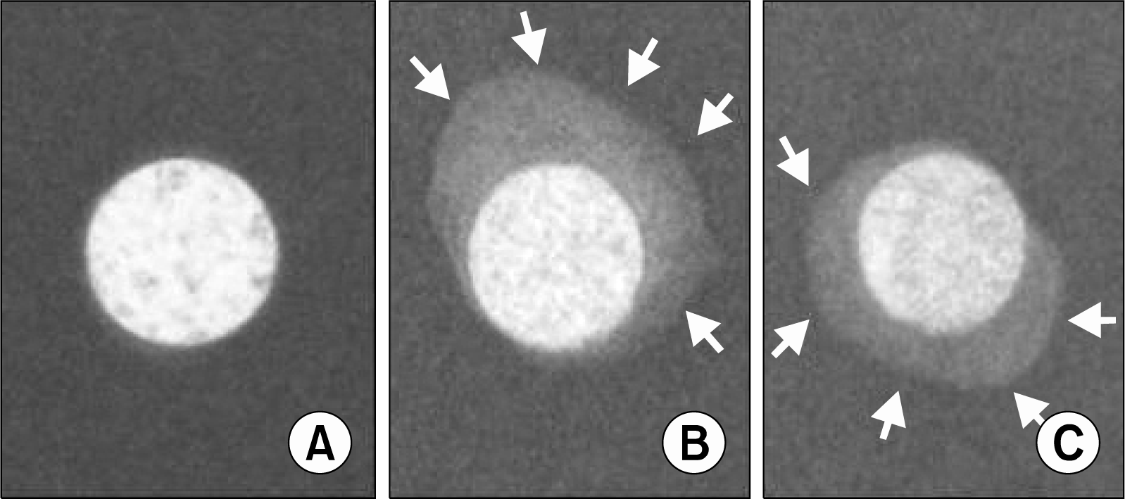 | Fig. 1.Representative X-ray images of harvested disks 4 weeks after the injection. (A) Control group: the disk received an injection of culture medium. (B) Immediate group: a cell sheet was injected immediately after implantation of the disk. (C) 1-wk group: a cell sheet was injected 1 week after the implantation. No obvious calcification was observed in the control group. The immediate group and the 1-wk group exhibited calcification around the disks. Arrows indicate areas of calcification.
|
The bone formation ratio evaluated by histology was 100% in the immediate group (8 disks exhibited bone formation: 8 disks received a cell sheet injection) and 50% in the 1-wk group (4 disks exhibited bone formation: 8 disks). No bone formation was observed in the control group (data not shown). Representative histological sections of the immediate and 1-wk group are shown in Fig. 2. Bone matrix, together with osteocytes, was seen in the sections of both the immediate (Fig. 2A, B) and 1-wk (Fig. 2D, E) groups, whereas no bone formation was observed in the control group (data not shown). The immediate group exhibited bone formation inside the pores and around the disk, as well as the 1-wk group. However, less bone formation in the central pores was observed in the 1-wk group compared with the immediate group. Although bone formation surrounding each disk was seen in both groups, soft tissue existed between disks and newly formed bone area in the 1-wk group (Fig. 2E). More vessels were observed within the harvested disks of the immediate group (Fig. 2C) than in those of the 1-wk group (Fig. 2F).
 | Fig. 2.Representative histological sections. (A) Immediate group. Obvious bone formation was observed in the pores, as well as on the surface of the disk. (B) A higher-magnification image of the rectangular area of B in A. Bone formation was observed on the disk surface. (C) A higher magnified image of the rectangular area of C in A. Several vessels were observed. (D) 1-wk group. Obvious bone formation was observed in the pores, as well as on the surface of the disk. (E) A higher magnification image of the rectangular area of E in D. A soft tissue interface was observed between the disk surface and the newly formed bone tissue. (F) A higher magnified image of the rectangular area of F in D. A few vessels were observed. Asterisks, arrows and arrowheads indicate bone tissue, vessels and the soft tissue interface, respectively.
|
Fig. 3 shows the OC mRNA expression levels evaluated by real-time PCR. The OC expression in the immediate group was significantly higher than that in the control and 1-wk group. Although average OC expression in the 1-wk group was higher than in the control group, there was no significant difference.
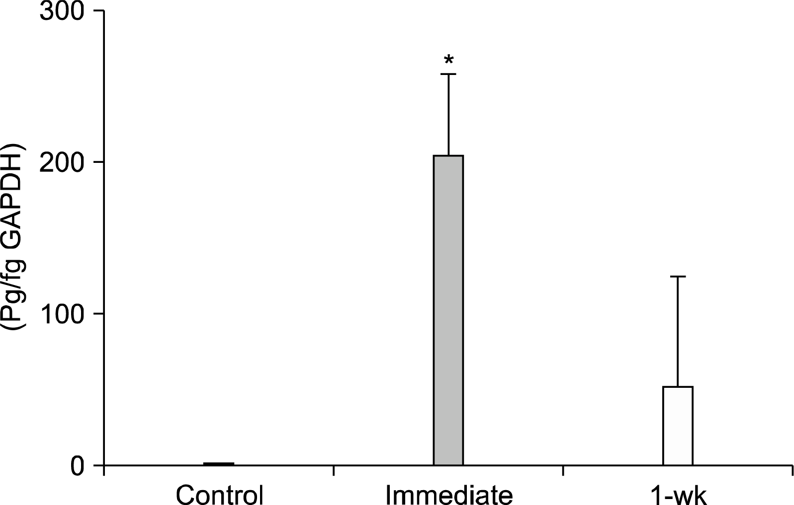 | Fig. 3.Real-time PCR of OC mRNA expression. The expression in the immediate group was significantly higher than in the control and 1-wk group. Although a similar tendency of increased OC expression was seen in the 1-wk group as compared with the control group, there was no significant difference. *p<0.05.
|
Go to :
Discussion
Recently, many researchers have reported methods for fabricating cell sheets. Generally, these methods have required special culture plates or multifunctional copolymers (13–15). Specifically, Okano et al. (16) reported culture dishes coated with thermosensitive polymers, which have been clinically applied for the treatment of corneal surface dysfunction. Similar to Ma et al. (17), we reported cell sheet fabrication using a scraping technique (8). Ma et al. (17) reported that a scaffold-free transplanted cell sheet, which was created by a scraping technique, formed bone tissue within a subcutaneous site, consistent with our previous results (8, 11, 18). However, they used an open transplantation method through a skin incision. In the present study, we used our original cell sheet fabrication method and then injected in vivo whole cell sheets fabricated in 10-cm diameter culture dishes, which resulted in bone formation around the implanted ceramics. Thus, we believe that cell sheet injection provides an osteogenic stimulus to the injection site.
Previously, we reported significantly greater staining of ALP and Alizarin red S within cell sheets in vitro, as well as expression of ALP mRNA and osteocalcin secretion into the culture medium as compared with undifferentiated cells (8). We have also reported advantages of our method, including the cells in the sheet can be differentiated into osteogenic cells during the culture period; the cell sheet can produce vascular endothelial growth factor-A (VEGFA), and the continuity of the whole cell sheet injection can prevent complete wash out from the target site of injection and guarantee a steady size of bone area (7, 8, 11, 18). We also described a method of cell sheet injections into subcutaneous sites and into dead bone without the use of a scaffold, resulting in bone formation and osteogenic rescue of the dead bone (17, 18). Our pilot study (data not published) indicated approximately 55×105 cells per cell sheet, a population similar to the cell number in a confluent 10 cm culture dish maintained under regular culture conditions. Therefore, we believe that our method of cell sheet injections can supply a high concentration of osteogenic cells that have already created an osteogenic matrix (ECM of the cell sheet) and have further potential for stimulating osteogenesis in vivo. MSCs can differentiate into adipocytes, chondrocytes, neurons and myogenic cells, as well as osteoblasts (1–4). Therefore, the potential exists for the cell sheet to differentiate into various cell types after in vivo injection. Histological sections did not show chondrocytes in the disks. However, the sections showed adipocytes that were surrounded with newly formed bone tissue. Thus, the adipocytes may have differentiated from the injected cell sheet. However, the total amount of bone area was larger than the adipocyte area; thus, we believe that our method is suitable for osteogenic cell supply. Blood vessels were observed in both the immediate and 1-wk groups, whereas the control group showed fewer vessels (data not shown). VEGF-A, which may be produced by the injected sheet, potentially affects vessel formation in the disks. To clarify furthermore, additional studies are needed to evaluate angiogenic effects of injected cell sheets.
Other researchers have reported a few methods of injectable osteogenic composites, such as cell/platelet-rich-plasma (PRP) mixtures (19, 20). A few centrifugation steps of whole blood are required to make PRP, which contains growth factors such as platelet-derived growth factor and transforming growth factor β (20). Thus, this technique may be more challenging compared with our method, which only requires culture expansion of MSCs. However, both methods require bone marrow cell harvests for culture expansion, which are usually obtained clinically from the iliac bone via aspiration.
In the present study, the bone formation patterns varied among disks that were implanted 1 week or immediately prior to the cell sheet injection. Bone tissue in the pores and around the disks was observed in both groups. However, soft tissue was observed between newly formed bone tissue and the surface of the disks in the 1-wk group, and the observed bone ratio was 50%. Because the observed bone ratio in the present study indicates the proportion of disks that exhibited bone formation among the implanted disks, we concluded that 50% of the implanted disks supported osteogenic cells by sheet injection in the 1-wk group, exhibiting newly formed bone tissue. It is likely that the soft tissue may have grown on the implanted disks in the 1-wk group prior to cell sheet injection, thus preventing the injected cells from adhering and colonizing the disks, resulting in a low bone ratio. The soft tissue interface between the small amounts of new bone and the disks in the 1-wk group likely contributed to the lower OC expression, which showed no significant difference compared with the control group. Thus, we speculate that, for clinical cases in which an injection of cell sheets into the implanted ceramics is delayed for 1 week or more, extra care needs to be taken to remove any soft tissue growth from the ceramics prior to the injection or to inject the cell sheets directly into the inner area of the ceramics. In the present study, we scratched the surface of the implanted disks using the tip of the needle to remove soft tissue; however, the soft tissue removal was most likely incomplete. If it had been complete, we would have potentially observed more bone tissue inside the ceramics due to migration of the osteogenic cells into the interior of the implant.
Cell-based treatments have advanced and have been applied for some diseases involving osteonecrosis and cartilage defects of joints (10, 21, 22). Recently, Kawate et al. (10) applied constructs of MSCs and β-TCP ceramics to a clinical case of femoral head osteonecrosis. They reported that the osteonecrosis did not progress any further, and early bone regeneration was observed, indicating that such a cell-based tissue engineering strategy has potential for clinical application. However, it has also been reported that new bone volume varies among the constructs of human MSCs and ceramics (9), indicating that the constructs may not always bring adequate bone formation in clinical cases. In light of our results, we believe that our method of cell sheet injection could supply osteogenic potential to implanted ceramics (particularly β-TCP) in such clinical cases, even though soft tissue removal of the ceramic surface is needed if the injection is delayed. Owing to its usage of a needle for cell sheet transplantation, this injection method can be applied repeatedly as a minimally invasive supply of osteogenic cells. However, more extensive research is needed to determine any clinical applications of cell sheet injections.
Go to :
 XML Download