

 PDF
PDF Citation
 Print
Print


Introduction
The development of scaffolds used to restore damaged bone tissue is an increasing field in bone tissue engineering (1–3). Desirable characteristics for a scaffold for bone regeneration include the following: an appropriate extra-cellular matrix (ECM) structure to promote cell adhesion, proliferation, and differentiation, an architecture similar to that of real bone tissue and made from a bio-degradable or biocompatible material, and an internal design with fully interconnected pores with high mechanical strength to support various loads (4, 5). Various fabrication methods for scaffold manufacture to replace bone tissue have evolved. Among these conventional methods, salt leaching, solvent casting, fiber bonding, phase-separation processes, and membrane lamination methods have been used to fabricate scaffolds with irregular pore sizes and porosity to date. However, uncontrollable pore size and porosity may obstruct enhanced bone tissue regeneration (6–8).
As a result, many researchers have recently turned to solid free-form fabrication (SFF) equipment to make porous, fully interconnected scaffolds for bone tissue engineering applications. The SFF method creates a three-dimensional (3D) architecture with controllable structures by stacking computer-aided design (CAD)/computer-assisted manufacturing (CAM) based on two-dimensional (2D) shapes. These techniques permit researchers to control scaffold parameters, such as pore size, porosity, and interconnectivity. SFF methods that are applicable to bone tissue engineering include stereolithography (SL), fused deposition modeling (FDM), selective laser sintering (SLS), and 3D printing (3DP) (9–12).
As 3D bone scaffolds are developed using SFF technology, the selection of appropriate biomaterials will be one of the most challenging problems for bone tissue regeneration (1, 2, 4). This paper discusses how SFF technologies have helped in creating scaffolds with control-lable architectures and how scaffolds with various bio-materials have affected bone tissue engineering.
Go to : 
SFF-based scaffold fabrication techniques
Stereolithography (SL)
Since Kodama and Marutani et al. of Japan developed stereolithography (SL) at the same time, but independently, many researchers have applied this technology. 3D Systems Inc. (Valencia, CA, USA) was the first to realize an economical SL system. Efforts have also been made to improve system performance and to develop an adequate photopolymer for the system (13, 14). The basic principles of the technology are as follows. An ultraviolet (UV) laser beam irradiates the surface of a UV-curable liquid photo-polymer and hardens the photopolymer. Many solidified lines created with a scanning UV laser are overlapped, and a cross-sectional structure is generated. By stacking these cross-sectional structures successively, the desired 3D shape is realized. Microstereolithography (MSTL) was developed from SL and it provides the highest precision of the micron-scale SFF technologies; the best resolution achieved has been approximately 1 μm. In MSTL, a laser beam is used to solidify very small areas of the photopolymer using a focusing lens (Fig. 1). Since Ikuta et al. introduced this technology in the early 1990s, many attempts have been made to apply it in various fields, such as lab-on-a-chip, micro-actuators, and prototype fabrication (15, 16). MSTL enables the fabrication of 3D free-form structures at the micron scale, and the inner architecture can also be precisely controlled using CAD/CAM technology such as with SL. As a result of these distinctive characters, SL including MSTL provides great opportunities for bone tissue scaffold fabrication.
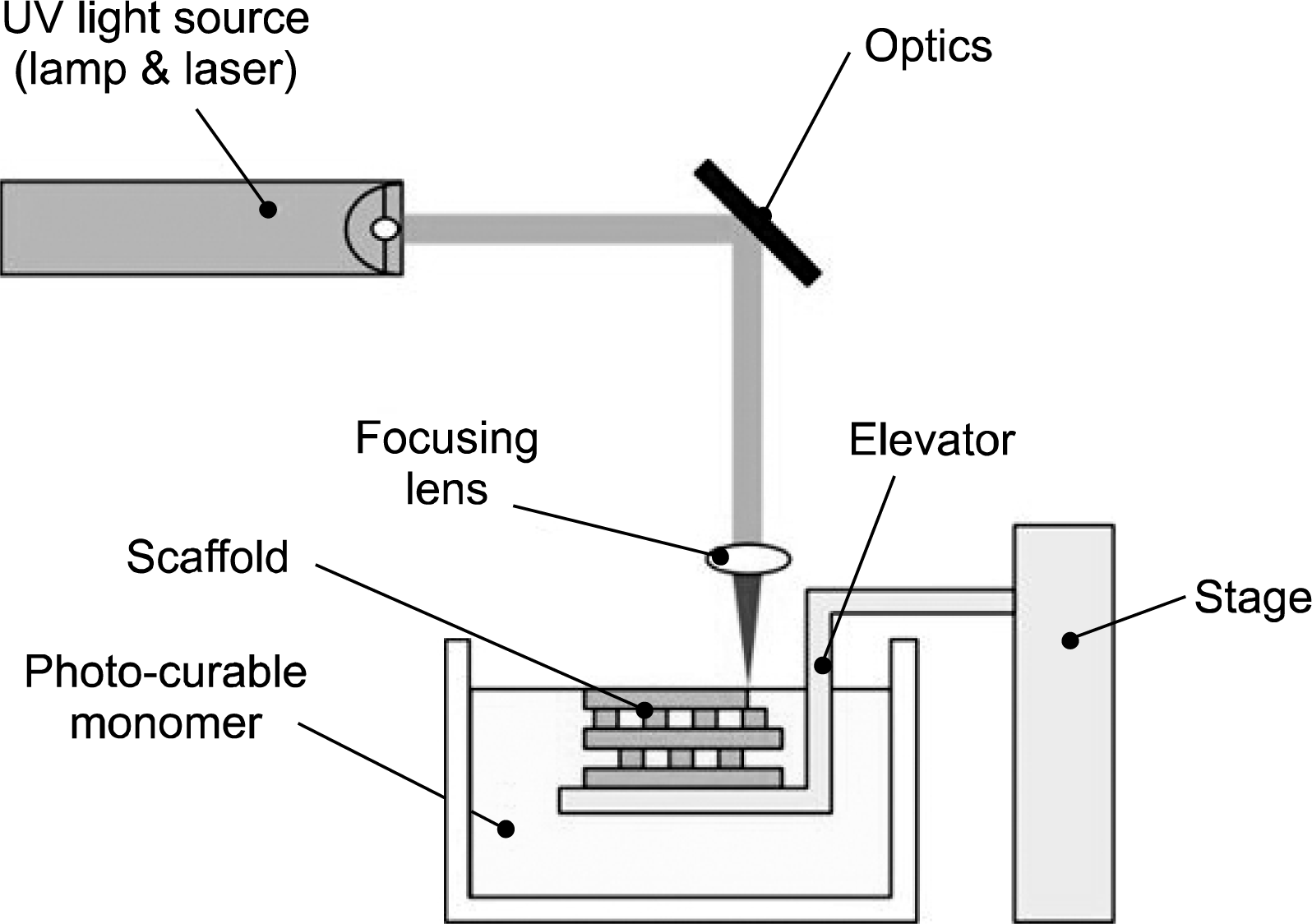
Fabrication of a scaffold using SL is occurs by radical generation through the absorption of photon energy by a photoinitiator and a series of monomer chain reactions referred to as initiation, propagation, and termination. Thus, the characteristics of the photo-polymerization reaction determine the performance of the final structure, such as the yield strength, elastic modulus, and shape accuracy. Thus, biomaterials with good photo-polymerization capabilities are essential in fabricating scaffolds using SL. Unfortunately, existing photo-curable biomaterials are limited and this has been a big hurdle in applying SL to bone tissue engineering. However, because of the importance of photocurable biomaterials in SL, the development of new materials has accelerated. Using the easiest approach, many researchers have tried to develop new bio-degradable materials by synthesizing or modifying existing materials to provide or improve photo-polymerization. As a result, polypropylene fumarate (PPF)-based materials, gelatin-based materials, and trimethylene carbonate (TMC)-based materials have been actively studied in recent years.
PPF was first synthesized in 1988 by Sanderson through a transesterification reaction between propylene glycol and diethyl fumarate (17). Since then, synthetic methods using various materials and processing conditions have been reported. Because structures using synthesized PPF possess better mechanical properties than human trabecular bone, it has gained attention as a biomaterial for bone tissue engineering. Mikos et al. built a very simple structure with PPF using a commercial rapid prototyping system (SLA-250, 3D Systems) and demonstrated that this material could be used for bone tissue reconstruction (18).
Cho et al. reported the fabrication of high-precision scaffolds for bone tissue engineering with a precision of a few tens of microns using PPF-based materials and MSTL technology (19, 20). In their study, the pores were completely interconnected with each other, and mechanical properties and cell affinity were also analyzed in the fabricated scaffolds (Fig. 2). Additionally, they suggested methods for enhancing cell adhesion and proliferation capability through surface modification processes, such as a bio-mimetic apatite coating method and a coating method using various peptides (21). They also fabricated 3D scaffolds that gradually released bone morphogenetic protein 2 (BMP-2) using PLGA microspheres as a protein carrier, while the scaffold degrades. Yaszemski et al. investigated the solidification characteristics and the manufacturability in controlling the amount of PPF and diethyl fumarate (DEF), which is a cross-linking agent (22).
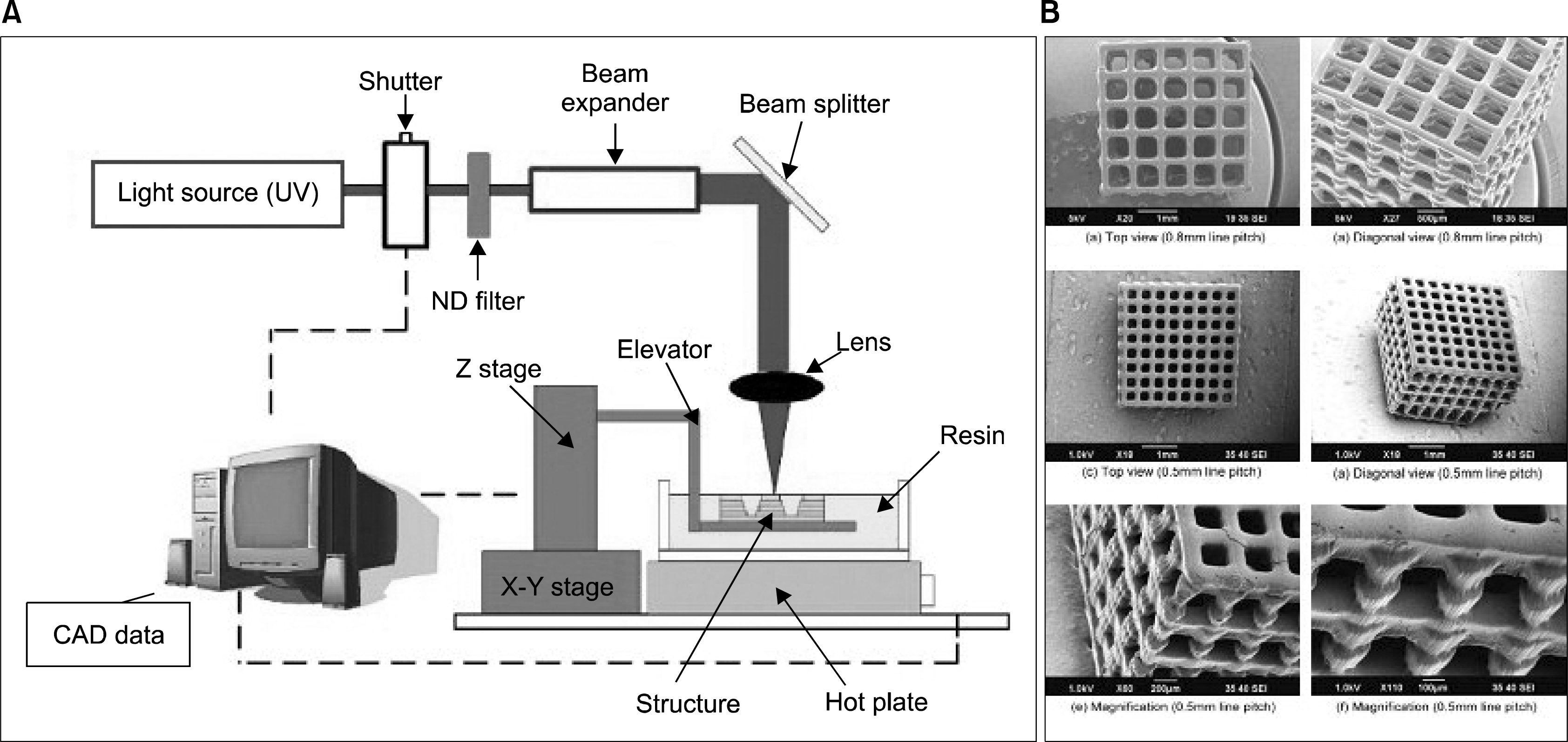 | Fig. 2.(A) MSTL system and (B) scanning electron microscope image of 3D high-accuracy scaffolds (19).
|
Liska et al. fabricated a 3D structure using a gelatin-based photo-polymer that was made by linking gelatin hydrolysate and an acrylated-based monomer. They mixed the gelatin hydrolysate with various reactive diluents, such as diisobutylacrylamide, dipentaerythritol pentaacrylate, urethane dimethacrylate (UDMA), pentaerythritol triacrylate, trimethylolpropane triacrylate, and glycol diacrylate, and added a commercial photoinitiator. Then, the photo-polymerization capability, storage modulus, and bending strength of each mixing condition were compared. The strength of the UDMA (87 Mpa) came close to that of natural bone (100–150 Mpa) (23). Cell adhesion and proliferation of MG63 osteoblast-like cells were evaluated on the material to determine its biocompatibility.
A TMC-based photopolymer is made by combining initiators, such as trimethylolpropane (TMP), ε-caprolactone (CL), or polyethylene glycol (PEG) of various molecular weights. Matsuda et al. fabricated various 3D structures, such as micro pillar arrays, micro cone arrays, banks, and tunnels, with TMC/TMP, TMC/PEG200 and TMC/PEG 1000 using their own MSTL (24). Additionally, using their fabricated structures, they observed the effects of various materials on cell adhesion and proliferation and investigated the in vivo hydrolytic degradation behavior.
Photo-curable polymers may possess inadequate mechanical properties. Studies to improve the mechanical properties are being conducted by mixing polymers with appropriate amounts of minerals. For example, Cho et al. fabricated a scaffold using a mixture of PPF/DEF polymer and hydroxyapatite (HA) nanoparticles (25). Nanoparticles inside the polymer scaffold reinforced the mechanical properties of the structure and nanoparticles on the surface improved cell affinity. In particular, while the surfaces of many synthetic polymers have low cell affinity, cell-friendly surface HA nanoparticles counteracted the disadvantages of the synthetic polymer PPF/DEF. Yaszemski et al. studied the physical properties and cellular responses of poly (propylene fumarate)/HA nanocomposites (26). Liska et al. investigated gelatin hydrolysate, which was mixed with reactive diluents, and HA to enhance mechanical strength (23). Furthermore, they fabricated a spongiosa-like structure that was built with an SL machine using the digital light processing principal. Cho et al. also developed a direct fabrication method for ceramic scaffolds using a mixture of commercial photo-curable photo-polymer and HA with a sintering process. During this process, a commercial photopolymer played a role as a binder and was removed by the sintering process at over 1000°C. They fabricated HA structures that were the same shapes as parts of human ear ossicles. Popov et al. also attempted the fabrication of scaffolds and prostheses using a commercial photocurable photopolymer with lower cytotoxicity (27).
Hollister et al. fabricated a high-strength scaffold made of HA by sintering a negative mold, which was fabricated using commercial epoxy photopolymer (28). They also successfully fabricated various shapes of scaffolds and confirmed its suitability for bone tissue engineering applications. Woesz et al. fabricated a scaffold using calcium phosphate and also conducted in vitro cell proliferation and differentiation tests using MC3T3-E1 cells (pre-osteoblasts) (29).
In cases of ceramic structure fabrication, the sintering process is essential for binding ceramic powders and removing the negative mold made by the polymer. However, this sintering process cannot be used for structural fabrication with synthetic and natural polymers, because negative mold materials are also polymers, and selective removal of negative mold materials between two polymers is impossible. Thus, new methods that can remove the negative mold materials have been developed for fabricating a 3D bone tissue engineering scaffold.
Liska et al. developed a photopolymer that could be dissolved in alkaline solution with a strength and viscosity appropriate for stereolithography (30). After fabricating the structure and filling it with synthetic and natural polymers, they fabricated the desired scaffolds by dipping the structure in an alkaline solution. Vallet-Regi et al. fabricated scaffolds for bone tissue regeneration using a negative mold and a commercial photopolymer (Accrura TM SI10, 3D Systems) and etching technology for removing the photopolymer (31, 32). They tested several different materials, such as HA, β-tricalcium phosphate, and aga-rose. Cho et al. also evaluated the fabrication characteristics of various biomaterials, such as PCL, PLGA, chitosan, and bone cement, for creating structures by filling precise soluble molds made by MSTL (33, 34). Dichen et al. fabricated 3D structures using layer-by-layer lamination and 2D molding technology (35). They prepared a polydimethylsiloxane 2D master pattern mold using MSTL technology. A 2D pattern was fabricated from the mold with chitosan/gelatin using the casting and freeze drying method. Finally, they formed the 3D shape by accumulating the patterns layer-by-layer.
Fused deposition modeling (FDM)
FDM is a manufacturing technology used for mechanical system modeling, fabrication, and production applications (36). The technology applies the melt extrusion method to fabricate a tissue scaffold, making use of a layer-by-layer thermoplastic polymer (37). The monofilament in a commercial FDM apparatus is moved by two rollers and activates as a piston to drive the semi-molten polymer. When each layer in the xy plane is finished, the base platform (z axis) is lowered and the procedure is repeated (8). The purpose of the liquefier is to heat and extrude the filament materials through a nozzle onto the base plate following a path, which is predefined by CAD and CAM (38, 39). 3D scaffolds with controllable pore size and porosity can be fabricated by changing the material deposition amount, the spacing between the material paths, and the height interval (z axis).
The main advantages of the FDM method are that it does not require a toxic solvent and offers flexibility in material handling and processing (8, 9, 40). Filament material also lessens the manufacturing time required in the heating compartment and allows for continuous production without the need for replacing feedstocks. However, the main difficulty of the FDM technique is the requirement for preformed fibers with a consistent size and material properties to feed through the rollers and nozzle (7–9). Furthermore, its application to biodegradable polymers, apart from polycaprolactone (PCL), may be limited. Thus, various modified FDM processes have been developed and evaluated to overcome the limitations of the traditional FDM process in tissue scaffold fabrication (41–45).
Hutmacher et al. investigated the mechanical properties and the responses of cells (human fibroblasts and primary human osteoprogenitor cells) cultured on PCL scaffolds manufactured using the FDM 3D Modeler RP system apparatus (Stratasys Inc., Eden Prairie, MN, USA). PCL scaffolds of 32.0×25.5×13.5 mm with 61% porosity were fabricated directly using Stratasys QuickSlice software. Two patterns of 0°/60°/120° and 0°/72°/144°/36°/108° were examined to give a honeycomb-like pattern of triangular and polygonal pores, respectively. The scaffolds with a 0°/60°/120° lay-down pattern had a compressive stiffness and yield strength of 41.9±3.5 and 3.1±0.1 Mpa, respectively. In comparison, the scaffolds with a 0°/72°/144°/36°/108° lay-down pattern had a compressive stiffness and yield strength of 20.2±1.7 and 2.4±0.1 Mpa, respectively. In vitro studies over a 3–4 weeks period showed that fibroblasts and osteoblast-like cells proliferated, differentiated, and produced a cellular tissue in a fully interconnected 3D PCL scaffold (46).
Cui et al. produced calcium phosphate-coated PCL scaffolds using commercial FDM apparatus. PCL scaffolds (50×50×5 mm3) were fabricated with regular triangular pores using a 0°/60°/120° lay-down pattern and seeded with human bone marrow osteogenic cells. The feasibility of using calcium phosphate-coated PCL scaffolds manufactured for bone tissue engineering application was evaluated using in vivo and in vitro studies (47).
Teoh et al. demonstrated successful in vitro coculturing of osteoblasts and chondrocytes on PCL scaffolds for more than 50 days using a commercial FDM system. Honeycomb-like scaffolds were fabricated with a lay-down pattern (0°/60°/120°). The porosity ranged from 60 to 65% and the pore size ranged from 300 to 580 μm. The 10×10×3.2 mm3 PCL scaffold was divided vertically into two halves with a space between them. One-half of the divided scaffold was allocated for bone marrow stromal cell seeding and the other half was allocated for chondrocyte seeding. Both osteoblasts and chondrocytes produced rich ECM in their scaffold region. At the boundary region, a mixture of cell types was observed. Thus, it was demonstrated that osteogenic and chondrogenic cells can grow, proliferate, distribute, and produce ECM in novel 3D FDM-fabricated PCL scaffolds under a coculture environment (48).
Kalita et al. fabricated an Mg/Zn-doped TCP scaffold using the indirect FDM process. Porous polymeric molds were first fabricated using an FDM machine with commercially available acrylonitrile butadiene styrene filaments. The molds were infiltrated with a ceramic slurry, followed by binder removal and a sintering process, to manufacture ceramic scaffolds. Compression tests on the scaffolds showed a failure strength of 3.8 Mpa for structures made of Mg-doped β-TCP scaffolds. By this approach, they fabricated 3D porous resorbable ceramic scaffolds as bone grafts via the indirect FDM process (49).
Sun et al. evaluated the feasibility of precision extruding deposition to fabricate PCL tissue scaffolds with designed micro-architectures. It was possible to manufacture scaffolds with a controlled pore size of 350 μm with designed structural orientations. An in vitro cell scaffold interaction study was conducted using primary fetal bovine osteoblasts. Additionally, an in vivo study was performed on nude mice to determine the capability of osteoblast-seeded PCL to induce osteogenesis. Experimental results showed multiple areas of osseous ingrowth suggesting that the osteoblast-seeded PCL scaffolds evoked osteogenesis (50).
Xiong et al. developed a novel low-temperature deposition manufacturing (LDM) technology to manufacture a bone tissue scaffold. Compared with other SFF based processes, the LDM process enhanced the bioactivities of scaffold materials because of the non-heating liquefying processing of the materials. Porous poly (L-lactic acid) (PLLA) and tricalcium phosphate (TCP) scaffolds fabricated for bone tissue engineering have a high porosity, up to 89.6% and a controlled interconnective macrocellular and microcellular morphology suitable for the repairing bone defects. An in vivo experiment showed that the scaffolds had good biocompatibility, good bone conductivity, and appropriate biodegradation properties for bone repair (51).
Cho et al. developed a precision deposition system for tissue engineering scaffold applications. The precision deposition system uses technology that enables the manufacture of 3D structures with complex, accurate shapes and precision of less than 100 μm. With four deposition heads, the system can fabricate various high-precision scaffolds using many biodegradable and biocompatible polymers more conveniently than other SFF-based fabrication methods (Fig. 3). PCL, poly-lactic-co-glycolic acid (PLGA), blended PCL/PLGA, and blended PCL/PLGA/TCP scaffolds were fabricated with controllable pore size, porosity and fully interconnected structures. The mechanical testing and cell affinity results showed that the blended PCL/PLGA/TCP scaffold was superior to the other scaffolds with respect to compressive strength, compressive modulus, cell adhesion, and proliferation. Moreover, they investigated the feasibility of using SFF-based scaffolds seeded with osteoblasts, derived from human adipose-derived stem cells, and human umbilical vein endothelial cells (HUVECs) to enhance osteogenesis. At 8 and 12 weeks after in-vivo implantation, scaffolds in the osteoblast-HUVEC group had the largest area of new bone tissue and were superior to the other groups for effective bone tissue regeneration (44, 52, 53).
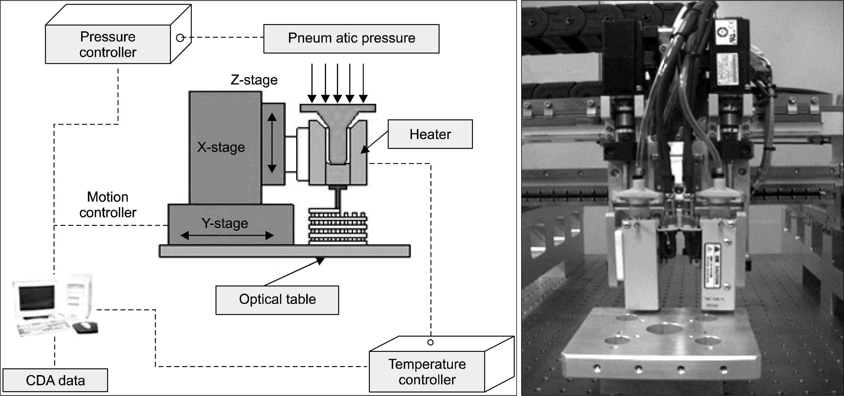 | Fig. 3.Schematic diagram and image of the precision deposition system (44).
|
Selective laser sintering (SLS)
The SLS uses a high-power laser, such as a carbon dioxide laser, to melt thin layers of plastics, ceramic, metal, or composite powder for scaffold fabrication (7, 54). During SLS operation, the laser selectively fuses powders following the cross-sectional information carried by the pre-defined CAD data (8). The interaction of the laser beam and powder increases the temperature to the glass-transition temperature, causing surfaces in contact to deform and fuse together. After each cross-section is scanned, the powder bed is lowered by one layer thickness, a new layer of material is exposed on top, and the process is repeated until the part is completed (55). The structure is supported by and embedded in the surrounding unprocessed powder and has to be extracted from the powder bed. Because the powders are maintained with low compaction forces after the sintering process to form new layers, structures have an internally porous structure suitable for a bone scaffold (Fig. 4) (56).
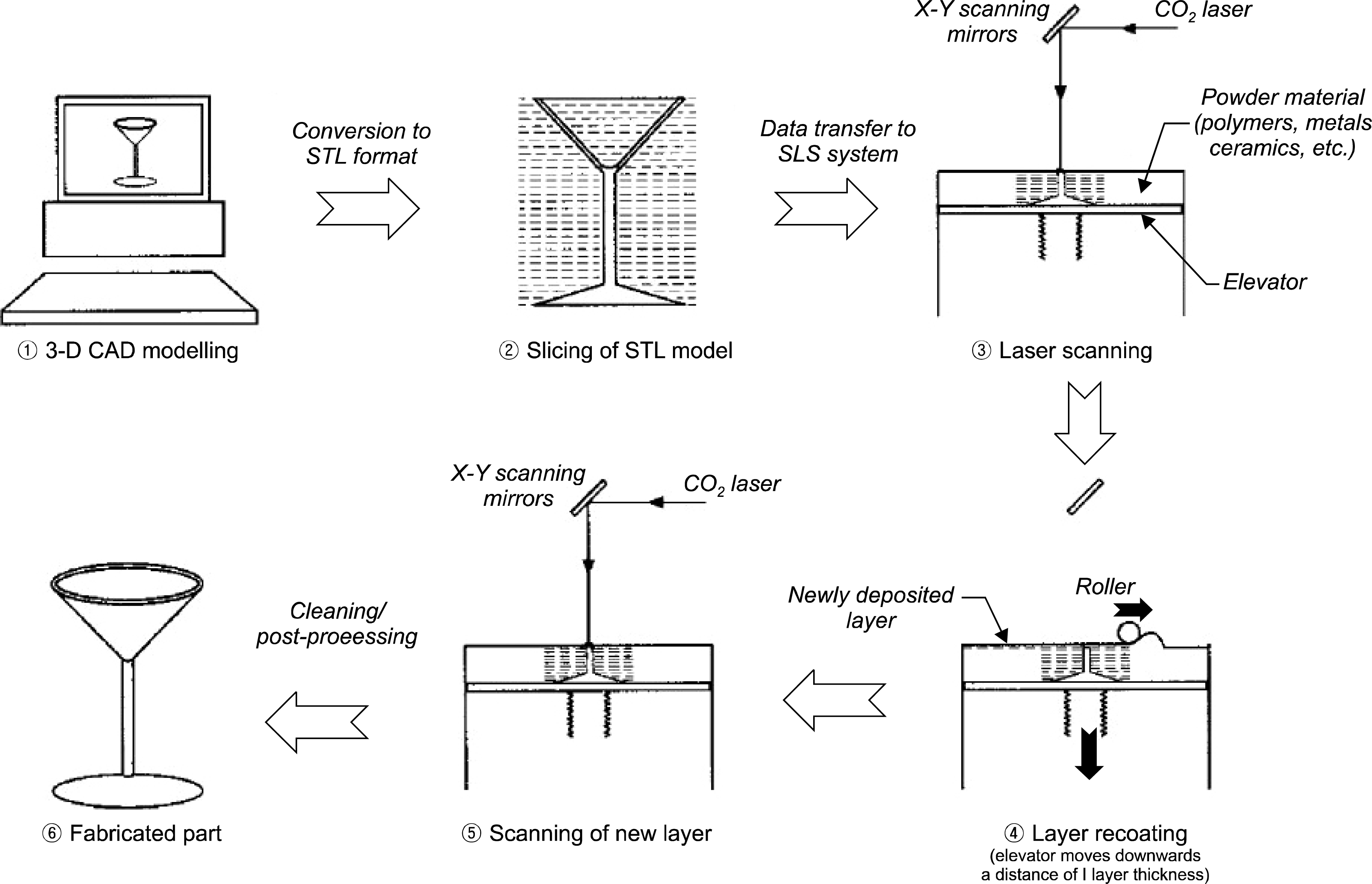 | Fig. 4.Schematic layout of the selective laser sintering process (56).
|
Compared with other SFF technologies, SLS can create complex structures, such as anatomically shaped scaffolds with controlled pore sizes, porosity, and topology more conveniently. SLS produces parts from a relatively wide range of powder materials such as bio-ceramics and titanium. Moreover, large numbers of scaffolds can be generated within the powder bed, allowing for mass production. However, this method may be limited in the manufacture of bio-polymers because the operating temperature is very high (5, 7, 8).
Das et al. designed and fabricated PCL scaffolds using of the SLS apparatus for clinical applications. SLS processing of the PCL powder was conducted by preheating the powder to 49.5°C and scanning the laser at 4.5 W power and a 1.257 m/s scan speed. The compressive modulus and yield strength of the scaffolds ranged from 52 to 67 Mpa and 2.0 to 3.2 Mpa, respectively. Scaffolds with BMP-7 were seeded with cells and implanted subcutaneously to evaluate biological properties and to demonstrate bone tissue in-growth. Additionally, they designed and fabricated a prototype mandibular condyle scaffold based on an actual pig condyle (57).
Chua et al. reported the possibility of using biocompatible polymers in SLS for developing bone scaffolds. Several biocompatible polymers, such as polyetheretherketone, poly-(vinyl alcohol), PCL, PLLA, and HA, were investigated for scaffold fabrication. The research showed that part bed temperature, laser power, and scan speed can control the micro-porosity of the specimens (58).
Liulan et al. fabricated cylindrical and spherical β-TCP scaffold microstructures as bone substitutes. Epoxy resin/nylon and β-TCP were used on a SLS system to fabricate bone scaffolds. During the fabrication, laser power was kept at 12 W, and the scaffolds were built layer-by-layer using a powder layer thickness of 100 μm, a preheating temperature of 50°C, and a scan speed of 2.5 m/s. After the sintering process, the temperature was increased to 600°C to remove the epoxy resin/nylon polymer, and a β-TCP only scaffold was successfully manufactured (59).
Wiria et al. demonstrated the potential of a PCL/HA biocomposite as a bone tissue scaffold, fabricated using SLS technology. Different compositions of PCL and HA with 10, 20 and 30 wt% content in the power blends were mixed with a mixing roller (CZ98174, US Stoneware, East Palastine, OH, USA) to fabricate the scaffold. Through cell culture experiments (osteoblast-like Saos-2 cells), they showed that the sintering and sterilization treatments prior to cell culture did not affect scaffold cell growth and the cells could proliferate well on the sintered PCL/HA scaffolds (60).
Zhou et al. focused on using bio-nano-composite micro-spheres, consisting of carbonated HA (CHAp) nanospheres within a PLLA matrix, to produce bone tissue engineering scaffolds using a modified SLS machine. The CHAp with a 20-nm mean particle size was synthesized in-house using a nano-emulsion method. A tetragonal porous scaffold (L×W×H=8×8×10 mm3) was designed and manufactured using an extrude-cut patterning method and SolidWorks (ver. 2005) and modified Sinterstation 2000 SLS machine (3D Systems), respectively. With this platform, porous bone tissue scaffolds were successfully manufactured from PLLA and PLLA/CHAp nanocomposite microspheres (61).
Kanczler et al. conducted in vitro and in vivo experiments to evaluate novel surface selective laser sintering (SSLS) scaffolds for their biocompatibility as templates and for human fetal femur-derived cell viability, growth, and osteogenesis. Using an experimental prototype SLS-80 laser, PLA scaffolds were fabricated with the desired architecture designed and produced by ILIT RAS (Troitsk/Shatura, Moscow, Russia). In the subcutaneous implant model, the SSLS-PLGA scaffolds provide a platform to differentiate human fetal femur-derived cells to generate new cartilage and osteogenic matrices, as evidenced by staining for Alcian blue/Sirius red and for collagen type I expression (62).
3D printing (3DP)
3DP employs inkjet printing technology to process powder materials for application on bone tissue scaffolds (63). The 3DP technology was developed at the Massachusetts Institute of Technology in 1995. Fundamentally, 3DP is also a layer-by-layer process in which the pre-defined 2D pattern of the target structure is printed on a first layer of powder via an inkjet printing head. This process is repeated until every layer is printed. After the binder has dried in the powder bed, the finished scaffold is retrieved and the unbound powder is removed (Fig. 5) (8, 64).
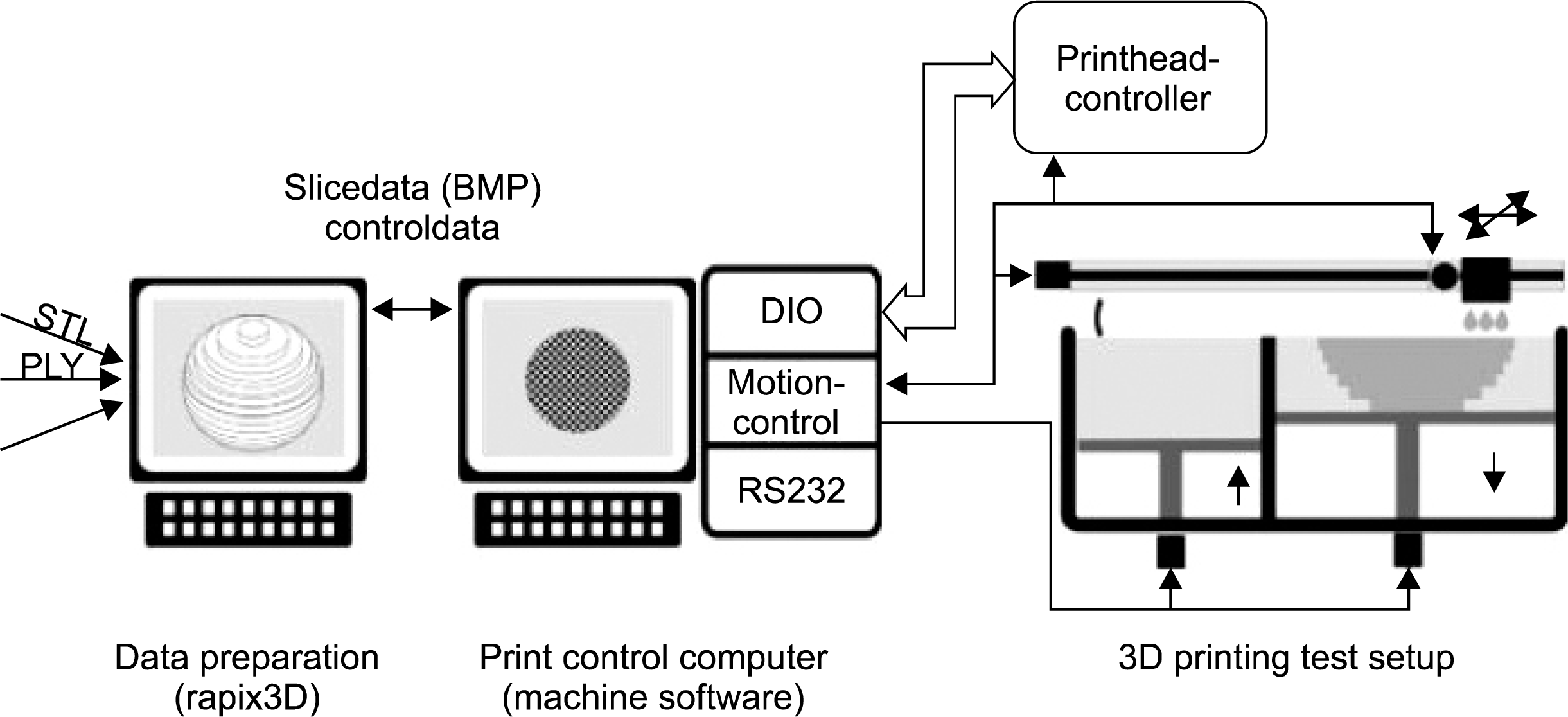 | Fig. 5.Process chain beginning from data preparation, process control to 3D printing (66).
|
3DP is generally faster, more affordable, and easier to use than other SFF technologies. 3DP offers scaffolds with several biomaterials for fabrication. A strong point of 3DP for scaffold fabrication is that commercial inkjet printers can be remodeled to deposit various polymers at precise positions with high throughput. Furthermore, bio-printing technology can be developed to perform computer-assisted deposition of natural polymers, and biomolecules, and even viable cells (65, 66). However, if the scaffold is designed to be porous, one problem with powder-supported and powder-filled structures is the difficulty removing the internal unbound powder. Moreover, scaffold architecture may be restricted by the nozzle size and the low positional accuracy of the inkjet printer (5, 7, 8).
Seitz et al. reported a new process for custom-made 3DP porous ceramic scaffolds for bone replacement with fully interconnected channel networks for repairing osseous from trauma or disease. A 3DP machine was designed and built in cooperation with Generis GmbH (Augsburg, Germany). After the sintering process (2 h at 1,240°C), a HA-based scaffold with a diameter of 7.8 mm, 447±37 μm in the z direction and 569±33 μm in x direction was successfully fabricated (64).
TCP scaffolds with controlled internal porosity have been fabricated with a suspension of TCP in diacrylate cross-linking monomers using a mold prepared by inkjet printing. Molds were fabricated using a commercial inkjet machine (Modelmaker II; Solidscape, Merrimack, NH, USA), developed for rapid prototype applications. They demonstrated that a lost-mold process can be successfully used to prepare a TCP with a controlled internal porosity. Despite a linear shrinkage of about 22%, the sintered ceramic showed a regular internal porous structure, as defined by the mold (67).
Wu et al. developed indirect 3DP technology for scaffold fabrication. In the indirect 3DP protocol, molds are printed with a commercial 3DP machine (Z402, Zcorp, Burlington, MA, USA) and the final materials are cast into the mold cavity to overcome the limitations of the direct technique. They demonstrated that a PLGA scaffold, made by indirect 3DP, supported IEC6 cell growth in culture (68).
Lelkes et al. developed a drop-on-demand printing process, which is a thermoplastic porogen-based injection molding manufacturing method. This process can be used to fabricate porous PCL and PCL-calcium phosphate (CaP) composite scaffolds with pore sizes as small as 200 μm. The compressive strengths of the 90:10 and 80:20 PCL-CaP scaffolds, according to ASTM standards, were 19.5±1.4 and 24.8±1.3 Mpa, respectively. The compressive strength of a pure PCL scaffold was 2.77±0.26 Mpa. Cytocompatibility tests using human embryonic palatial mesenchymal (HEPM) cells indicated that all porogen-based scaffolds facilitated attachment and supported HEPM cell proliferation in vitro. It was also suggested that the presence of CaP in the PCL-CaP composite enhanced the proliferation of HEPM cells and reduced spreading in favor of a multi-layer assembly (69).
Schnabelrauch et al. developed a new CaP powder-binder system for the 3D printing of patient-specific implants. A β-TCP (70 wt%)/TTCP (30 wt%) scaffold of about 12 mm in diameter and 4 mm in height was fabricated using a commercial Z 402 printer (Z Corp.). A preliminary examination of relevant application properties, including in vitro cytocompatibility testing, indicated that the new powder-binder system represents an efficient approach to patient-specific ceramic bone substitutes and scaffolds for bone tissue engineering (70).
Go to :
Conclusion
This paper has described various SFF-based scaffold fabrication technologies for bone tissue engineering. SFF equipment permits scaffold fabrication with controllable pore size, porosity, interconnectivity, and high mechanical strength. Because SFF technologies are designed and fabricated by a CAD/CAM, these methods have advantages in fabricating a 3D scaffold with anatomical shapes. Further investigations of appropriate biomaterials for each SFF system are required for the fabrication of optimal scaffolds for bone tissue engineering. Future developments in SFF require the design of new materials, optimal scaffold fabrication systems, and cell biology studies, including determining optimal cell adhesion, proliferation, and differentiation in SFF-based scaffolds.
Go to :
 XML Download