

 PDF
PDF Citation
 Print
Print


Introduction
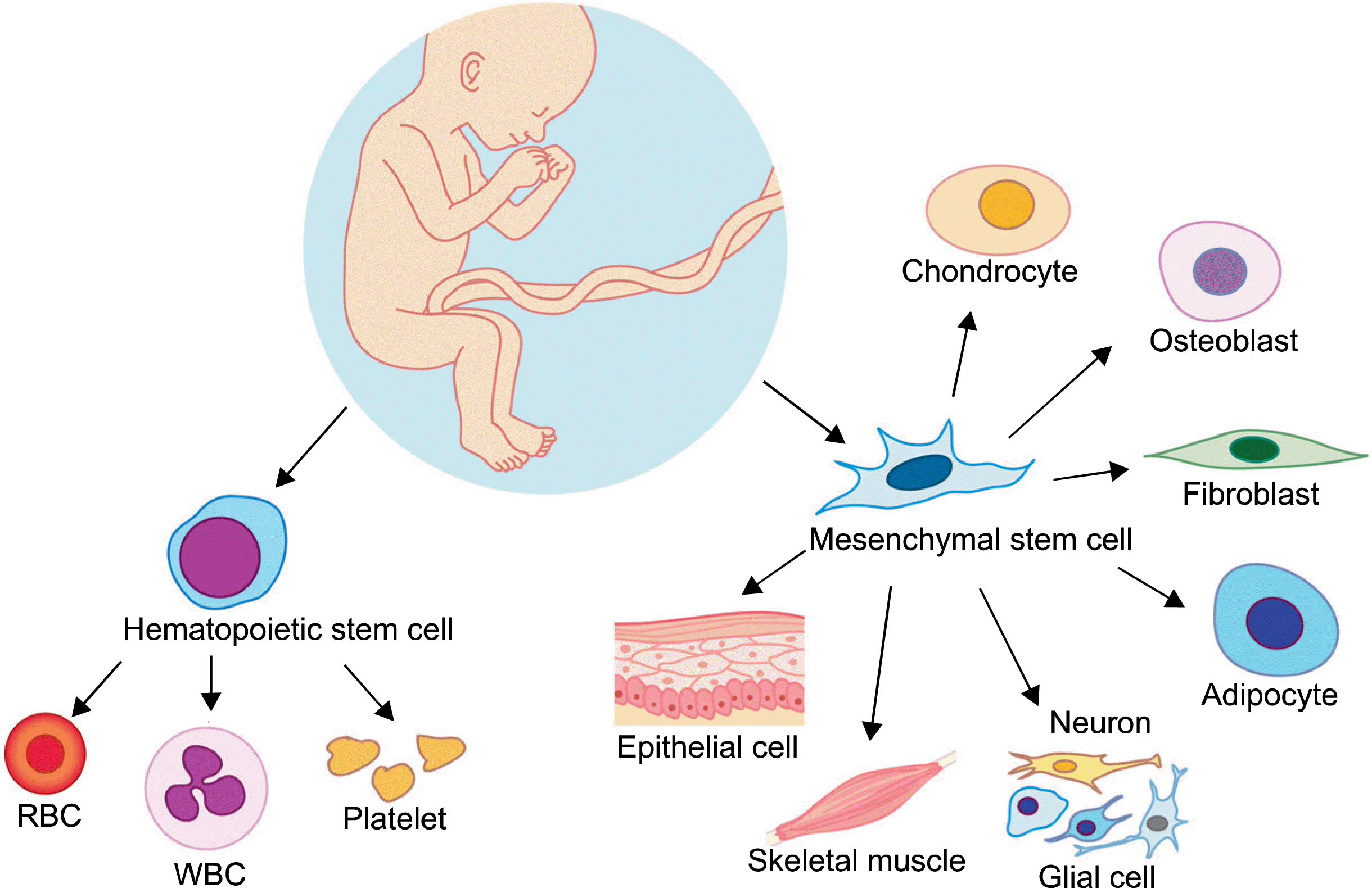
The cord blood (CB) was introduced for the first time in human to reconstitute the hematopoietic system in patient with Fanconi anemia (1). Since the first cord blood transplantation (CBT), more than 20,000 CBTs have been reported worldwide and more than 400,000 CB units have been stored in more than 100 CB banks (2). The first report of CBT in Korea was introduced in 1998 (3), since then more than 500 CBTs have been performed and more than 20,000 CB units have been stored for public purposes (4). Efficacy of unrelated CBT has been demonstrated in children and adults with hematological malignancies and children with a variety of nonmalignant hematologic disorders, including hemoglobinopathies (5), immunodeficiencies (6, 7). The clinical use of CB has expanded into various areas such as treatment of inherited metabolic disorders. Since CB contains hematopoietic stem cells as well as a mixture of multipotent stem cells such as unrestricted somatic stem cells, mesenchymal stem cells, and endothelial colony-forming cells, CB has the ability to regenerate numerous tissue types with functional improvements (8) (Fig. 1). Recently, the use of CB in several regenerative medicine applications has expanded its clinical utility. The application of CB for regenerative medicine is different from typical hematopoietic stem cell transplantation (HSCT) which has been performed for inherited metabolic disorders (IMD) requiring pre-conditioning chemotherapy regimes. This review describes clinical and pre-clinical application of CB cell-based therapy for IMDs and tissue regenerations, and discusses current status and future directions of this promising field.
Go to : 
Inherited metabolic disease
Inborn errors of metabolism are now often referred to as congenital metabolic diseases or IMD, and these terms are considered synonymous. The majority are due to defects of single genes that code for enzymes that facilitate conversion of various substances into other products. In most of the disorders, problems arise due to accumulation of substances which are toxic or interfere with normal function, or to the effects of reduced ability to synthesize essential compounds. The evidence for and potential usefulness of HSCT in IMD relate only to diseases that belong to the family of lysosomal and peroxisomal storage disorders (PSD). In general, lysosomal storage disease (LSD) and PSD are progressive in nature and frequently fatal in childhood.
LSDs are caused by lysosomal dysfunction usually as a consequence of deficiency of a single enzyme required for the metabolism of mucopolysaccharides, glycoproteins (sugar containing proteins), lipids. The symptoms of LSD vary, depending on the particular disorder and other variables like the age of onset, and can be mild to severe. They can include developmental delay, movement disorders, seizures, dementia, deafness and/or blindness. Some people with LSD have enlarged liver and spleen, pulmonary and cardiac problems, and bones that grow abnormally. A major function of the peroxisome is the breakdown of fatty acid molecules, in a process called beta-oxidation. The formation of plasmalogen, the most abundant phospholipid in myelin, also occurs in peroxisomes. Deficiency of plasmalogens causes profound abnormalities in the myelination of nerve cells, which is one of the reasons that many PSDs lead to neurological disease, like adrenoleukodystrophy (ALD).
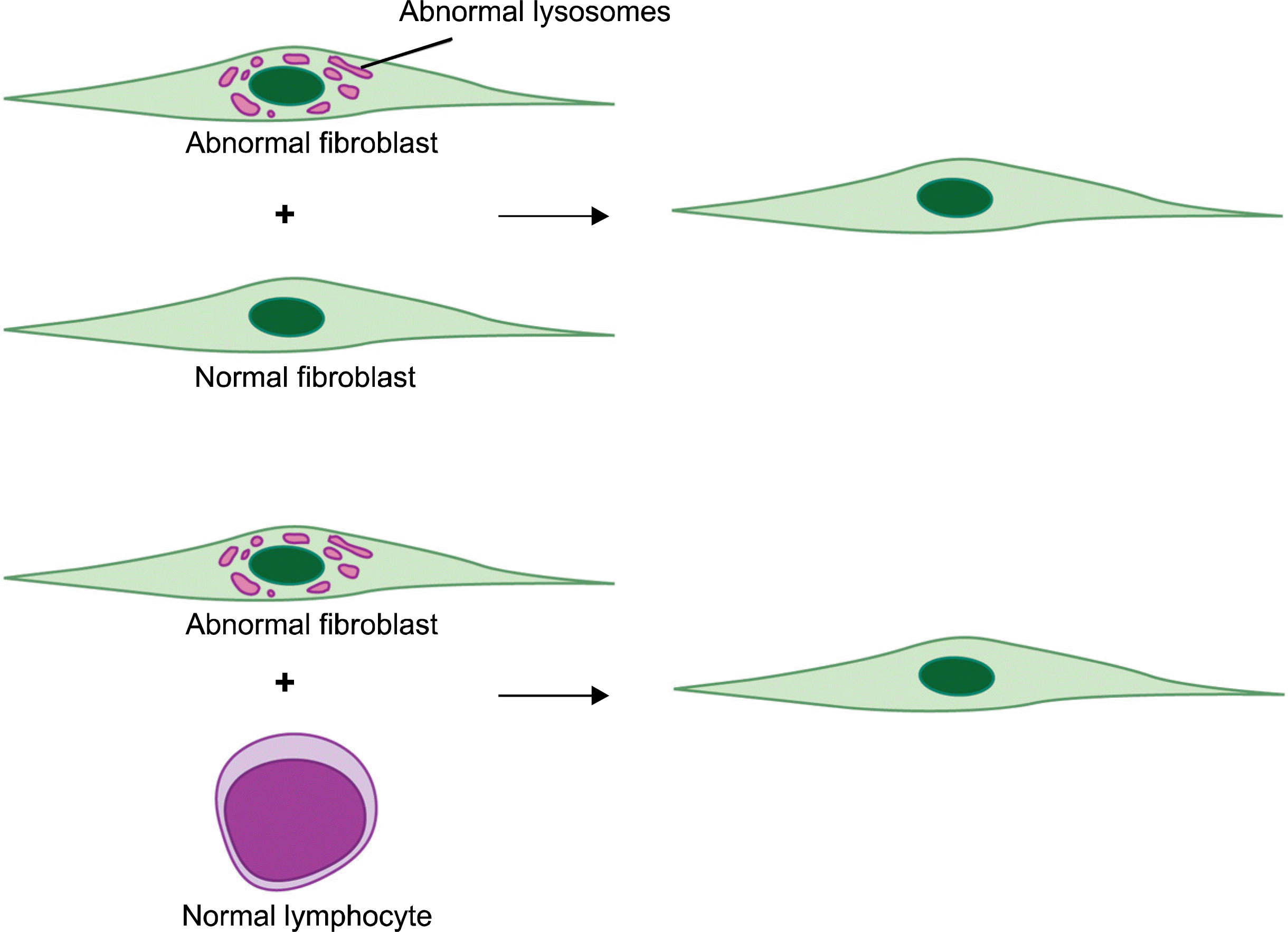
The important 2 basic concepts for correction of IMD by HSCT are as follows: First, the microglial cells in the brain, alveolar macrophages in the lungs, and Kupffer cells in the liver are derived from monocytes in the hematopoietic system (Fig. 2). After unrelated HSCT has been performed in patients with IMD, engrafted HSCs in BM have the ability to differentiate to blood monocytes and cross the blood-brain barrier (9). Donor-derived cells containing normal levels of enzymes migrate to and engraft in non-hematopoietic organs in close proximity to the patients’ enzyme-deficient cells (Fig. 3). Second, the consequent inter-cellular enzyme transport is mediated by cross-correction, a phenomenon by which close proximity of normal cells leads to the correction of the biochemical consequences of enzymatic deficiency within the neighboring cell. This phenomenon was proved in cultured fibroblasts from Hurler and Hunter patients, whose biochemical problem was corrected if cells of these two genotypes were mixed with each other or with normal cells (10) (Fig. 4). In these patients, durably engrafted CB cells of donor origin provide a form of cell-based enzyme replacement therapy. Donor cells provide continuous, life-long production of the missing or defective enzyme (11).
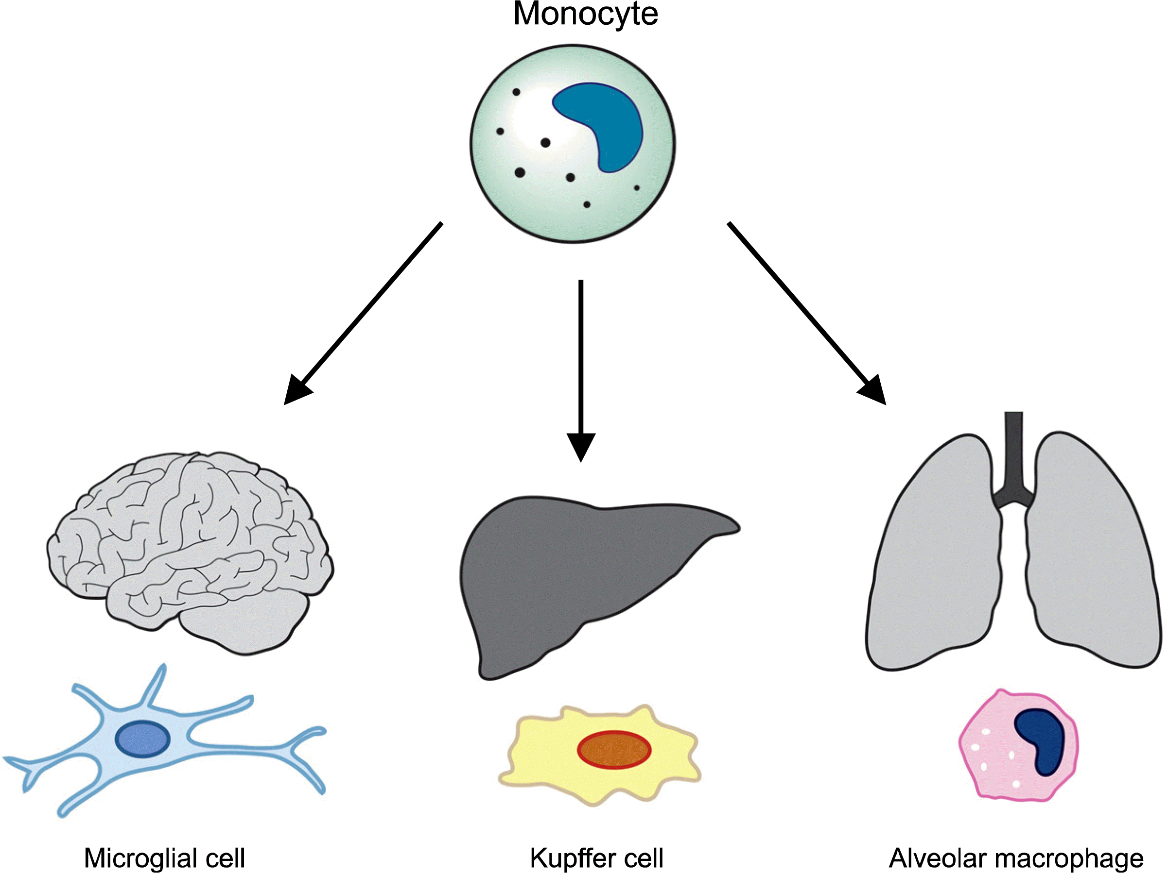 | Fig. 2.The microglial cells in the brain, alveolar macrophages in the lungs, and Kupffer cells in the liver are derived from monocytes in the hematopoietic system.
|

To date, more than 2,000 patients with almost 20 different LSD and PSD have been treated with HSCT following chemotherapy-based conditioning. Of these, severe mucopolysaccharidosis (MPS) type I (Hurler syndrome), ALD, metachromatic leukodystrophy (MLD), and globoid leukodystrophy (Krabbe disease) have accounted for more than 80% of the cases (12). The graft sources for IMD have been bone marrow (BM) and CB. However, most of reports provide that CBT is an appropriate option for HSCT for infants and children with IMD. The outcomes following transplantation with matched related or unrelated BM, or sibling donor BM who are carriers of the disease showed inferior results as compared to CBT (13–15). In comparison to previously published BMT experience, CBT study demonstrated higher near-total chimerism, better enzyme recovery in the blood, and superior engrafted and alive rates (16). One of the advantages of CBT for IMD could be explained by containing a greater dose of nonhematopoietic progenitor cells in CB rather than in BM. It was demonstrated in the autopsied brain of a Krabbe disease, which showed the CB-derived cells in blood vessels, periventricular tissues, white matter, cerebellum, choroid plexus, and forebrain parenchyma (17). The donor cells were differentiated to microglia and choroid plexus, but not into neuroectodermal cells (e.g., neurons, astrocytes, or oligodendrocytes).
The clinical benefits of CBT for Hurler syndrome demonstrated either stabilization or improvement of neuro-cognitive function and continued to gain new skills (18). There were also an improvement in the somatic features, linear growth and bone disease in these patients. In most patients, organ dysfunction from accumulation of glycosaminoglycan in the heart, liver and spleen are reduced. The outcome of CBT for infantile Krabbe disease showed dramatic efficacy if the patient was transplanted early in the course of the disease (19). In these patients, CBT can prevent demyelination in the central and, often, the peripheral nervous systems, extending life and improving the overall quality of life. However, survival was only 45% in infants who were symptomatic at transplant and, while their disease stabilized, no appreciable gains were seen in neurological development. In later, more slowly progressive forms of Krabbe disease, patients show improvements even if they are transplanted after developing neurological symptoms. BMT has also been shown to arrest progression of the disease after stabilization 3∼6 months post transplant in the most severe infantile form of Krabbe disease (globoid-cell leukodystrophy) as well as in the milder juvenile and adult types (20). Analysis of the outcomes of CBT for ALD showed lower survival and neurological deterioration in symptomatic patients. However, the children transplanted before the onset of clinical symptoms continue to develop at a normal rate 5∼7 years post-transplant (21).
In summary, although no individual or combination therapy can cure all aspects of any IMD, CBT could offer promising and effective therapy for many patients with IMD. At the present time, many reports recommend the following categories as a standard of care for IMD (22): severe type of Hurler syndrome in MPS; mucolipidosis, aspartyglucosaminuria in glycoproteinosis; Krabbe disease, MLD, Wolman disease in lipidosis; ALD in PSD. Every effort should be made to perform transplantation early in the course of disease before extensive damage to nervous system and other organs occur.
Go to :
Neonatal hypoxic-ischemic encephalopathy
Despite major advances in monitoring technology and knowledge of fetal and neonatal pathologies, perinatal asphyxia or, more appropriately, hypoxic-ischemic encephalopathy (HIE), remains a serious condition that causes significant mortality and long-term morbidity. HIE is characterized by clinical and laboratory evidence of acute or sub-acute brain injury due to asphyxia (e.g., hypoxia, acidosis) (23). Most often, the exact timing and underlying cause remain unknown. At the cellular level, neuronal injury in HIE is an evolving process. The magnitude of the final neuronal damage depends on duration and severity of the initial insult combined to the effects of reperfusion injury, and apoptosis. In severe HIE, the mortality rate is reportedly 25∼50%. Most deaths occur in the first week of life due to multiple organ failure or redirection of care. Some infants with severe neurologic disabilities die in their infancy from aspiration pneumonia or systemic infections. As many as 80% of infants who survive severe HIE develop serious complications, 10∼20% develop moderately serious disabilities, and as many as 10% are healthy (24). Although it is one of the most common causes of death and long-term neurological impairment in full-term neonates worldwide, up to now, treatment for HIE is mainly supportive.
Recently, since CB, BM, and peripheral blood (PB) contain a population of stem cells that can enter the central nervous system and differentiate into the brain macrophages and microglial cells, the potential use of various stem cells to reduce brain damage or to promote regeneration has been performed in animal model. Although stem cells from BM and PB have been introduced, the use of CBs in animal models of HIE was first reported in 2006 (25). Most of reported studies have shown that CB administration in stroke models resulted in some degree of therapeutic benefit with no adverse effects. Even donor cells were not found in the brain, a functional improvement was documented. In these experiments, donor cells produced several neurotrophic factors and cytokines, and these factors are released into the circulation, stimulating regenerative events such as angiogenesis and neural plasticity (26, 27).
Studies demonstrated that direct injection of the stem cells into the brain was not required (28), and beneficial effects could be observed even if the stem cells did not actually home into the target organ (29, 30). The beneficial effects seemed to be dose-dependent and could reduce the size of the infarcted tissue (31). Unlike current pharmacological interventions that require treatment within the first few hours after stroke, CB stem cell therapies were effective upto 48 hours after the thrombotic event (32). The administration of CB stem cells immediately after the ischemic event may be detrimental in that the inflammatory milieu may be toxic to the administered stem cells. Further studies would be warranted to determine the best cell type, the route of administration, the number of cells, and the timing for administration of the cells.
In the model of neonatal HIE, modulation of the endogenous capacity to form new neurons is a new challenge, and promising treatments may be developed to overcome the limitations of cell-based therapy. In this regards, it was shown that intracerebroventricular administration of β fibroblast growth factor increased the number of newly born neurons in the subventricular zone (33). Hypothermia is also the most successful treatment to reduce long-term effects of moderate and severe HIE in preclinical and clinical studies (34). It was also reported that erythropoietin had a neuroprotective effect in several preclinical studies and improved the neurological outcome of children with moderate HIE in a recent clinical trial (35).
At present, there have been no reported data regarding clinical trials of stem cell therapy for neonatal HIE in human. Currently, the administration of autologous CBs in term newborns with HIE is being evaluated in a clinical trial being conducted at Duke University. Since a greater dose of non-hematopoietic progenitor cells are contained in CB rather than in BM, CB has a greater potential to be differentiated into the blood vessels and microglial cells. Therefore, CB could be a more appropriate and potential source for cell-based therapy when further guidelines regarding appropriate cell type, dose, timing, and route of administration of CB cells for engraftment in the brain tissues have been developed in the model of neonatal HIE.
Go to :
Cerebral palsy
Cerebral palsy (CP) is a group of non-progressive brain diseases which produce chronic motor disability in children. CP affects 2/1,000 live-born children. Due to the increasing survival of the very preterm and very low birth weight infant secondary to improvements in neonatal care, the incidence of CP may be increasing. While premature infants and term infants who have suffered neonatal hypoxic-ischemic injury represent only a minority of the total cerebral palsy population, this group demonstrates easily identifiable clinical findings, and much of their injury is to oligodendrocytes and the cerebral white matter.
Currently, clinical trials of autologous CB infusion for CP are being conducted by Dr. Kurtzberg. She said her hypothesis that autologous CBTs might be beneficial in children with CP is based on research using allogeneic CBTs for children with infantile Krabbe’s disease. Her team found that donor cells could get into the brain, that remyelination and improvement in neurologic disabilities in demyelinating diseases. While the use of stem cell therapy is promising and the Medical College of Georgia is also starting a randomized, double-blind clinical trials using autologous CB to treat children with CP, there are no controlled trials yet in humans with CP (36). However, these current clinical trials, including author’s, using autologous CB for children with CP could be justified, because there have been lots of studies suggesting that stem cells can modify the brain after injury and promote behavioral recovery. Moreover, autologous CB infusion is very safe and ethical.
Go to :
Juvenile diabetes mellitus (Type 1 DM)
Type 1 diabetes is an autoimmune disorder characterized by T-cell-mediated destruction of insulin-producing β-cells and lifelong dependence on exogenous insulin administration. To date, unfortunately, there is presently no permanent cure for diabetes. Whole pancreas or islet cell transplantation is available only to a very limited number of patients and necessitates potential lifelong immunosuppressive therapy.
In animal model with type 1 DM, treatment with CB stem cells demonstrated lower blood glucose level, reduced insulitis and increased lifespan compared to control group (37, 38). It is postulated that the infused CB stem cells differentiate into new islets cells based on the in vitro data. In addition, cord blood contains a large number of immune cells called regulatory T cells, these regulatory T cells may be helpful in diminishing autoimmunity. The need to re-establish tolerance in patients with established autoimmunity provides another potential mechanism for CB as a therapy for type 1 diabetes (39).
Recently, Haller et al conducted an open-label phase I study using autologous CB infusion to ameliorate type 1 diabetes (39). Fifteen patients diagnosed with type 1 diabetes and for whom autologous CB was stored underwent a single intravenous infusion of autologous cells and completed 1 year of postinfusion follow-up. Intensive insulin regimens were used to optimize glycemic control. They experienced no harm with autologous CB infusion but did not demonstrate efficacy in preserving C-peptide. The potential of CB to participate in the future of type 1 diabetes interventional therapies exists. Therefore, further studies will likely need to achieve the dream of safely and permanently reversing or preventing type 1 diabetes.
Go to :
Conclusion
CB may be a rich source of non-hematopoietic stem and progenitor cells as well as of cells capable of transdifferentiation. Therefore, CB may be a better graft source than BM for patients who need enzyme replacement therapy or tissue regeneration. Particularly, in the pediatric age including neonatal period, a lot of clinical trials using autologous CBs for these categories would be encouraged, because many CBs are stored in private banks worldwide and autologous CB infusion is very safe and ethical.
Go to :
 XML Download