Journal List > Int J Stem Cells > v.2(2) > 1158890

Zheng, Cheng, Yu, Liu, and Cheng: The Primary Study of CD90+CD34−and Sca-1+ Stem Cells Mobilized by EPO Plus G-CSF in Mice

Abstract

To investigate effects of recombinant human erythropoietin (rhEPO) plus recombinant human granulocyte colony stimulating factor (rhG-CSF) on mobilization of CD90+CD34 and Sca-1+ stem cells in mice. rhEPO 1000 IU·kg−1·d−1 per mice alone or plus rhG-CSF 250 μg·kg−1·d-1−1per mice was administered to mice for five days, the peripheral blood was collected at 6 hours, 1, 3,5 and 7 days after the last administration. The number of white blood cells (WBC) and mononuclear cells (MNC) was counted. The level of CD34, CD90 and Sca-1 mRNA were detected by reverse transcription-polymerase chain reaction (RT-PCR).The expressions of CD90+CD34 in absolute nuclear cells of peripheral blood was detected by flow cytometry. The results indicate that compared to mobilizing by rhG-CSF, rhEPO or plus rhG-CSF did not significantly enhance the number of WBC and MNC, on the fifth and the seventh day after the last administration, both the expression of CD34,CD90 and Sca-1 mRNA and the proportion of CD90+CD34 cells dramatically raised in rhEPO or in addition to rhG-CSF groups. We conclude that rhEPO or plus rhG-CSF had a strong capacity of mobilization of stem cells. The alteration of expression of CD34, CD90 and Sca-1 mRNA after the administration of rhEPO or combined with rhG-CSF indicated that this agents might potentially alter the peripheral blood graft content.

Introduction

Cytokine-mediated mobilization of hematopoietic stem cells (HSCs) has become an established method in the field of autologous and allogenic stem cell transplantation. Furthermore, it presents a new concept in tissue repair and regenerative medicine. Currently, G-CSF remains the gold standard mobilizing agent for peripheral blood stem cell transplantation (PBSCT). However, there are many areas for improvement that can have significant clinical impact: specifially, a significant proprtion (10∼20%) of healthy allogeneic donors mobilize poorly in response to G-CSF and this proportion dramatically increases (up to 40%) in autologous donors; inability to predict optimal mobilization times; inability to expanse bone marrow stem cell pool and graft composition (13. Thus, intense efforts have been made to alter mobilization strategies, such as Pegfilgrastim, the bicyclam AMD3100, chemokines and their receptors. In the present study, we studied the efficiency of rhEPO plus rhG-CSF on mobilization of peripheral blood stem cells (PBSCs), and also focused on the content of CD34+, CD90+, CD90+ CD34−and Sca-1+ stem cells in the mobilized peripheral blood graft.

Materials and Methods

Animals

The expriment was performed on male mice (KM strain, n=60) weighing about 22±2 g. Animals were provided by Laboratory Animal Center of Yunyang Medical College, China. Animals were housed in polypropylene cages in 12 h dark/12 h light cycles with temperature of the laboratory animal house ranging from 20°C to 25°C, and humidity between 50% to 65%. Saw dust was used as the bedding, which was changed during cleaning of cages on alternate days. All the animals were produced from the Laboratory Animal Center of Yunyang Medical College, China. And all the animals were received care in accordance with Guide for the Care and Use of Laboratory Animals published by the US National Institutes of Health (NIH publication No. 85-23).

Adiministration of rhG-CSF or rhEPO

Mice were divided into 4 groups, each comprising 15 mice:1) adiministration of saline daily for 5 days. 2) adiministration of rhG-CSF (Lot:20090206, produced by Shandong Qilu Phaimaceutical Co., Ltd.) daily for 5 days (250 μg per kg body weight per day, intraperitoneally). 3) adiministraion of rhEPO (Lot: 20081109S, produced by Sansheng Shenyang Pharmaceutical Co., Ltd.) daily for 5 days (1,000 IU per kg body weight per day, intraperitoneally). 4) adiministraion of rhG-CSF in combination with rhEPO for 5 consecutice days (dosage as above). All mice were anaesthetized with 10% Chloral Hydrate and sacrificed at 6 hours, day 1, day 3, day 5 and day 7 after the last injection drugs.

Hematologic parameters

Mobilized-peripheral blood samples were analyzed using Automatic Hematology Analyzer (Cell Dyn 3700, Abbott, United States).

Reverse transcriptase-polymerase chain reaction (RT-PCR) analysis

Total RNA was extracted from WBC or bone marrow cells using the TrizolTM kit (GIBCO BRL, Invitrogen, Paisley, UK) according to the manufacturer's instructions. For PCR, the primers were designed to exclude detection of genomic DNA. As positive controls, total RNA was extracted from mice bone marrow. Primers were used as indicated in Table 3. The PCR was performed using a Hot Star Taq Master Mix Kit (Qiagen, Hilden, Germany) according to the manufacturer's instructions and was carried out in a programmable Applied Biosystems Veriti 96 Well Thermal Cycler (Applied Biosystems, America) with the following conditions: 94°C for 5 min and then 30 cycles, each comprising 94°C for 30s, annealing for 1 min with: GAPDH 62°C, CD34 62°C, CD90 62°C, Sca-1 55°C and then 72°C for 1 min. After the PCR was completed, the tubes were kept for 5 min at 72°C. Negative controls were performed without template. The difference of the expression level of a specific RNA was determined by normalization to the expression level of GAPDH and quantification was made using a standard curve assay (Table 1).

Flow cytometry analysis

At 6 hour, day 1, day 3, day 5 and day 7 after the last adiministration, 1mL peripheral blood was harvested from each mouse by aspiration from heart. Bone marrow cells were obtained by flushing tibias and femurs with phosphate-buffered saline. To determine CD90+/CD34+ and CD90+/CD34 subpopulations, cells were incubated for 40 minutes in the dark at 4°C with the following fluorescein isothiocyanate-(FITC) and phycoerythrin-(PE) conjugated monoclonal antibodies: CD90-FITC and CD34-PE (all from eBioscience, San Diego, USA). Isotype-identical antibodies: IgG2α-FITC and IgG2α-PE (eBioscience) served as controls. Cells were analyzed by two-color flow cytometry using a Coulter Epics XL- MCLTM flow cytometer (Beckman Coulter). Each analysis included 10000 events.

Statistical analysis

Data are shown as mean±standard error of mean. Multiple group comparison was performed by one-way analysis of variance followed by the Bonferroni procedure for comparison of means, and performed by K Independent Samples Test for comparison of rate by way of SPSS13.0 statistical package program for Windows. Comparisons between two groups were performed using the unpaired t-test and Chi-Square test. Values of P less than 0.05 were considered statistically significance.

Results

Hematologic parameters

At first, hematologic parameters were analyzed to compare the effects of rhEPO, rhG-CSF+rhEPO with standard rhG-CSF treatment as a positive control according to established mobilizing strategies for PBSCT. The number of leukocytes and mononuclear cells was summarized in Table 2 and Table 3. These data showed leukocytes significantly increased values at the 1st day of rhEPO stimulation (1.67- fold; p<0.001) and at the 3rd day of rhG-CSF+rhEPO stimulation (1.52-fold; p<0.01) compared to the positive control group. Increased levels were also detected for mononuclear cells at different time of rhEPO and rhG-CSF+rhEPO stimulation, but there was no significant compared to the positive control group.

FACS analyses of CD90+ cells populations in peripheral blood

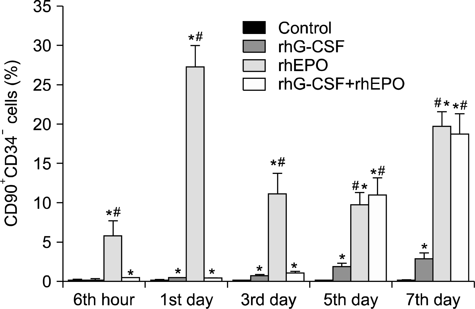
In order to know whether defined populations of bone marrow stem cells circulate in peripheral blood with the stimulation of rhG-CSF, rhEPO or rhG-CSF+rhEPO, fluorescein-activated cell sortiong (FACS) analyses were performed. We found a significant increase of the defined subtypes of CD90+ cells (CD90+/CD34+ and CD90+/CD34). Especially, CD90+/CD34 cells were stronger increased than CD90+/CD34+ cells(the percentage of CD90+/CD34+ cells was very low and the data was not shown). After rhEPO application, a higher increase of CD90+/CD34 cells at different time excluded at hour 6 than adiminstration of rhG-CSF and rhG-CSF+rhEPO. At day 5 and day 7, CD90+/CD34 cells were mobilized with the stimulation of rhG-CSF+rhEPO more than rhG-CSF (p=0.000, p=0.025, However, this phenomenon wasn't seen at day 1 and day 3 (Fig. 1).

Graft composition during mobilization

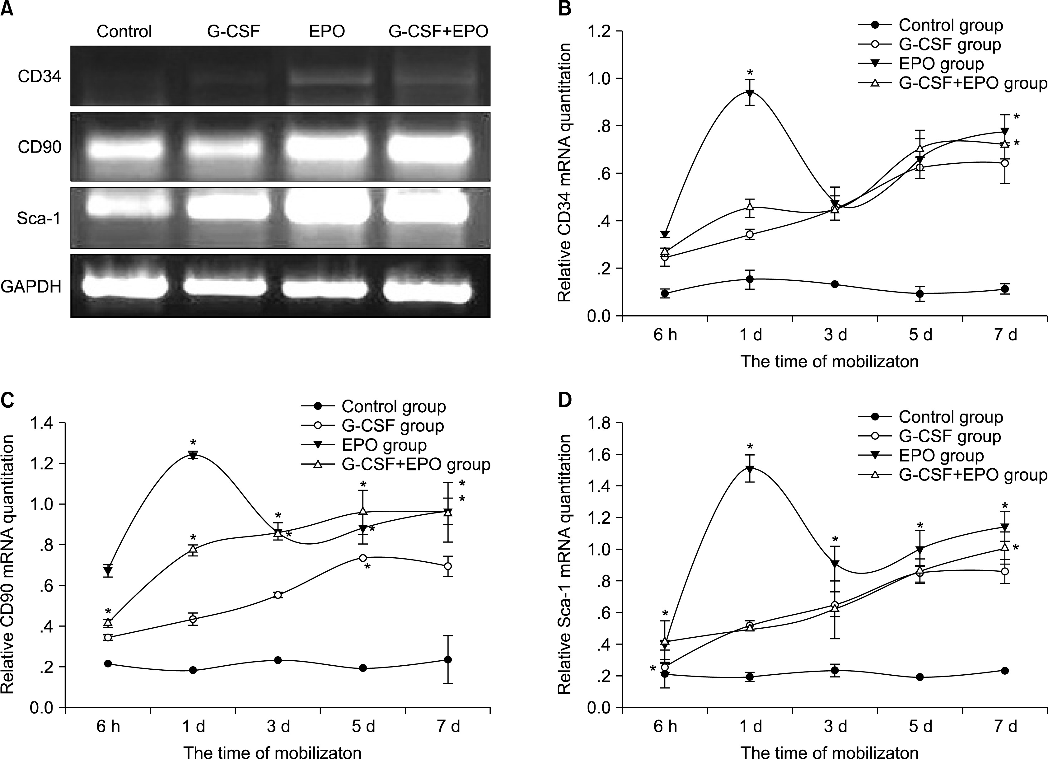
To analyze the graft composition in mobilized peripheral blood, the expression of stem cells surface makers: CD34, CD90 and Sca-1 were investigated by RT-PCR. As show in Fig. 2, mRNA expression of CD34, CD90 and Sca-1 at hour 6, day 1 of rhEPO stimulation was significantly higher than adiministration of rhG-CSF and rhG-CSF+rhEPO. At day 3, mRNA expression of CD34 was no significant difference among the three groups; mRNA expression of CD90 of rhEPO or rhG-CSF+rhEPO stimulation was more higher than adiministration of rhGCSF, but there was no significant difference between two groups; Sca-1 mRNA expression in rhEPO group was higher than rhG-CSF or rhG-CSF+rhEPO group (p= 0.003). At day 5 and day 7, the changes of three types mRNA levels showed consistency, rhEPO or rhG-CSF+rhEPO group was higher than rhG-CSF group, but there was no difference between rhEPO and rhG-CSF+rhEPO group (p=0.16).

Discussion

Recent data from the International Bone Marrow Transplant Registry (IBMTR) show PBSCs are used in more than 90% of autologous stem-cell transplantation and 69% of allogeneic transplantation, and G-CSF is the predominant mobilizer used clinically (14). Although the success of G-CSF, there are many areas for improvement that can have significant clinical impact: Meisel et al. (5) showed the survival of G-CSF-based mobilization in pediatric siblings donating for children is shorter than bone marrow transplantation, it mainly beacuse rare HSCs in mobilized peripheral blood; many other studies (68) reported PBSCs grafts contained 3∼4 times the dose of CD34+ cells/kg, 10∼50 times total number of T cells, which seriously impact the incidence of graft-versus-host disease, infectious complications and even survival post transplant. These studies raise the issue of what is the optimal graft content.
Adult bone marrow is the primary source of stem cells that regenerate hematopoiesis, and also harbors other stem and progenitor cells including endothelial progenitor cells (EPCs) and mesenchymal stem cells (MSCs). MSCs and EPCs can be used as accessory cells of hematopoietic stem and progenitors cells (HSPCs) in transplantation to promote hematopoietic reconstitution process, reduce complications of post-transplantation and speed up the repair of damaged organs such as heart, liver, kidney and vascular (9, 10). As we konw multiple agents induce PBSCs mobilization, each with different kinetics and efficiencies. Whether combination agents can be developed that differentially mobilize adult stem cells in order to obtain the optimal graft. In our study, we wanted to know whether rhEPO+rhG-CSF can efficiently mobilize bone marrow-derived progenitor cells into peripheral blood and which types of cells are mainly released from bone marrow.
Our data showed that the peripheral blood was collected on hours 6, days 1, 3, 5 and 7, the number of WBC and MNC in peripheral blood gradually increased in cytokines treated mice, and the peak have occurred during the 7 th day after the last adiministration. The high amount of leukocytes indicated a mobilizing effect of cytokines. However, the addition of rhEPO to the treatment protocol did not further increase the mobilizing effect of rhG-CSF as reported by Grassinger et al. (11). The counts of WBC and MNC in KM mice is consistently lower than the study reported (12). This could be explained by the characteristics of agents: electrical conductivity, pH, osmotic pressure, ionic strength, hemolytic agents and concentration used in automatic hematology analyzer are the same as human blood cells, while leukocytes of mice have unique characteristics which are different with human in size, internal structure and so on.
Kang et al. (13) showed G-CSF+EPO enhance the mobilization of EPCs, benefit to angiogenesis and improve the myocardial fuction, this protocol would be a promising option to overcome the significant but limited efficacy of cytokine-based cell therapy for patients with myocardial infarction. This suggest that the graft content has changed with the mobilizing of EPO+G-CSF. According to HSCs in mouse bone marrow have the phenotype of Sca-1+CD34+/CD45+Lin and c-kit+CD90low Lin-Sca-1high, and Sca-1, c-kit, CD34 and CD90 are commonly used (14). We found that mRNA expression of CD34, CD90 and Sca-1 increased higher with the stimulation of rhEPO than rhG-CSF alone. This could be explained by more HSCs mobilized by rhEPO.
Although the dosage of CD34+ cells is used in humans to indicate the mobilization of HSPCs, this population is heterogeneous, which containing primitive as well as more mature progenitors. In contrast to humans, CD34+ is a less useful marker in the mouse, and there is good evidence that murine HSCs can also be CD34 negative. For example, Linc-Kit+CD34+ is the major phenotype of HSCs in fetal and newborn mice, whereas CD34 HSPCs emerge in 7-week-old mice and gradually increase thereafter, and after 10 weeks there is mainly CD34 HSPCs, only 20% of HSPCs are CD34+ cells (15). Brunner, et al. (16) reported about 70% of “HSC” purified as of LinSca1+c-Kit+ express CD45 + CD34, only 17% express CD45+ CD34 + . All these datas show CD34 is the major of HSPCs in mice BM. Our study found the expression of CD34 is lower than CD90 and Sca-1, which may be explained CD34HSCs are more than CD34+cells. Using FACS analyses that CD90+ CD34 cells increased more significantly after the administration of rhEPO or rhEPO +rhG-CSF than rhG-CSF alone. According to the phenotypes of mice HSCs, our datas suggest more HSCs mobilized by rhEPO.
Currently, many techniques can be used to identify HSPCs, such as the capable of forming colonies, flow cytometric assays and in vivo transplantation. However, the phenotypes and biological characteristics of MSCs remain controversy. The International Society for Cellular Therapy (ISCT) in 2006 have provided a set of standards identifying MSCs: MSCs must express CD105, CD73 and CD90; must lack the expression of hematopoietic antigens like CD45, CD34 and markers for monocytes, macrophages and B cells (17). However, MSCs has a strong heterogeneity. For example, Yamamoto et al. (18) showed the phenotypes of MSCs from adipose tissue in mice as follows: (94.6±2.6)% CD90+, (89.4±4.5)% Sca-1+ and (4.6±2.4)% CD34+. Obviously, some phenotypes of MSCs and HSCs are consistent. In addition, whether MSCs can be mobilized or not has also aroused controversy. While Kassis et al. (19) successfully isolated MSCs by fibrin microspheres in G-CSF mobilized peripheral blood. Therefore, in our experiment, FACS analyses that CD90+CD34 cells increased more significantly after the administration of rhEPO or rhEPO+rhG-CSF than rhG-CSF alone, may also mobilize a certain number of MSCs.
The importance of this study is that exploring rhEPO or combined with rhG-CSF had a strong capacity of mobilization of CD90+CD34and Sca−1+stem cells, which may enhance the content of HSCs or MSCs or both in graft. As a result of this experiment only identified cells phenotypes and lack of detecting other biological characteristics such as the potential of self-renewal and self-differentiation, it is difficult to accurately definite the CD90+ CD34and Sca-1+ cells in the mobilized-peripheral blood, which is the next subject we study in the future.

ACKNOWLEDGMENTS

Authors thank the staffs of the Instiute of Heamtology and the students research groups of Yunyang Medical college for their participation. This work was supported by the Funds for Outstanding Youth in Hubei Province (QJX 2008-40, Hubei, China), the Funds for Natural Science in Hubei Province (200800156, Hubei, China) and Outstanding Youth of Scientific Innovation Projects in Yunyang Medical College (2008CXX02, Hubei, China).

Notes

Potential Conflict of Interest

The authors have no conflicting financial interest.

References

1. Levesque JP, Winkler IG. Mobilization of hematopoietic stem cells: state of the art. Curr Opin Organ Transplant. 2008. 13:53–58.
crossref
2. MacMillan ML, Davies SM, Nelson GO, Chitphakdithai P, Confer DL, King RJ, Kernan NA. Twenty years of unrelated donor bone marrow transplantation for pediatric acute leukemia facilitated by the National marrow donor program. Biol Blood Marrow Transplant. 2008. 14:16–22.
crossref
3. Pelus LM. Peripheral blood stem cell mobilization: new regimens, new cells, where do we stand. Curr Opin Hematol. 2008. 15:285–292.
crossref
4. Cashen AF, Lazarus HM, Devine SM. Mobilizing stem cells from normal donors: is it possible to improve upon G-CSF? Bone Marrow Transplant. 2007. 39:577–588.
crossref
5. Meisel R, Laws HJ, Balzer S, Bernbeck B, Kramm C, Schönberger S, Sinha K, Tröger A, Schmitz M, Fischer J, Göbel U, Enczmann J, Dilloo D. Comparable long-term survival after bone marrow versus peripheral blood progenitor cell transplantation from matched unrelated donors in children with hematologic malignancies. Biol Blood Marrow Transplant. 2007. 13:1338–1345.
crossref
6. Luo XH, Chang YJ, Xu LP, Liu DH, Liu KY, Huang XJ. The impact of graft composition on clinical outcomes in un-manipulated HLA-mismatched/haploidentical hematopoietic SCT. Bone Marrow Transplant. 2009. 43:29–36.
crossref
7. Dey BR, Shaffer J, Yee AJ, McAfee S, Caron M, Power K, Ting DT, Colby C, Preffer F, Ballen K, Attar E, Saidman S, Tarbell N, Sachs D, Sykes M, Spitzer TR. Comparison of outcomes after transplantation of peripheral blood stem cells versus bone marrow following an identical nonmyeloablative conditioning regimen. Bone Marrow Transplant. 2007. 40:19–27.
crossref
8. Vela-Ojeda J, Garcia-Ruiz Esparza MA, Reyes-Maldonado E, Jiménez-Zamudio L, Garcia-Latorre E, Moreno-Lafont M, Estrada-Garcia I, Montiel-Cervantes L, Tripp-Villanueva F, Ayala-Sánchez M, Garcia-León LD, Borbolla- Escoboza JR, Mayani H. Clinical relevance of NK, NKT, and dendritic cell dose in patients receiving G-CSF-mobilized peripheral blood allogeneic stem cell transplantation. Ann Hematol. 2006. 85:113–120.
crossref
9. Dome B, Dobos J, Tovari J, Paku S, Kovacs G, Ostoros G, Timar J. Circulating bone marrow-derived endothelial progenitor cells: characterization, mobilization, and therapeutic considerations in malignant disease. Cytometry A. 2008. 73:186–193.
crossref
10. Lévesque JP, Winkler IG, Larsen SR, Rasko JE. Mobilization of bone marrow-derived progenitors. Handb Exp Pharmacol. 2007. 3–36.
crossref
11. Grassinger J, Mueller G, Hart C, Nilsson SK, Haylock DN, Andreesen R, Hennemann B. Detection and quantification of functionally defined hematopoietic progenitor cells and tissue specific mRNA within the peripheral blood of myeloma patients after administration of granulocyte colony-stimulating factor and erythropoietin. Eur J Haematol. 2008. 80:20–30.
crossref
12. Liu B, Yang M, Li X, Qian X, Shen Z, Ding Y, Yu L. Enhanced efficiency of thermally targeted taxanes delivery in a human xenograft model of gastric cancer. J Pharm Sci. 2008. 97:3170–3181.
crossref
13. Kang HJ, Kim HS. G-CSF- and erythropoietin-based cell therapy: a promising strategy for angiomyogenesis in myocardial infarction. Expert Rev Cardiovasc Ther. 2008. 6:703–713.
crossref
14. Ratajczak MZ. Phenotypic and functional characterization of hematopoietic stem cells. Curr Opin Hematol. 2008. 15:293–300.
crossref
15. Matsuoka S, Ebihara Y, Xu M, Ishii T, Sugiyama D, Yoshino H, Ueda T, Manabe A, Tanaka R, Ikeda Y, Nakahata T, Tsuji K. CD34 expression on long-term re-populating hematopoietic stem cells changes during developmental stages. Blood. 2001. 97:419–425.
crossref
16. Brunner S, Zaruba MM, Huber B, David R, Vallaster M, Assmann G, Mueller-Hoecker J, Franz WM. Parathyroid hormone effectively induces mobilization of progenitor cells without depletion of bone marrow. Exp Hematol. 2008. 36:1157–1166.
crossref
17. Dominici M, Le Blanc K, Mueller I, Slaper-Cortenbach I, Marini F, Krause D, Deans R, Keating A, Prockop Dj, Horwitz E. Minimal criteria for defining multipotent mesenchymal stromal cells. The International Society for Cellular Therapy position statement. Cytotherapy. 2006. 8:315–317.
crossref
18. Yamamoto N, Akamatsu H, Hasegawa S, Yamada T, Nakata S, Ohkuma M, Miyachi E, Marunouchi T, Matsunaga K. Isolation of multipotent stem cells from mouse adipose tissue. J Dermatol Sci. 2007. 48:43–52.
crossref
19. Kassis I, Zangi L, Rivkin R, Levdansky L, Samuel S, Marx G, Gorodetsky R. Isolation of mesenchymal stem cells from G-CSF-mobilized human peripheral blood using fibrin microbeads. Bone Marrow Transplant. 2006. 37:967–976.
crossref

Fig. 1.
Expressions of CD90+ CD34-cells of peripheral blood nucleated cells in each group at different time after mobilization. *p<0.05, Compared with the control group, *p<0.05, Compared with the G-CSF group.
ijsc-02-129f1.tif
Fig. 2.
RT-PCR analyses of mobilized-peripheral blood for surface makers gene expression. (A) Examples for RT-PCR from four different mobilized groups and on day 7 after mobilization. (B) Qualitative PCR analysis the expression of CD34-mRNA (Mean±S.E.). (C) Qualitative PCR analysis the expression of C90-mRNA (Mean±S.E.). (D) Qualitative PCR analysis the expression of Sca-1-mRNA (Mean±S.E.). *p<0.05, Compared with the G-CSF group.
ijsc-02-129f2.tif
Table 1.
Primer pairs of GAPDH, CD34, CD90 and Sca-1
mRNA Primers
GAPDH Forward: 5′-GAGAATGGGAAGCTTGTCATC
Reverse: 5′-GTCCACCACCCTGTTGCTGTA
CD34 Forward: 5′-TACTGTGTGAAAAGGAGGAGGCTGAGGC
Reverse: 5′-ACGGTTCATCAGGAAATAACCAGTGGT
CD90 Forward: 5′-GTCCCGAGGGCAGAAGGT
Reverse: 5′-TCCAGGCGAAGGTTTTGG
Sca-1 Forward: 5′-GAGGATGGACACTTCT
Reverse: 5′-GGTCTGCAGGAGGACTGAGC
Table 2.
WBC counts at different time after mobilization (109/L) (Mean±S.E.)
Group Sixth hour Firth day Third day Fifth day Seventh day
control 1.98±0.91 2.00±0.87 1.82±0.51 1.76±0.26 1.88±0.56
G-CSF 2.50±1.09 3.99±1.32 3.50±0.99* 6.67±3.82* 9.17±3.22
EPO 3.33±1.44 6.67±1.75* 5.00±1.44* 5.17±1.98* 7.50±1.76*
G-CSF+EPO 2.83±1.11 3.50±1.44 5.33±1.25* 4.17±1.44 7.17±1.52*

G-CSF: granulocyte colony stimulating factor; EPO: erythropoietin,

* p<0.05,

p<0.001, compared with the control group.

p<0.05, there is difference between G-CSF group.

Table 3.
MNC counts at different time after mobilization (109/L) (Mean±S.E.)
Group Sixth hour Firth day Third day Fifth day Seventh day
control 0.67±0.46 1.04±0.50 1.13±0.47 1.07±0.19 0.99±0.32
G-CSF 1.24±0.71 2.82±1.23 2.28±0.76 4.18±1.49* 6.88±2.89
EPO 2.59±1.48* 4.84±1.82* 3.28±1.15* 3.56±1.62* 5.53±1.33*
EPO+G-CSF 1.93±0.72 2.59±1.25 3.63±1.29 2.88±1.38 5.22±1.94*

G-CSF: granulocyte colony stimulating factor; EPO : erythropoietin.

* p<0.05,

p<0.001, compared with the control group.

TOOLS
Similar articles