Journal List > Int J Stem Cells > v.13(1) > 1158822

Baharlooi, Azimi, Salehi, and Izad: Mesenchymal Stem Cell-Derived Exosomes: A Promising Therapeutic Ace Card to Address Autoimmune Diseases

Abstract

With the development of novel treatments for autoimmune disorders, it has become a popular research focus which mesenchymal stem cells (MSCs) have the capacity to counteract with autoimmune diseases progression. One of the underlying mechanisms behind their activities is the release of extracellular vesicles especially exosomes. MSC-derived exosomes are hypoimmunogenic nanocarriers which contain numerous immunoregulatory factors and similar to other exosomes, are able to pass through boundaries like the blood-brain barrier (BBB). Accumulating evidence provided by animal studies has demonstrated that MSC-derived exosomes, as a novel therapy, can re-induce self-tolerance, without subsequent complications reported for other treatments. Therefore, therapeutic applications of MSC-derived exosomes are contributing to core advances in the field of autoimmune diseases. Here, we briefly describe the biological characteristics of MSC-derived exosomes and review the experimentally verified outcomes for autoimmune disease therapy purposes.

Graphical Abstract.

IJSC-13-013-abs.tif
Mesenchymal exosomes have been newly used to address impaired immune responses in some animal models of autoimmune diseases. Clinical experiments demonstrated that these vesicles successfully recover immune balance of autoreactive cells. The current picture highlights the main beneficial outcomes achieved from in vivo evaluation of MSC-derived exosomes in the mouse models.

Introduction

Mesenchymal stem cells (MSCs) are multipotent, self-renewable, and spindle shape cells (1) which were firstly isolated from guinea-pig bone marrow and spleen by Friedenstein et al. in 1970 (2). Now, MSCs can be isolated from cord blood, umbilical cord, bone marrow, adipose tissue, and many other tissues (3, 4). The single connecting feature of all these tissues is the presence of perivascular space around the blood vessels where pericytes come off and differentiate into MSCs after vessels got inflamed or injured. According to their common origin, pericytes and MSCs both are negative for CD34, CD45, CD56 and positively express CD29, CD44, CD73, CD90, CD105, CD146 (5-7). MSCs are functionally defined based on their capacity to produce molecules that provide a regenerative and immunoregulatory milieu. Moreover, these multipotent cells can differentiate into a closely related family of cells in vitro or following transplantation and subsequently recover the damages. However, recent findings have reported that MSCs do not significantly differentiate into other lineages in vivo under normal condition (8). Consequently in a new paradigm, Arnold Caplan coined “Medical Signaling Cell” for MSCs to precisely express that mesenchymal stem cells dock at the sites of injury and produce regenerative and immunoregulatory components (9, 10). Afterwards, a great number of clinical trials have exploited MSC-based modalities in clinical conditions such as Type 1 diabetes mellitus, Rheumatoid Arthritis (RA), Multiple Sclerosis (MS), Crohn’s disease, Lupus, kidney transplant, Graft versus Host Disease (GvHD) and many cancers (clinicaltrials.gov).
Unlike other stem cells (embryonic stem cells as well as induced pluripotent stem cells), MSCs have a dynamic immunomodulatory profile which lies on their ability to sense the injury or inflammation and then switch to the required response (11). For instance, MSCs produce transforming growth factor beta (TGF-β), indoleamine 2,3-dioxygenase (IDO), nitric oxide (NO), soluble HLA-G, and prostaglandin E2 (PGE2) in response to high concentration of interferon gamma (IFNγ), tumor necrosis factor alpha (TNF-α), and toll like receptor 3 (TLR3) agonists (11-13). This reaction activates CD4CD25hiFoxP3 regulatory T cells (Tregs) while inhibits natural killer cell and T cell responses (11-13). In contrast, low level of inflammatory agents and TLR4 agonists, decrease IDO, NO, PGE2 production and increase expression of pro-inflammatory cytokines (CXCL9, CXCL10, MIP-1α, MIP-1β, CCL5), indicating their double-edged sword function within the immune system (14). Accumulating investigations have demonstrated that these factors are mainly delivered through direct secretions of soluble molecules and release of extracellular vesicles particularly exosomes.

Exosomes

As shown in Table 1 (15-20), exosomes are one of the nanoscale cell-derived extracellular vesicles (EV) with 30∼100 nm diameter (15). Exosomes were first thought to be required for discarding unnecessary proteins out of the cells and termed as “garbage cans” (21). Later, they were introduced as gulps of cytosol delivering bioactive molecules especially proteins and small RNAs (like microRNAs) in intercellular communications. Recent investigation have shown that exosomes transfer bioactive molecules among cells and therefore is able to play an essential role in pathological conditions including Alzheimer’s disease (22), transmissible spongiform encephalopathies (23), Parkinson’s disease (24), and amyotrophic lateral sclerosis (25) or could be employed as nanoscale platforms for drug delivery.
Constitutive shedding of exosomes is mostly reported for immune cells (i.e. MSCs, lymphocytes, mast cells and dendritic cells) and non-immune cells particularly tumor cells (26, 27). Additionally, interaction between T lymphocytes and antigen presenting cells strongly induce the release of the exosomes bearing TCR/CD3/zeta complex (28) and MHC II-peptide (29), respectively. It was lately demonstrated that in hypoxic preconditioning and presence of lipopolysaccharide, MSCs significantly enhance the release of exosomes which shuttle anti-inflammatory agents to polarize macrophages into the M2 phenotype (30, 31).
Generally, extracellular vesicles affect the neighboring cells via two different strategies: 1) fusion dependent 2) fusion independent. The first mechanism takes place when vesicle is taken up by a recipient cell. There are two types of fusions: 1) direct fusion of the extracellular vesicle with the plasma membrane of recipient cell that requires receptor-ligand interactions 2) Back fusion which confers to the endocytosis of vesicle by the recipient cell and subsequently incorporation of vesicle with endosome’s membrane (32, 33). In fusion independent manner, the vesicle and recipient cells interact with each other, like when exosomes bearing MHC-peptide are being specifically attached to the T cell receptor (TCR) on T cells (34).

MSC-Derived Exosome Therapy: Cons and Pros

As shown in Table 2 (35-46), recent findings depicted that MSC-derived exosomes deliver various cytoplasmatic constituents of the MSC secretomes, relevant to the stemness, angiogenesis, and particularly inflammatory factors. They were first used in 2010 for regeneration of tissues in a mouse model of myocardial ischemia/reperfusion injury (47) and are extensively on investigations at present. As a cell-free therapeutic tools provided a potential opportunity to unlock existing boundaries of cell therapy for autoimmune diseases. Exosomes introduce several advantages including capacity to cross the barriers (blood-brain barrier, capillaries) (48) and small enough to evade from being cleared by reticuloendothelial system (RES) (49). In addition to their general characteristics, MSC-derived exosomes are normally hypo-immunogenic due to the lack of MHC-II and low expression of MHC-I similar to their parental cells (50). It has also been revealed that MSCs can produce higher amount of exosomes compared with human cell lines like human embryonic kidney (HEK) and human acute monocytic leukemia (THP-1); Therefore, MSCs appear to be one of the best sources for high amount of immunoregulatory exosomes (51, 52).
Despite therapeutic advantages of MSC-derived exosomes, quantification of their protein profile and more importantly durability of cryopreserved exosomes as well as their in vivo tracking are still effortful. Regarding frozen exosomes, Cosenza et al. (53) analyzed functions of cryopreserved MSC-derived exosomes compared to freshly isolated exosomes and demonstrated that storage destabilize the integrity of membrane, leading to leakage of its content, aggregation of vesicles, and decrease in their immunosuppressive capacity. MSC-derived exosomes obviously express both the common MSC markers, such as CD44, CD90, and CD73 and markers of exosomes, CD9, CD63, and CD81 (51). These markers are used to track exosomes especially for drug delivery. Alternatively, cell tracing dyes can also help to chase exosomes in vivo. For instance, intravenous (i.v.) administration of Vybrant Cell Tracer DiD- or DiI-labeled exosomes to the mouse model of acute kidney injury, were used to monitor accumulation of MSC-derived exosomes. In this investigation, exosomes were mostly found in kidney, spleen, liver, and lung as filtering organs of the body (54). Apart from utilization of fluorescent molecules to chase organ localization of EVs, lower sensitivity of fluorophores negatively affect pharmacokinetic analysis of their applications (especially for quantification of exosome accumulated in a target tissue) (55). Radiolabeling with iodine-125 and bioluminescence emitted from luciferase provide a more practical solution for whole-body imaging of exosome-treated animals with significantly higher sensitivity of detection (56, 57). Also, stability of B16-BL6 melanoma cell-derived exosomes in blood circulation were evaluated using exosomes expressing a fusion protein of Gaussia luciferase-Lactadherin, a reporter protein attached to one of the exosome’s trophic protein, respectively. The results indicated exosomal half-life of about 2 minutes following i.v. injection. This finding suggests short half-life of exosomes is because of their fast uptake by liver and spleen resident macrophages (56).

MSC-Derived Exosome Therapy for Autoimmune Diseases

Utilization of the MSC-derived exosomes for autoimmune diseases therapy is at early stages. Most of studies are focused on immunomodulatory effects of these small vesicles in animal models. Here we will discuss the roles of these exosomes in the treatment of autoimmune diseases.

Multiple Sclerosis

Multiple Sclerosis (MS) is a chronic autoimmune disease characterized by neuroinflammation and demyelination in the central nervous system (CNS) (58). Numerous studies have revealed activation and recruitment of autoaggressive CD4 and CD8 T cells into the CNS as the main pathogenic players of MS (59, 60). Disease-modifying treatments (DMTs) especially IFN-β are commonly prescribed to treat the affected individuals (61, 62). Despite their beneficial impacts, DMTs increase the risk of adverse effects including lymphopenia, lymphadenopathy, dyspnea and lipoatrophy (63-65). Hence, identification and evaluation of alternative approaches with lower side effects are necessary.
We have already demonstrated that MSCs and their activity have been altered after DMT consumption in relapsing-remitting MS patients (66). Indeed, patients own stem cells may have more deficiencies which not efficiently prevent for MS initiation and progression (67). However, MSCs can differentiate into neural cells and release neurotrophic molecules, indicating their strong neuroprotective effects for treating MS (68). Accumulating evidence has shown that MSCs can regulate autoreactive cells in a paracrine manner through secretion of inhibitory cytokines or molecules as well (69-71). MSC-derived exosomes can also induce peripheral tolerance toward autoreactive T cells via transferring the tolerogenic molecules to the autoreactive immune cells.
Since exosomes are lipid-bound nano-vesicles, they can cross the blood-brain barrier. Therefore, incubation of exosomes in presence of neurotrophic and remyelination factors elevate their therapeutic profits. Additionally, expression of receptor(s) with ability to recognize neural markers in the MSCs increase efficiency of the delivery system. In this case, Zhuang et al. (72) have administered exosomes bearing anti-inflammatory compounds such as curcumin to the EAE mouse and demonstrated that clinical symptoms of the diseases were improved.
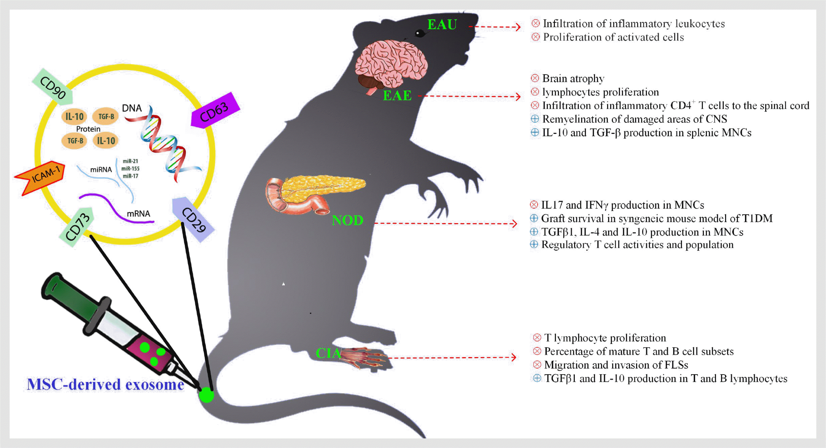
In 2012, Mokarizadeh et al. (73) showed that MSC-derived extracellular vesicles exert immunomodulatory effects on splenic mononuclear cells (MNCs) of EAE mouse. These small vesicles not only reduced autoreactive lymphocytes proliferation, but also potentially induced MNCs to secrete significant amount of IL-10. They also demonstrated that MSC-derived vesicles contain PD-L1, galectin-1, and TGF-β, which help them with their immunoregulatory functions. This investigation proposed that MSC-derived vesicles including exosomes are capable of transferring anti-inflammatory molecules into autoreactive cells and re-inducing a self-tolerance. Later in 2018, Laso-García et al. (74) revealed that i.v. administration of MSC-derived exosomes to a progressive model of MS, Theiler’s murine encephalomyelitis virus (TMEV)-induced demyelinating disease, recovered the disabilities observed before treatment. They indicated that administrated MVs prompt remyelination, reduce the brain atrophy, and infiltration of inflammatory CD4 T cells to the spinal cord of TMEV-infected mice.
Recently, Li et al. (75) demonstrated that MSC-derived exosomes polarize microglia cells mainly into the M2 phenotype and consequently alleviate clinical scores of EAE. In this study, the balance of M1/M2 in EAE rates significantly deviated toward M2 phenotype and their cytokines profile (IL-10 and TGF-β), while frequency and activity of M1 cells were decreased. Interestingly for the first time, Hosseini Shamili et al. (76) bioconjugated the MSC-derived exosomes to an aptamer which targets oligodendrocyte markers. The armed exosome improved proliferation of oligodendroglia cell line (OLN93) in vitro and reduced both inflammatory responses and demyelinated lesions in the CNS of EAE mice. These methodologies may provide a new perspective toward indirect applications of MSCs in the treatment of MS.

Rheumatoid Arthritis

Rheumatoid arthritis (RA) is an inflammatory disease with unknown etiology that causes articular destruction and subsequently functional loss in joints. RA immunopathogenesis is characterized by disordered innate and adaptive immunity that result in immune complex-mediated complement activation, autoreactive lymphocyte response against self-antigens, and also dysregulated cytokine networks (77, 78).
As with MS, there are various biological disease-modifying anti-rheumatic drugs (bDMARDs) available to control the symptoms of RA. For example, blockade of proinflammatory cytokines including GM-CSF, TNF-α, and IL-6 with antibodies or soluble decoy receptors have been mainly utilized for patients suffering from RA for many years (79). Along with beneficial effects of immunosuppressive and immunomodulatory drugs, they present various side effects including nausea, liver problems, lymphopenia, high risk of infection, and etc. (79). There is indeed a crucial need for new treatment modules with fewer side effects. MSC-derived exosomes have been considered to exert immunosuppressive activities, supporting their application in rheumatic diseases. Although the use of exosomes derived from various origins have been studied extensively to date (80, 81), to date, the role of MSC-derived exosomes is still in the beginning.
In 2018, Cosenza et al. (53) showed that MSC-derived exosomes reveal anti-inflammatory impacts on T and B lymphocytes in collagen-induced arthritis (CIA) mice model. In this study, MSCs-derived exosomes effectively inhibit T lymphocyte proliferation in a dose-dependent manner and decreased the percentage of mature T and B cell subsets. Interestingly, exosomes increased regulatory T cell populations much better than MSCs and MSC-derived microvesicles. Further investigations confirmed that microvesicles derived from MSCs are less effective to induce TGF-β and IL-10 production in T and B lymphocytes compared to MSCs alone (82). In 2018, Chen et al. (83) demonstrated that modified MSC-derived exosomes which contain miRNA-150, target the matrix metalloproteinase 14 (MMP14) and thereafter, decrease migration and invasion of fibroblast-like synoviocytes (FLS). They also indicated that injection of these engineered exosomes to the CIA mouse can effectively reduce the hind paw thickness and the clinical arthritic scores of animals. Another study evaluated beneficial impacts of human umbilical cord mesenchymal stem cells-derived exosomes (hUCMSC-derived exosomes) on bone destruction in CIA rats. Interestingly, the treatment reduced recruitment of inflammatory cell to the joints and prevent from joint synovial hyperplasia. The authors proposed that the underlying mechanism could be due to regulating of the imbalance of RANKL/OPG. To be clear, they showed that RANKL was decreased in serum and synovial tissues of the CIA rats, while the treatment raised the OPG concentration (84). In consensus with this study, another group demonstrated that intra-articular injection of hUCMSC-derived exosomes significantly decreased autoreactive infiltering chemokines CCL2 and CXCL12 in serum and synovial fluid (85).

Type 1 Diabetes Mellitus (Insulin-Dependent Diabetes Mellitus)

Type 1 diabetes mellitus (T1DM) is an incurable and chronic autoimmune disease which is classically identified by reduced level of insulin due to autoimmune destruction of insulin-producing pancreatic β cell, leading to hyperglycemia (86). Although underlying mechanism of disease is unknown, disruption of immune tolerance in autoreactive B cells, CD4 T cells and particularly CD8 cytotoxic T lymphocytes (CTL) are believed to contribute to the pathogenesis of type 1 diabetes. As a result, diabetic patients need exogenous insulin regiment to control their blood glucose level and avoid from consequences caused by hyperglycemia like fatigue, cataract, and also diabetic coma. Repetitive injection of insulin point to the fact that insulin titer is not stable in affected subjects and patients require a durable cure for diabetes.
In order to develop novel therapies for T1DM, researchers have taken different approaches so far. Since first whole pancreas transplantation in 1966, long-term normoglycemia has been achieved with a graft survival rate of 50 to 70 % after 5 years (87, 88). But sequential complications including pancreatitis, thrombosis, and pseudocyst appeared in graft are major disadvantages of this surgery (89). Eventually, transplantation of endocrine part of pancreas (known as islet transplantation) is a well-developed procedure to minimize complications of insulin therapy and pancreas transplantation (90). last two methods prevent from the disease progression for a limited period of time, but can not correctify autoimmune response against the β cells. The cure must be in the region where autoimmune response is originated.
Various strategies have been applied to recover immune tolerance at critical points of regulatory pathways. Of the different approaches, anti-CD3 antibodies nonspecifically induce T cell anergy by co-stimulation blockade. Thereafter, beta cells are preserved in disease established non-obese diabetic (NOD) mice (91). Traditional αCD3 antibodies like OKT3 were provided from mouse and could not be administered continuously for chronic diseases. This drawback is overwhelmed by humanizing the antibody sequence (92). Moreover, mitogenic effect of CD3 specific antibodies on T cells, owning to their cross-linking ability to bind to the Fc receptors on natural killer cells and monocytes, brings about a notable cytokine release (93). This problem can be solved via engineering of C-terminal sequence of antibody to stop its binding possibility to Fcγ receptors.
MSC therapy has also shown antidiabetic effects to restore immune response balance in pancreas. Ezquer et al. (94) injected GFP expressing MSCs to diabetic mouse and reported normalized Th1/Th2 response in diabetic group compared to control. They also traced labeled MSCs after 7 and 65 days to be sure if MSCs differentiate into beta cells and lead to normoglycemia. Surprisingly, they found MSCs in heart and secondary lymphoid organs (like Payer’s patch, inguinal lymph node, and pancreatic lymph nodes) not in pancreas. It suggests that the antidiabetic effect of MSC is due to its endocrine activity not differentiation capacity. In 2016, Nakano et al. (95) have used MSC-derived exosomes to diminish cognitive impairments of diabetic rats by repairing damaged astrocytes and more importantly neurons. Furthermore, administration of MSC-derived exosomes simultaneously with islet transplantation, suppressed proliferation of PBMCs, improved regulatory T cell activities and increased graft survival in syngeneic mouse model of T1DM (96). Similar to previous studies, a group of scientists also evaluated immunomodulatory effects of MSC-derived exosomes compared with control and reported significant increase of regulatory T cell population among splenic MNCs, accompanied by upregulation of IL-4, IL-10, TGF-β plus downregulation of IL-17 and IFNγ cytokines after treatment. Accordingly treatment stabilized the blood glucose and vanished the signs of diabetes (97).

Uveitis

Uveitis is an autoimmune disorder which causes severe visual disability (98). Current clinical treatment includes corticosteroids, other immunosuppressive drugs, and newly developed biologics. The long-term utilization of each is followed by serious systemic side effects and the local risk of cataracts and glaucoma (99, 100). Hence, most investigation intended to find localized and specific therapies. Although, it has been recently revealed that i.v. injection of MSCs improved experimental autoimmune uveitis (EAU) in animal model of uveitis (101-103), the underlying mechanisms of MSCs is not clear yet. In 2017, Bai et al. (104) showed that, human umbilical cord MSC-derived exosomes can reduce the severity of EAU. They demonstrated that MSC-derived exosomes inhibited the infiltration of inflammatory leukocytes to the eyes and these outcomes could, at least partly, be due to expression of CD73 on the MSCs. As expected, exosomes bearing CD73 interact with CD39 on activated immune cells and induce production of adenosine which result in proliferation inhibition. This study is the only study on the role of exosomes uveitis, indicating the importance of further investigation in this field.

Conclusions

Until last decade, the contribution of MSC-derived exosomes in the control of inflammatory responses has been less clear. As a functional vesicle derived from MSCs, exosomes have shown same beneficial impacts of the cell of origin, to suppress several autoreactive cells in autoimmune diseases. Moreover, although mesenchymal-derived exosomes expose immunomodulatory functions by self, it seems that engineering these vesicles through adding anti-inflammatory molecules and certain receptors, they can specifically target tissue/organ of interest. This attitude might consider as a potentially novel candidate to treat other autoimmune disorders like spondylitis ankylosing and scleroderma as well.
With respect to remaining challenges, more investigations are required to elucidate unclear aspects of cell-free therapy using MSC-derived exosomes in ongoing studies.

Notes

Potential Conflict of Interest

The authors have no conflicting financial interest.

References

1. Crisan M, Corselli M, Chen CW, Péault B. 2011; Multilineage stem cells in the adult: a perivascular legacy? Organogenesis. 7:101–104. DOI: 10.4161/org.7.2.16150. PMID: 21593599. PMCID: PMC3142446.
2. Friedenstein AJ, Chailakhjan RK, Lalykina KS. 1970; The development of fibroblast colonies in monolayer cultures of guinea-pig bone marrow and spleen cells. Cell Tissue Kinet. 3:393–403. DOI: 10.1111/j.1365-2184.1970.tb00347.x. PMID: 5523063.
crossref
3. Murray IR, West CC, Hardy WR, James AW, Park TS, Nguyen A, Tawonsawatruk T, Lazzari L, Soo C, Péault B. 2014; Natural history of mesenchymal stem cells, from vessel walls to culture vessels. Cell Mol Life Sci. 71:1353–1374. DOI: 10.1007/s00018-013-1462-6. PMID: 24158496.
crossref
4. Hassan G, Kasem I, Soukkarieh C, Aljamali M. 2017; A Simple method to isolate and expand human umbilical cord derived mesenchymal stem cells: using explant method and umbilical cord blood serum. Int J Stem Cells. 10:184–192. DOI: 10.15283/ijsc17028. PMID: 28844128. PMCID: PMC5741200.
crossref
5. Caplan AI. 2008; All MSCs are pericytes? Cell Stem Cell. 3:229–230. DOI: 10.1016/j.stem.2008.08.008. PMID: 18786406.
crossref
6. Pham H, Tonai R, Wu M, Birtolo C, Chen M. 2018; CD73, CD90, CD105 and Cadherin-11 RT-PCR screening for mesenchymal stem cells from cryopreserved human cord tissue. Int J Stem Cells. 11:26–38. DOI: 10.15283/ijsc17015. PMID: 29843192. PMCID: PMC5984056.
crossref
7. Geevarghese A, Herman IM. 2014; Pericyte-endothelial crosstalk: implications and opportunities for advanced cellular therapies. Transl Res. 163:296–306. DOI: 10.1016/j.trsl.2014.01.011. PMID: 24530608. PMCID: PMC3976718.
crossref
8. Guimarães-Camboa N, Cattaneo P, Sun Y, Moore-Morris T, Gu Y, Dalton ND, Rockenstein E, Masliah E, Peterson KL, Stallcup WB, Chen J, Evans SM. 2017; Pericytes of multiple organs do not behave as mesenchymal stem cells in vivo. Cell Stem Cell. 20:345–359.e5. DOI: 10.1016/j.stem.2016.12.006. PMID: 28111199. PMCID: PMC5337131.
crossref
9. Caplan AI. 2010; What's in a name? Tissue Eng Part A. 16:2415–2417. DOI: 10.1089/ten.tea.2010.0216. PMID: 20412005.
crossref
10. Caplan AI. 2017; Mesenchymal stem cells: time to change the name! Stem Cells Transl Med. 6:1445–1451. DOI: 10.1002/sctm.17-0051. PMID: 28452204. PMCID: PMC5689741.
crossref
11. Bernardo ME, Fibbe WE. 2013; Mesenchymal stromal cells: sensors and switchers of inflammation. Cell Stem Cell. 13:392–402. DOI: 10.1016/j.stem.2013.09.006. PMID: 24094322.
crossref
12. Di Nicola M, Carlo-Stella C, Magni M, Milanesi M, Longoni PD, Matteucci P, Grisanti S, Gianni AM. 2002; Human bone marrow stromal cells suppress T-lymphocyte proliferation induced by cellular or nonspecific mitogenic stimuli. Blood. 99:3838–3843. DOI: 10.1182/blood.V99.10.3838. PMID: 11986244.
crossref
13. Bruno S, Deregibus MC, Camussi G. 2015; The secretome of mesenchymal stromal cells: role of extracellular vesicles in immunomodulation. Immunol Lett. 168:154–158. DOI: 10.1016/j.imlet.2015.06.007. PMID: 26086886.
crossref
14. Kyurkchiev D, Bochev I, Ivanova-Todorova E, Mourdjeva M, Oreshkova T, Belemezova K, Kyurkchiev S. 2014; Secretion of immunoregulatory cytokines by mesenchymal stem cells. World J Stem Cells. 6:552–570. DOI: 10.4252/wjsc.v6.i5.552. PMID: 25426252. PMCID: PMC4178255.
crossref
15. Edgar JR, Eden ER, Futter CE. 2014; Hrs- and CD63-dependent competing mechanisms make different sized endosomal intraluminal vesicles. Traffic. 15:197–211. DOI: 10.1111/tra.12139. PMID: 24279430. PMCID: PMC4253088.
crossref
16. Bobrie A, Colombo M, Krumeich S, Raposo G, Théry C. 2012; Diverse subpopulations of vesicles secreted by different intracellular mechanisms are present in exosome preparations obtained by differential ultracentrifugation. J Extracell Vesicles. 1:18397. DOI: 10.3402/jev.v1i0.18397. PMID: 24009879. PMCID: PMC3760636.
crossref
17. Théry C, Boussac M, Véron P, Ricciardi-Castagnoli P, Raposo G, Garin J, Amigorena S. 2001; Proteomic analysis of dendritic cell-derived exosomes: a secreted subcellular compartment distinct from apoptotic vesicles. J Immunol. 166:7309–7318. DOI: 10.4049/jimmunol.166.12.7309. PMID: 11390481.
crossref
18. Cocucci E, Meldolesi J. 2015; Ectosomes and exosomes: shedding the confusion between extracellular vesicles. Trends Cell Biol. 25:364–372. DOI: 10.1016/j.tcb.2015.01.004. PMID: 25683921.
crossref
19. Akers JC, Gonda D, Kim R, Carter BS, Chen CC. 2013; Biogenesis of extracellular vesicles (EV): exosomes, microvesicles, retrovirus-like vesicles, and apoptotic bodies. J Neurooncol. 113:1–11. DOI: 10.1007/s11060-013-1084-8. PMID: 23456661. PMCID: PMC5533094.
crossref
20. Hess C, Sadallah S, Hefti A, Landmann R, Schifferli JA. 1999; Ectosomes released by human neutrophils are specialized functional units. J Immunol. 163:4564–4573.
crossref
21. Johnstone RM. 1992; The Jeanne Manery-Fisher memorial lecture 1991. Maturation of reticulocytes: formation of exosomes as a mechanism for shedding membrane proteins. Biochem Cell Biol. 70:179–190. DOI: 10.1139/o92-028. PMID: 1515120.
crossref
22. Rajendran L, Honsho M, Zahn TR, Keller P, Geiger KD, Verkade P, Simons K. 2006; Alzheimer's disease beta-amyloid peptides are released in association with exosomes. Proc Natl Acad Sci U S A. 103:11172–11177. DOI: 10.1073/pnas.0603838103. PMID: 16837572. PMCID: PMC1544060.
crossref
23. Fevrier B, Vilette D, Archer F, Loew D, Faigle W, Vidal M, Laude H, Raposo G. 2004; Cells release prions in association with exosomes. Proc Natl Acad Sci U S A. 101:9683–9688. DOI: 10.1073/pnas.0308413101. PMID: 15210972. PMCID: PMC470735.
crossref
24. Emmanouilidou E, Melachroinou K, Roumeliotis T, Garbis SD, Ntzouni M, Margaritis LH, Stefanis L, Vekrellis K. 2010; Cell-produced alpha-synuclein is secreted in a calcium-dependent manner by exosomes and impacts neuronal survival. J Neurosci. 30:6838–6851. DOI: 10.1523/JNEUROSCI.5699-09.2010. PMID: 20484626. PMCID: PMC3842464.
crossref
25. Gomes C, Keller S, Altevogt P, Costa J. 2007; Evidence for secretion of Cu,Zn superoxide dismutase via exosomes from a cell model of amyotrophic lateral sclerosis. Neurosci Lett. 428:43–46. DOI: 10.1016/j.neulet.2007.09.024. PMID: 17942226.
crossref
26. Tamai K, Tanaka N, Nakano T, Kakazu E, Kondo Y, Inoue J, Shiina M, Fukushima K, Hoshino T, Sano K, Ueno Y, Shimosegawa T, Sugamura K. 2010; Exosome secretion of dendritic cells is regulated by Hrs, an ESCRT-0 protein. Biochem Biophys Res Commun. 399:384–390. DOI: 10.1016/j.bbrc.2010.07.083. PMID: 20673754.
crossref
27. Haney MJ, Klyachko NL, Zhao Y, Gupta R, Plotnikova EG, He Z, Patel T, Piroyan A, Sokolsky M, Kabanov AV, Batrakova EV. 2015; Exosomes as drug delivery vehicles for Parkinson's disease therapy. J Control Release. 207:18–30. DOI: 10.1016/j.jconrel.2015.03.033. PMID: 25836593. PMCID: PMC4430381.
crossref
28. Blanchard N, Lankar D, Faure F, Regnault A, Dumont C, Raposo G, Hivroz C. 2002; TCR activation of human T cells induces the production of exosomes bearing the TCR/CD3/zeta complex. J Immunol. 168:3235–3241. DOI: 10.4049/jimmunol.168.7.3235. PMID: 11907077.
crossref
29. Buschow SI, Nolte-'t Hoen EN, van Niel G, Pols MS, ten Broeke T, Lauwen M, Ossendorp F, Melief CJ, Raposo G, Wubbolts R, Wauben MH, Stoorvogel W. 2009; MHC II in dendritic cells is targeted to lysosomes or T cell-induced exosomes via distinct multivesicular body pathways. Traffic. 10:1528–1542. DOI: 10.1111/j.1600-0854.2009.00963.x. PMID: 19682328.
crossref
30. Lo Sicco C, Reverberi D, Balbi C, Ulivi V, Principi E, Pascucci L, Becherini P, Bosco MC, Varesio L, Franzin C, Pozzobon M, Cancedda R, Tasso R. 2017; Mesenchymal stem cell-derived extracellular vesicles as mediators of anti-inflammatory effects: endorsement of macrophage polarization. Stem Cells Transl Med. 6:1018–1028. DOI: 10.1002/sctm.16-0363. PMID: 28186708. PMCID: PMC5442783.
crossref
31. Ti D, Hao H, Tong C, Liu J, Dong L, Zheng J, Zhao Y, Liu H, Fu X, Han W. 2015; LPS-preconditioned mesenchymal stromal cells modify macrophage polarization for resolution of chronic inflammation via exosome-shuttled let-7b. J Transl Med. 13:308. DOI: 10.1186/s12967-015-0642-6. PMID: 26386558. PMCID: PMC4575470.
crossref
32. Bissig C, Gruenberg J. 2014; ALIX and the multivesicular endosome: ALIX in wonderland. Trends Cell Biol. 24:19–25. DOI: 10.1016/j.tcb.2013.10.009. PMID: 24287454.
crossref
33. Abrami L, Lindsay M, Parton RG, Leppla SH. , van der Goot FG. 2004; Membrane insertion of anthrax protective antigen and cytoplasmic delivery of lethal factor occur at different stages of the endocytic pathway. J Cell Biol. 166:645–651. DOI: 10.1083/jcb.200312072. PMID: 15337774. PMCID: PMC2172425.
crossref
34. Denzer K, van Eijk M, Kleijmeer MJ, Jakobson E, de Groot C, Geuze HJ. 2000; Follicular dendritic cells carry MHC class II-expressing microvesicles at their surface. J Immunol. 165:1259–1265. DOI: 10.4049/jimmunol.165.3.1259. PMID: 10903724.
crossref
35. Krasnodembskaya A, Song Y, Fang X, Gupta N, Serikov V, Lee JW, Matthay MA. 2010; Antibacterial effect of human mesenchymal stem cells is mediated in part from secretion of the antimicrobial peptide LL-37. Stem Cells. 28:2229–2238. DOI: 10.1002/stem.544. PMID: 20945332. PMCID: PMC3293245.
crossref
36. Baglio SR, Rooijers K, Koppers-Lalic D, Verweij FJ, Pérez Lanzón M, Zini N, Naaijkens B, Perut F, Niessen HW, Baldini N, Pegtel DM. 2015; Human bone marrow- and adipose-mesenchymal stem cells secrete exosomes enriched in distinctive miRNA and tRNA species. Stem Cell Res Ther. 6:127. DOI: 10.1186/s13287-015-0116-z. PMID: 26129847. PMCID: PMC4529699.
crossref
37. Spaggiari GM, Capobianco A, Becchetti S, Mingari MC, Moretta L. 2006; Mesenchymal stem cell-natural killer cell interactions: evidence that activated NK cells are capable of killing MSCs, whereas MSCs can inhibit IL-2-induced NK-cell proliferation. Blood. 107:1484–1490. DOI: 10.1182/blood-2005-07-2775. PMID: 16239427.
crossref
38. Di Trapani M, Bassi G, Midolo M, Gatti A, Kamga PT, Cassaro A, Carusone R, Adamo A, Krampera M. 2016; Differential and transferable modulatory effects of mesenchymal stromal cell-derived extracellular vesicles on T, B and NK cell functions. Sci Rep. 6:24120. DOI: 10.1038/srep24120. PMID: 27071676. PMCID: PMC4829861.
crossref
39. Eirin A, Zhu XY, Puranik AS, Tang H, McGurren KA, van Wijnen AJ, Lerman A, Lerman LO. 2017; Mesenchymal stem cell-derived extracellular vesicles attenuate kidney inflammation. Kidney Int. 92:114–124. DOI: 10.1016/j.kint.2016.12.023. PMID: 28242034. PMCID: PMC5483390.
crossref
40. Tan CY, Lai RC, Wong W, Dan YY, Lim SK, Ho HK. 2014; Mesenchymal stem cell-derived exosomes promote hepatic regeneration in drug-induced liver injury models. Stem Cell Res Ther. 5:76. DOI: 10.1186/scrt465. PMID: 24915963. PMCID: PMC4229780.
crossref
41. Okunishi K, Dohi M, Nakagome K, Tanaka R, Mizuno S, Matsumoto K, Miyazaki J, Nakamura T, Yamamoto K. 2005; A novel role of hepatocyte growth factor as an immune regulator through suppressing dendritic cell function. J Immunol. 175:4745–4753. DOI: 10.4049/jimmunol.175.7.4745. PMID: 16177122.
crossref
42. Garo LP, Murugaiyan G. 2016; Contribution of MicroRNAs to autoimmune diseases. Cell Mol Life Sci. 73:2041–2051. DOI: 10.1007/s00018-016-2167-4. PMID: 26943802.
crossref
43. Harding C, Heuser J, Stahl P. 1983; Receptor-mediated endocytosis of transferrin and recycling of the transferrin receptor in rat reticulocytes. J Cell Biol. 97:329–339. DOI: 10.1083/jcb.97.2.329. PMID: 6309857. PMCID: PMC2112509.
crossref
44. Bigagli E, De Filippo C, Castagnini C, Toti S, Acquadro F, Giudici F, Fazi M, Dolara P, Messerini L, Tonelli F, Luceri C. 2016; DNA copy number alterations, gene expression changes and disease-free survival in patients with colorectal cancer: a 10 year follow-up. Cell Oncol. 39:545–558. DOI: 10.1007/s13402-016-0299-z. PMID: 27709558.
crossref
45. Liu Y, Lou G, Li A, Zhang T, Qi J, Ye D, Zheng M, Chen Z. 2018; AMSC-derived exosomes alleviate lipopolysaccharide/d-galactosamine-induced acute liver failure by miR-17-mediated reduction of TXNIP/NLRP3 inflammasome activation in macrophages. EBioMedicine. 36:140–150. DOI: 10.1016/j.ebiom.2018.08.054. PMID: 30197023. PMCID: PMC6197728.
crossref
46. Ti D, Hao H, Fu X, Han W. 2016; Mesenchymal stem cells-derived exosomal microRNAs contribute to wound inflammation. Sci China Life Sci. 59:1305–1312. DOI: 10.1007/s11427-016-0240-4. PMID: 27864711.
crossref
47. Lai RC, Arslan F, Lee MM, Sze NS, Choo A, Chen TS, Salto-Tellez M, Timmers L, Lee CN, El Oakley RM, Pasterkamp G, de Kleijn DP, Lim SK. 2010; Exosome secreted by MSC reduces myocardial ischemia/reperfusion injury. Stem Cell Res. 4:214–222. DOI: 10.1016/j.scr.2009.12.003. PMID: 20138817.
crossref
48. Choi H, Lee DS. 2016; Illuminating the physiology of extracellular vesicles. Stem Cell Res Ther. 7:55. DOI: 10.1186/s13287-016-0316-1. PMID: 27084088. PMCID: PMC4833943.
crossref
49. Li SD, Huang L. 2009; Nanoparticles evading the reticuloendothelial system: role of the supported bilayer. Biochim Biophys Acta. 1788:2259–2266. DOI: 10.1016/j.bbamem.2009.06.022. PMID: 19595666. PMCID: PMC2757503.
crossref
50. Klyushnenkova E, Mosca JD, Zernetkina V, Majumdar MK, Beggs KJ, Simonetti DW, Deans RJ, McIntosh KR. 2005; T cell responses to allogeneic human mesenchymal stem cells: immunogenicity, tolerance, and suppression. J Biomed Sci. 12:47–57. DOI: 10.1007/s11373-004-8183-7. PMID: 15864738.
crossref
51. Yeo RW, Lai RC, Zhang B, Tan SS, Yin Y, Teh BJ, Lim SK. 2013; Mesenchymal stem cell: an efficient mass producer of exosomes for drug delivery. Adv Drug Deliv Rev. 65:336–341. DOI: 10.1016/j.addr.2012.07.001. PMID: 22780955.
crossref
52. Ma ZJ, Wang YH, Li ZG, Wang Y, Li BY, Kang HY, Wu XY. 2019; Immunosuppressive effect of exosomes from mesenchymal stromal cells in defined medium on experimental colitis. Int J Stem Cells. doi: 10.15283/ijsc18139. [Epub ahead of print]. DOI: 10.15283/ijsc18139. PMID: 31242720. PMCID: PMC6881044.
crossref
53. Cosenza S, Toupet K, Maumus M, Luz-Crawford P, Blanc-Brude O, Jorgensen C, Noël D. 2018; Mesenchymal stem cells-derived exosomes are more immunosuppressive than microparticles in inflammatory arthritis. Theranostics. 8:1399–1410. DOI: 10.7150/thno.21072. PMID: 29507629. PMCID: PMC5835945.
crossref
54. Grange C, Tapparo M, Bruno S, Chatterjee D, Quesenberry PJ, Tetta C, Camussi G. 2014; Biodistribution of mesenchymal stem cell-derived extracellular vesicles in a model of acute kidney injury monitored by optical imaging. Int J Mol Med. 33:1055–1063. DOI: 10.3892/ijmm.2014.1663. PMID: 24573178. PMCID: PMC4020482.
crossref
55. Meng F, Wang J, Ping Q, Yeo Y. 2018; Quantitative assessment of nanoparticle biodistribution by fluorescence imaging, revisited. ACS Nano. 12:6458–6468. DOI: 10.1021/acsnano.8b02881. PMID: 29920064. PMCID: PMC6105334.
crossref
56. Morishita M, Takahashi Y, Nishikawa M, Sano K, Kato K, Yamashita T, Imai T, Saji H, Takakura Y. 2015; Quantitative analysis of tissue distribution of the B16BL6-derived exosomes using a streptavidin-lactadherin fusion protein and iodine-125-labeled biotin derivative after intravenous injection in mice. J Pharm Sci. 104:705–713. DOI: 10.1002/jps.24251. PMID: 25393546.
crossref
57. Badr CE, Tannous BA. 2011; Bioluminescence imaging: progress and applications. Trends Biotechnol. 29:624–633. DOI: 10.1016/j.tibtech.2011.06.010. PMID: 21788092. PMCID: PMC4314955.
crossref
58. Geurts JJ, Barkhof F. 2008; Grey matter pathology in multiple sclerosis. Lancet Neurol. 7:841–851. DOI: 10.1016/S1474-4422(08)70191-1.
crossref
59. Popescu BF, Lucchinetti CF. 2012; Pathology of demyelinating diseases. Annu Rev Pathol. 7:185–217. DOI: 10.1146/annurev-pathol-011811-132443. PMID: 22313379.
crossref
60. Salehi Z, Doosti R, Beheshti M, Janzamin E, Sahraian MA, Izad M. 2016; Differential frequency of CD8+ T cell subsets in multiple sclerosis patients with various clinical patterns. PLoS One. 11:e0159565. DOI: 10.1371/journal.pone.0159565. PMID: 27467597. PMCID: PMC4965085.
crossref
61. Tanasescu R, Ionete C, Chou IJ, Constantinescu CS. 2014; Advances in the treatment of relapsing-remitting multiple sclerosis. Biomed J. 37:41–49. DOI: 10.4103/2319-4170.130440. PMID: 24732658.
62. Gholamzad M, Ebtekar M, Ardestani MS, Azimi M, Mahmodi Z, Mousavi MJ, Aslani S. 2019; A comprehensive review on the treatment approaches of multiple sclerosis: currently and in the future. Inflamm Res. 68:25–38. DOI: 10.1007/s00011-018-1185-0. PMID: 30178100.
crossref
63. Nakhaei-Nejad M, Barilla D, Lee CH, Blevins G, Giuliani F. 2017; Characterization of lymphopenia in patients with MS treated with dimethyl fumarate and fingolimod. Neurol Neuroimmunol Neuroinflamm. 5:e432. DOI: 10.1212/NXI.0000000000000432. PMID: 29296636. PMCID: PMC5746425.
crossref
64. Comi G, Martinelli V, Rodegher M, Moiola L, Bajenaru O, Carra A, Elovaara I, Fazekas F, Hartung HP, Hillert J, King J, Komoly S, Lubetzki C, Montalban X, Myhr KM, Ravnborg M, Rieckmann P, Wynn D, Young C, Filippi M. PreCISe Study Group. 2009; Effect of glatiramer acetate on conversion to clinically definite multiple sclerosis in patients with clinically isolated syndrome (PreCISe study): a randomised, double-blind, placebo-controlled trial. Lancet. 374:1503–1511. DOI: 10.1016/S0140-6736(09)61259-9.
crossref
65. Wang M, Yuan Q, Xie L. 2018; Mesenchymal stem cell-based immunomodulation: properties and clinical application. Stem Cells Int. 2018:3057624. DOI: 10.1155/2018/3057624. PMID: 30013600. PMCID: PMC6022321.
crossref
66. Emamnejad R, Sahraian M, Shakiba Y, Salehi Z, Masoomi A, Imani D, Najafi F, Laribi B, Shirzad H, Izad M. 2019; Circulating mesenchymal stem cells, stromal derived factor (SDF)-1 and IP-10 levels increased in clinically active multiple sclerosis patients but not in clinically stable patients treated with beta interferon. Mult Scler Relat Disord. 35:233–238. DOI: 10.1016/j.msard.2019.08.013. PMID: 31421626.
crossref
67. Sargent A, Shano G, Karl M, Garrison E, Miller C, Miller RH. 2018; Transcriptional profiling of mesenchymal stem cells identifies distinct neuroimmune pathways altered by CNS disease. Int J Stem Cells. 11:48–60. DOI: 10.15283/ijsc17062. PMID: 29699382. PMCID: PMC5984058.
crossref
68. Johnson TV, Bull ND, Hunt DP, Marina N, Tomarev SI, Martin KR. 2010; Neuroprotective effects of intravitreal mesenchymal stem cell transplantation in experimental glaucoma. Invest Ophthalmol Vis Sci. 51:2051–2059. DOI: 10.1167/iovs.09-4509. PMID: 19933193. PMCID: PMC2868400.
crossref
69. Herrero C, Pérez-Simón JA. 2010; Immunomodulatory effect of mesenchymal stem cells. Braz J Med Biol Res. 43:425–430. DOI: 10.1590/S0100-879X2010007500033. PMID: 20490429.
crossref
70. Nasri F, Mohtasebi MS, Hashemi E, Zarrabi M, Gholijani N, Sarvestani EK. 2018; Therapeutic efficacy of mesenchymal stem cells and mesenchymal stem cells-derived neural progenitors in experimental autoimmune encephalomyelitis. Int J Stem Cells. 11:68–77. DOI: 10.15283/ijsc17052. PMID: 29699380. PMCID: PMC5984060.
crossref
71. Marti LC, Ribeiro AA, Hamerschlak N. 2011; Immunomodulatory effect of mesenchymal stem cells. Einstein (Sao Paulo). 9:224–228. DOI: 10.1590/s1679-45082011rw1843. PMID: 26760821.
crossref
72. Zhuang X, Xiang X, Grizzle W, Sun D, Zhang S, Axtell RC, Ju S, Mu J, Zhang L, Steinman L, Miller D, Zhang HG. 2011; Treatment of brain inflammatory diseases by delivering exosome encapsulated anti-inflammatory drugs from the nasal region to the brain. Mol Ther. 19:1769–1779. DOI: 10.1038/mt.2011.164. PMID: 21915101. PMCID: PMC3188748.
crossref
73. Mokarizadeh A, Delirezh N, Morshedi A, Mosayebi G, Farshid AA, Mardani K. 2012; Microvesicles derived from mesenchymal stem cells: potent organelles for induction of tolerogenic signaling. Immunol Lett. 147(1-2):47–54. DOI: 10.1016/j.imlet.2012.06.001. PMID: 22705267.
crossref
74. Laso-García F, Ramos-Cejudo J, Carrillo-Salinas FJ, Otero-Ortega L, Feliú A, Gómez-de Frutos M, Mecha M, Díez-Tejedor E, Guaza C, Gutiérrez-Fernández M. 2018; Therapeutic potential of extracellular vesicles derived from human mesenchymal stem cells in a model of progressive multiple sclerosis. PLoS One. 13:e0202590. DOI: 10.1371/journal.pone.0202590. PMID: 30231069. PMCID: PMC6145506.
crossref
75. Li Z, Liu F, He X, Yang X, Shan F, Feng J. 2019; Exosomes derived from mesenchymal stem cells attenuate inflammation and demyelination of the central nervous system in EAE rats by regulating the polarization of microglia. Int Immunopharmacol. 67:268–280. DOI: 10.1016/j.intimp.2018.12.001. PMID: 30572251.
crossref
76. Hosseini Shamili F, Alibolandi M, Rafatpanah H, Abnous K, Mahmoudi M, Kalantari M, Taghdisi SM, Ramezani M. 2019; Immunomodulatory properties of MSC-derived exosomes armed with high affinity aptamer toward mylein as a platform for reducing multiple sclerosis clinical score. J Control Release. 299:149–164. DOI: 10.1016/j.jconrel.2019.02.032. PMID: 30807806.
crossref
77. Malmström V, Catrina AI, Klareskog L. 2017; The immunopathogenesis of seropositive rheumatoid arthritis: from triggering to targeting. Nat Rev Immunol. 17:60–75. DOI: 10.1038/nri.2016.124. PMID: 27916980.
crossref
78. McInnes IB, Schett G. 2017; Pathogenetic insights from the treatment of rheumatoid arthritis. Lancet. 389:2328–2337. DOI: 10.1016/S0140-6736(17)31472-1.
crossref
79. Benjamin O, Lappin SL. 2019. Jan. Disease Modifying Anti-Rheumatic Drugs (DMARD). 2019 Oct 2. StatPearls [Internet]. StatPearls Publishing;Treasure Island: Available from: http://www.ncbi.nlm.nih.gov/books/NBK507863/. cited 2019 Jan 2.
80. Cosenza S, Ruiz M, Maumus M, Jorgensen C, Noël D. 2017; Pathogenic or therapeutic extracellular vesicles in rheumatic diseases: role of mesenchymal stem cell-derived vesicles. Int J Mol Sci. 18:E889. DOI: 10.3390/ijms18040889. PMID: 28441721. PMCID: PMC5412468.
crossref
81. Withrow J, Murphy C, Liu Y, Hunter M, Fulzele S, Hamrick MW. 2016; Extracellular vesicles in the pathogenesis of rheumatoid arthritis and osteoarthritis. Arthritis Res Ther. 18:286. DOI: 10.1186/s13075-016-1178-8. PMID: 27906035. PMCID: PMC5134070.
crossref
82. Conforti A, Scarsella M, Starc N, Giorda E, Biagini S, Proia A, Carsetti R, Locatelli F, Bernardo ME. 2014; Microvescicles derived from mesenchymal stromal cells are not as effective as their cellular counterpart in the ability to modulate immune responses in vitro. Stem Cells Dev. 23:2591–2599. DOI: 10.1089/scd.2014.0091. PMID: 24937591. PMCID: PMC4201301.
crossref
83. Chen Z, Wang H, Xia Y, Yan F, Lu Y. 2018; Therapeutic potential of mesenchymal cell-derived miRNA-150-5p-expressing exosomes in rheumatoid arthritis mediated by the modulation of MMP14 and VEGF. J Immunol. 201:2472–2482. DOI: 10.4049/jimmunol.1800304. PMID: 30224512. PMCID: PMC6176104.
crossref
84. Gao J, Xu K, Zhang G, Han J, Liu Y, Zhang L. 2019; FRI0510 the effect and mechanism of human umbilical cord mesenchymal stem cells-derived exosomes on bone destruction of collagen induced arthritis rats. Ann Rheum Dis. 78:950. DOI: 10.1136/annrheumdis-2019-eular.7325.
85. He P. 2019; AB0291E the effect of human umbilical cord mesenchymal stem cells-derived exosomes on chemokines in collagen-induced arthritis rats. Ann Rheum Dis. 78:1606. DOI: 10.1136/annrheumdis-2019-eular.7165.
86. Bluestone JA, Herold K, Eisenbarth G. 2010; Genetics, pathogenesis and clinical interventions in type 1 diabetes. Nature. 464:1293–1300. DOI: 10.1038/nature08933. PMID: 20432533. PMCID: PMC4959889.
crossref
87. Cohen DJ, St Martin L, Christensen LL, Bloom RD, Sung RS. 2006; Kidney and pancreas transplantation in the United States, 1995-2004. Am J Transplant. 6:1153–1169. DOI: 10.1111/j.1600-6143.2006.01272.x. PMID: 16613593.
crossref
88. Gruessner AC, Sutherland DE. 2008; Pancreas transplant outcomes for United States (US) cases as reported to the United Network for Organ Sharing (UNOS) and the International Pancreas Transplant Registry (IPTR). Clin Transpl. 45–56.
crossref
89. Johnson PR, Jones KE. 2012; Pancreatic islet transplantation. Semin Pediatr Surg. 21:272–280. DOI: 10.1053/j.sempedsurg.2012.05.012. PMID: 22800980.
crossref
90. Ballinger WF, Lacy PE. 1972; Transplantation of intact pancreatic islets in rats. Surgery. 72:175–186.
91. Chatenoud L, Primo J, Bach JF. 1997; CD3 antibody-induced dominant self tolerance in overtly diabetic NOD mice. J Immunol. 158:2947–2954.
92. Hirsch R, Gress RE, Pluznik DH, Eckhaus M, Bluestone JA. 1989; Effects of in vivo administration of anti-CD3 monoclonal antibody on T cell function in mice. II. In vivo activation of T cells. J Immunol. 142:737–743.
93. Ferran C, Dautry F, Mérite S, Sheehan K, Schreiber R, Grau G, Bach JF, Chatenoud L. 1994; Anti-tumor necrosis factor modulates anti-CD3-triggered T cell cytokine gene expression in vivo. J Clin Invest. 93:2189–2196. DOI: 10.1172/JCI117215. PMID: 8182150. PMCID: PMC294360.
crossref
94. Ezquer F, Ezquer M, Contador D, Ricca M, Simon V, Conget P. 2012; The antidiabetic effect of mesenchymal stem cells is unrelated to their transdifferentiation potential but to their capability to restore Th1/Th2 balance and to modify the pancreatic microenvironment. Stem Cells. 30:1664–1674. DOI: 10.1002/stem.1132. PMID: 22644660.
crossref
95. Nakano M, Nagaishi K, Konari N, Saito Y, Chikenji T, Mizue Y, Fujimiya M. 2016; Bone marrow-derived mesenchymal stem cells improve diabetes-induced cognitive impairment by exosome transfer into damaged neurons and astrocytes. Sci Rep. 6:24805. DOI: 10.1038/srep24805. PMID: 27102354. PMCID: PMC4840335.
crossref
96. Wen D, Peng Y, Liu D, Weizmann Y, Mahato RI. 2016; Mesenchymal stem cell and derived exosome as small RNA carrier and Immunomodulator to improve islet transplantation. J Control Release. 238:166–175. DOI: 10.1016/j.jconrel.2016.07.044. PMID: 27475298.
crossref
97. Nojehdehi S, Soudi S, Hesampour A, Rasouli S, Soleimani M, Hashemi SM. 2018; Immunomodulatory effects of mesenchymal stem cell-derived exosomes on experimental type-1 autoimmune diabetes. J Cell Biochem. 119:9433–9443. DOI: 10.1002/jcb.27260. PMID: 30074271.
crossref
98. Rosenbaum JT. 2015; Nibbling away at the diagnosis of idiopathic uveitis. JAMA Ophthalmol. 133:146–147. DOI: 10.1001/jamaophthalmol.2014.4272. PMID: 25356928.
crossref
99. Yang P. 2015; Editorial: uveitis: pathology, molecular mechanisms and therapy. Curr Mol Med. 15:510. DOI: 10.2174/1566524015999150804104144. PMID: 26242392.
crossref
100. Rosenbaum JT. 2010; Future for biological therapy for uveitis. Curr Opin Ophthalmol. 21:473–477. DOI: 10.1097/ICU.0b013e32833f00b3. PMID: 20829688.
crossref
101. Zhang X, Ren X, Li G, Jiao C, Zhang L, Zhao S, Wang J, Han ZC, Li X. 2011; Mesenchymal stem cells ameliorate experimental autoimmune uveoretinitis by comprehensive modulation of systemic autoimmunity. Invest Ophthalmol Vis Sci. 52:3143–3152. DOI: 10.1167/iovs.10-6334. PMID: 21296818.
crossref
102. Tasso R, Ilengo C, Quarto R, Cancedda R, Caspi RR, Pennesi G. 2012; Mesenchymal stem cells induce functionally active T-regulatory lymphocytes in a paracrine fashion and ameliorate experimental autoimmune uveitis. Invest Ophthalmol Vis Sci. 53:786–793. DOI: 10.1167/iovs.11-8211. PMID: 22232435. PMCID: PMC3317420.
crossref
103. Li G, Yuan L, Ren X, Nian H, Zhang L, Han ZC, Li X, Zhang X. 2013; The effect of mesenchymal stem cells on dynamic changes of T cell subsets in experimental autoimmune uveoretinitis. Clin Exp Immunol. 173:28–37. DOI: 10.1111/cei.12080. PMID: 23607419. PMCID: PMC3694532.
crossref
104. Bai L, Shao H, Wang H, Zhang Z, Su C, Dong L, Yu B, Chen X, Li X, Zhang X. 2017; Effects of mesenchymal stem cell-derived exosomes on experimental autoimmune uveitis. Sci Rep. 7:4323. DOI: 10.1038/s41598-017-04559-y. PMID: 28659587. PMCID: PMC5489510.
crossref

Table 1
Commonalities and diversities of EVs
Features\Type of vesicle Exosome Ectosome or Microvesicle or Microparticle Apoptotic body
The most frequent biomarkers CD9, CD63, CD81 (15), Alix (16), Tsg101 (16, 17) No common marker (18) Annexin V, Phosphatidylserine, Thrombospondin, C3b (19)
Size (nm) 30∼100 100∼1000 500∼4000
Stemed from Intraluminal microvesicles which are originated from endosomal pathway, pass through plasma membrane via exocytosis Plasma membrane budding of activated cells including human neutrophils, tumor cells, erythrocytes, and polymorphonuclear leukocytes (20) Plasma membrane of apoptotic cells containing cell fragments
Table 2
The content of mesenchymal-derived exosomes
Component(s) Function(s) Source(s) Reference(s)
hCAP-18/LL-37 Antibacterial hMSC (35)
SOX2, Nanog, POU5F1A/B Critical role in multipotency of MSC hMSC (36)
Transforming Growth Factor β1 (TGF-β1) Inhibits lymphocyte proliferation and differentiation hMSC (37, 38)
Interleukin 6 (IL-6) IL-10 and inducer hMSC (38)
Acute phase reaction enhancer
Interleukin 10 (IL-10) T cell proliferation suppressor pMSC (39)
Regulatory T cell inducer
Hepatocyte Growth Factor (HGF) Decrease antigen presenting capacity of Dendritic cells and suppress T cell response hMSC (40, 41)
PGE-2 T cell proliferation suppressor hMSC (42)
NK cell proliferation suppressor
miR-155 Pro and anti-inflammatory miRNA based depending on stimulant involved mMSC (43)
miR-146a Anti-inflammatory miRNA hMSC (36)
miR-181c Negatively affects NF-κB importin on nuclear membrane hMSC (44)
miR-17 Anti NLRP3 inflammasome signaling pathway hMSC, mMSC (36, 45)
Targets E-selectin
miR-21 Anti TLR4 signaling pathway hMSC (36, 46)
miR-10a Endothelial inflammation and senescence regulator hMSC (36)
miR-92a Targets MKK4 kinase upon TLR signaling pathway in macrophages hMSC (36)
miR-99b Downregulates expression of IL-1β, IL-6, and IL-12 by targeting either TNF-α and TNF-α superfamily member 4 receptor genes hMSC (36)

hMSC: human-derived MSC, mMSC: mouse-derived MSC, pMSC: porcine-derived MSC.

TOOLS
Similar articles