Journal List > J Rhinol > v.28(2) > 1158742

비중격에 발생한 청색모반

Abstract

Blue nevus (BN) is a blue- or black-colored benign melanocytic proliferation that commonly occurs on the dorsal surface of the hands and feet or in the head and neck region. However, BN arising from the sinonasal mucosa is extremely rare. BN is usually asymptomatic and small in size. Based on the pigmentation of BN, sinonasal malignant melanoma should be considered in the differential diagnosis of BNs in this region. We herein report a case of BN of the nasal septum. Histopathologic examination revealed the presence of heavily pigmented, spindle-shaped melanocytic cells within the stromal tissue. On immunohistochemical analysis, BN cells were positive for S-100 and HMB-45. Due to the few reported cases of sinonasal BN and challenging location, complete resection, biopsy, and adequate follow-up are recommended.

서 론

청색모반은 임상적으로 푸른색을 띄는 양성 멜라닌세포 병변이다. 진피에 위치한 멜라닌 색소에 의해 긴 파장의 빛은 흡수하고, 푸른색과 같은 짧은 파장의 빛은 반사하는 ‘Tyndall effect’로 인해 푸른색 또는 검은색으로 나타나게 된다[1]. 청색모반은 모든 연령에서 발생할 수 있으며, 아시아 인에서 더 흔하다고 알려져 있다[2]. 청색모반은 경계가 비교적 명확하고, 단독으로 발생하고, 살짝 융기 된 구진형태를(papule) 보이며, 두경부, 엉덩이, 사지 등에서 흔하게 발생한다[3]. 조직학적으로는 청색모반 내에 진하게 염색되는 수지상의 멜라닌세포가 특징적이다. 대부분 손등이나 발등의 피부에 발생하며, 두피, 림프절, 눈 주위에서도 생긴다. 간혹, 청색모반이 구강 점막이나 결막 등 점막조직에서 발생한 사례들도 보고 되었다[3,4]. 특히, 비강 점막에 발생한 경우는 극히 드물어 보고된 사례가 많지 않다[5,6]. 최근 저자들은 무증상의 비강내의 색소침착성 병변을 주소로 내원한 환자에게 절제 및 생검술을 시행하였으며 조직학적 검사 결과 청색모반으로 진단되었기에 이를 보고하는 바이다.

증 례

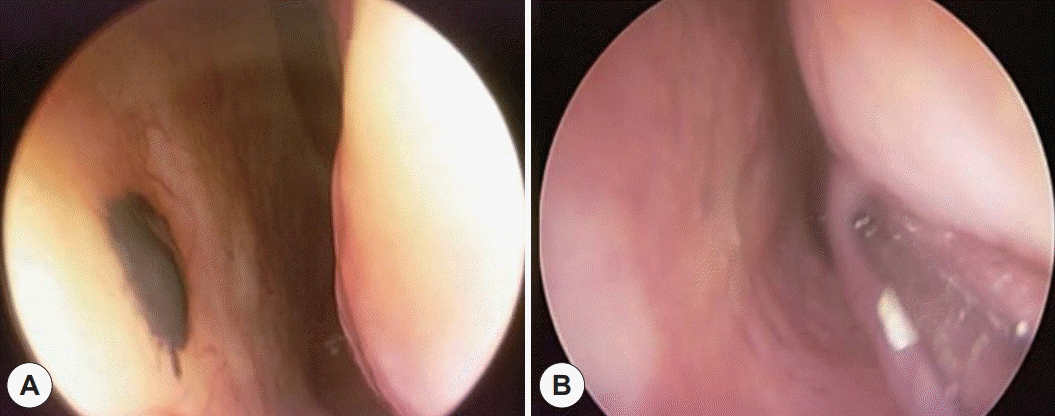
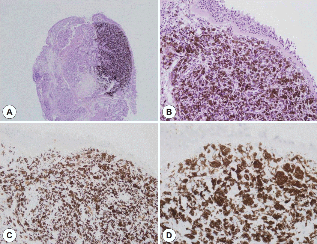
44세 여자환자가 우연히 발견된 좌측 비강의 색소성 병변으로 내원하였다. 코막힘이나 비출혈같은 증상은 없었다. 비내시경에서 좌측 비중격 중간-하단부 점막에 약 지름 5 mm 크기의 타원형의 흑색 점막병변이 관찰되었다(Fig. 1). 감별을 위해 시행한 조영 증강 부비동 전산화 단층 촬영에서 비중격 점막 병변부위의 조영 증강은 관찰되지 않았고, 연골이나 뼈 등의 주위기관으로의 침범은 없었다. 진단을 위하여 병변에 대한 부분마취를 시행하고, 내시경하에 2 mm 정도 주변 정상 조직과 연골막을 포함하여 절제를 시행하였다. 환자는 수술 후 합병증 없이 회복하여 당일 퇴원하였으며, 절제한 검체는 조직검사를 시행하였다. 조직학적으로 핵의 다형성이나, 핵소체의 두드러짐, 유사분열은 관찰되지 않았으며, 면역조직화학 염색 중 HMB-45, Melan A, S100 protein에서 양성 반응을 보여 최종적으로 청색모반으로 확진하였다(Fig. 2). 환자는 수술 후 6개월까지 재발을 포함한 합병증 없이 추적관찰 중이다 (Fig. 1).

고 찰

청색모반은 대부분 피부에 발생하는 멜라닌세포의 색소침착성 증식으로, 비강 점막에 발생한 경우는 극히 드물다고 알려져 있다. 멜라닌세포는 신경능선(neural crest)에서 기원하여 배아형성기에 피부나 점막으로 이동한다고 알려져 있다[7]. 비부비동에 발생한 청색모반은 호흡기점막의 간질이나 상피에 자리잡고 있던 멜라닌세포가 이동해서 발생한 것으로 추정하고 있다[4]. 비부비동에 발생한 청색모반의 처음 보고된 증례는 상악동근치술을 시행 받은 28세 여성에게 보고되었으며, 절제된 상악동 점막에서 병리학적 검사 중 우연히 발견되었다[8].
청색모반은 다발성으로 발현될 수 있는 ‘Carney complex’ 또는 가족성 다발 청색모반증(Familial multiple blue nevi) 같은 증후군 같은 질환에서도 나타날 수 있다. Carney complex는 상염색체 우성유전으로 심장이나 피부 점액종, 반점상 피부 색소증, 내분비계 과활동성(Cushing syndrome, acromegaly, and sexual precocity), 신경초종(Schwannomas)을 특징으로 한다[11]. 가족성 다발 청색모반증은 출생시부터 다발성 색소성 병변이 두경부, 체간, 사지, 공막에 나타나며 역시 상염색체 우성으로 유전된다[12]. 그러나 비부비동에 발생한 청색모반이 이러한 증후군들과 연관성은 아직까지 보고되지 않았으며, 우리 증례에서도 연관된 임상증상은 발견되지 않았다.
피부에서 발생한 청색모반은 대부분 양성 경과를 보이나, 드물게 악성 흑색종으로 진행하는 경우가 보고되었다[13]. 입술에서 발생한 청색모반에서 악성 흑색종이 진행한 것으로 추정되는 증례 도 보고되었는데, 3년간 윗입술의 검푸른 반점이 수개월사이 침윤성 변화와 궤양성 결절과 병변 경계가 불명확해지는 변화를 보여 조직검사상 악성 흑색종으로 판명되었으며, 기존의 청색모반 일부에서만 비전형 멜라닌세포가 상피세포의 기저층에서 확인되어 기존의 청색모반에서 진행하였거나 연관되어 있다고 추정하였다[14]. 그러나, 비부비동 영역에서는 청색모반이 재발하거나, 악성 흑색종으로 진행한 경우는 아직 보고되지 않았다. 비점막에 발생한 청색모반은 보고된 케이스가 매우 적어서 확립된 치료법이 나와 있지 않은 실정이다. 비점막에 발생한 청색모반을 보고한 사례에서는 대부분 병변의 완전절제를 시행하였다[5,6]. 또한, 피부나 입술에서 발생한 청색모반에서 악성 흑색종으로 진행된 경우에, 주변 정상 조직을 포함한 병변의 완전절제와 관련 림프절 절제술을 시행한 것을 고려해 볼 때[13,14], 정상 주변 조직을 포함한 병변의 완전한 절제와 조직학적 진단과 적절한 추적관찰이 필요할 것으로 사료된다.
저자들은 이번 증례를 통해서 비부비동 점막에 발생한 색소성 병변의 드문 원인으로 청색 모반도 고려해야 한다고 생각한다.

References

1. Phadke PA, Zembowicz A. Blue nevi and related tumors. Clin Lab Med. 2011; 31(2):345–58.
2. Murali R, McCarthy SW, Scolyer RA. Blue nevi and related lesions: a review highlighting atypical and newly described variants, distinguishing features and diagnostic pitfalls. Adv Anat Pathol. 2009; 16(6):365–82.
3. Sugianto JZ, Ralston JS, Metcalf JS, McFaddin CL, Smith MT. Blue nevus and “malignant blue nevus:” A concise review. Semin Diagn Pathol. 2016; 33(4):219–24.
4. Ojha J, Akers JL, Akers JO, Hassanein AM, Islam NM, Cohen DM, et al. Intraoral cellular blue nevus: report of a unique histopathologic entity and review of the literature. Cutis. 2007; 80(3):189–92.
5. Chuang WY, Hao SP, Yeh CJ, Jung SM. Blue nevi of the sinonasal mucosa: a report of two cases and review of the literature. Laryngoscope. 2007; 117(2):371–2.
6. Lim LM, Tan KB, Petersson F, Thong M. Sinonasal blue naevus: case report and clinicopathological review. J Laryngol Otol. 2013; 127(9):939–41.
7. Prasad ML. Update on pigmented lesions of the sinonasal tract. Head Neck Pathol. 2007; 1(1):50–4.
8. Aneiros J, O’Valle F, del Moral RG, Morales MG, Alvaro T. Blue nevus of the maxillary sinus. An immunohistochemical and electron microscopic study. Acta Otorhinolaryngol Belg. 1989; 43(2):163–7.
9. Thompson LDR, Wieneke JA, Miettinen M. Miettinen, Sinonasal tract and nasopharyngeal melanomas: a clinicopathologic study of 115 cases with a proposed staging system. Am J Surg Pathol. 2003; 27(5):594–611.
10. Smith K, Germain M, Williams J, Skeleton H. CD34-positive cellular blue nevi. J Cutan Pathol. 2001; 28(3):145–50.
11. Carney JA. Carney complex: the complex of myxomas, spotty pigmentation, endocrine overactivity, and schwannomas. Semin Dermatol. 1995; 14(2):90–8.
12. Blackford S, Roberts DL. Familial multiple blue naevi. Clin Exp Dermatol. 1991; 16(4):308–9.
13. Martinez-Peñuela A, Iglesias ME, Mercado MR, Martinez-Peñuela JM. Malignant transformation of a nevus of Ito: description of a rare case. Actas Dermosifiliogr. 2011; 102(10):817–20.
14. Lee HY, Na SY, Son YM, Kang HK, Baek JO, Lee JR, et al. A malignant melanoma associated with a blue nevus of the lip. Ann Dermatol. 2010; 22(1):119–24.

Fig. 1.
Blue nevus of the nasal septum. A: Endoscopic finding of blue nevus on the nasal septum preoperatively. B: Postoperative view of the nasal septum 6 months after surgery.
jr-2021-00345f1.tif
Fig. 2.
Photomicrographs showing nasal septal tissue with stromal presence of heavily pigmented spindle cells. A: H&E stain, ×40, B: H&E stain, ×200. Immunohistochemical stains in blue nevus of the nasal septum. Blue nevus cells are positive for S100 protein and HMB-45, respectively (C) and (D).
jr-2021-00345f2.tif
TOOLS
Similar articles