INTRODUCTION
Identifying the correct lumbar spine level is very important for performing interventional procedures for back pain patients with radicular pain of the lower limb. Injection at the wrong level can produce unwanted or no therapeutic effect on the patient. Even though many pain physicians use fluoroscopy to identify the segment of the lumbar vertebrae, an anatomical deformity in the lumbar spine, such as lumbosacral transitional vertebra (LSTV [lumbarization or sacralization]) is often observed, with a reported incidence of 9–30% [
1–
3]. LSTV found in the fifth lumbar vertebra may be shown to be similar to the sacrum (sacralization), and those affecting the first sacral vertebra may be shown to the features of the lumbar spine (lumbarization) [
1,
2]. In these circumstances, many clinicians designate the L1 vertebra based upon the location of the 12th rib. Generally, the vertebra bearing the most caudal rib is identified as T12, and the vertebra just below T12 is identified as the first lumbar vertebra. However, this method is prone to errors because there may be a significant morphological variation in the thoracolumbar junction [
1,
3–
5].
The thoracolumbar transitional vertebra (TLTV) is a relatively unknown congenital spinal anomaly in the thoracolumbar junction [
2,
6]. The TLTV may exist in the 19th or 20th vertebrae, and it may present with a hypoplastic rib, a transverse process or an accessory ossification center, either unilaterally or bilaterally [
6]. However, the accurate identification of the TLTV is not easy in clinical practice when only a simple X-ray or fluoroscopic image is obtained during interventional procedures, because the morphology of TLTV can’t be easily figured out especially in the geriatric patients with severe osteoporosis. Furthermore, the existence of concurrent LSTV or abnormal rib count (11 or 13 ribs) may lead to be more difficult to distinguish the first lumbar vertebra from TLTV even with imaging the entire spine [
6,
7]. Therefore, it is helpful to understand the clinical characteristics of the patients with spinal variation such as TLTV and LSTV or abnormal rib count.
In the present study, we investigated the prevalence of TLTV and LSTV (lumbarization and sacralization) by evaluating whole spine spiral three-dimensional (3-D) computed tomography (CT) images. And the authors evaluated the relationships between the existence of TLTV and the existence of abnormal rib count as the primary outcome and the correlation between the existence of thoracolumbar and lumbosacral transitional vertebra as the secondary outcome.
Go to :

MATERIALS AND METHODS
This study retrospectively reviewed 1,553 consecutive adult patients, aged 18 to 80 years, who underwent whole spine spiral 3-D CT due to trauma or diseases from January 2018 to December 2018. Our Institutional Review Board approved this retrospective study (no. CUH 2019-03-028), and the requirement for informed consent was waived.
The following cases were excluded from the study: 1) Severe fracture of the body or transverse process of transitional vertebrae, 2) Advanced Ankylosing spondylitis, 3) Severe scoliosis, 4) Severe degenerative change, 5) Others where the distinction is ambiguous in the simple images or 3-D CT images.
CT machines
Multi-slice Computed Tomography scanning was performed on a 128-slice dual-source CT scanner (Somatom definition flash, Siemens, Germany). We obtained images from the head to the proximal femur by automatically selecting the patient’s optimal kV setting in 10 kV increments by balancing the radiation dose and the image contrast by Siemens’ CARE kV function.
Nomenclature of ribs and the definition of transitional vertebrae (TLTV and LSTV)
The vertebral levels were counted craniocaudally, using whole-spine spiral 3D-CT images, starting from C1, based on the assumption of existing 7 cervical, 12 thoracic, and 5 lumbar vertebrae. The 20th and 25th vertebrae were defined as L1 and S1, respectively.
A rib is defined as a separated bone that articulates with the facet or the body of the vertebrae [
8]. Several investigators proposed that most caudal ribs can be classified into one of four types: normal rib, hypoplastic rib (or short rib), unfused transverse process (or accessory ossification center) and mixed type [
6,
8–
10]. A short rib has been defined as a separated bone, less than 3.8 cm in length that articulates with the facet or body of the vertebra [
6,
8]. Park et al. [
6] noted the difficulty in differentiating most caudal ribs. Therefore, a separated bone that did not meet the criteria for a short rib, a normal rib (> 3.8 cm in length) or an accessory ossification center at the thoracolumbar junction was defined as a mixed-type rib in the study. Thirteenth ribs, which are also known as lumbar ribs, were defined as ribs articulating with L1.
The TLTV is defined as a vertebra with partially retained features of the thoracic and lumbar segments at the thoracolumbar junction [
6]. Wigh [
8] suggested that the TLTV includes segments with a short rib or with an accessory ossification center. Lumbarization was defined as non-fusion of S1 and S2 (26th vertebra), meaning that there was one additional articulated vertebra. Sacralization was defined as anomalous fusion of L5 (24th vertebra) and S1 [
1,
3,
11,
12].
For the radiologic evaluation, two expert physicians, who performs spinal pain management, independently evaluated the whole spine spiral 3-D CT images for the final selected cases (n = 1,340). Each expert physician assessed the ribs counting, and the types of vertebral segments at the thoracolumbar or lumbosacral junction by classifying each vertebral segment as TLTV or non-TLTV (a thoracic or lumbar segment) and LSTV or non-LSTV (lumbar or sacral segment). Although accessory ossification centers sometimes resemble fracture fragments, these anomalies are radiologically different from fractures because they show smooth and round borders [
13]. In patients with inconsistent judgments regarding the number of vertebrae and ribs, TLTV and LSTV, the two physicians discussed and made a final decision. The types of TLTV and LSTV were identified to determine the existence of TLTV and LSTV but was not used as statistical data.
Statistical analysis
All statistical analyses were performed using SigmaPlot version 12.5. (Systat Software Inc., USA). All descriptive statistics are expressed as mean ± standard deviation, median (1Q, 3Q), percentage or the number of patients. First, we compared the patient demographics and other spinal morphological features between the TLTV and non-TLTV group, according to the existence of TLTV. The proportion of abnormal rib count (11 or 13 ribs) and the co-existent LSTV were compared between the two groups. Continuous variables were analyzed with the Student’s t-test or Mann–Whitney rank-sum test after normality test. Categorical variables were analyzed using the chi-square test. Second, we defined the relationship between the existence of TLTV and LSTV. A multiple logistic regression model was used to estimate the odds ratios (ORs) and their 95% confidence intervals (95% CIs) of the explanatory variables. P values < 0.05 were considered statistically significant.
Go to :

RESULTS
A total of 1,340 patients, who were 857 males (64.0%) and 483 females (36.0%) were included in this study, and the consort diagram is described in
Fig. 1. Patient demographics and spinal morphological features are described in
Table 1. The results from the two expert physicians differed in distinguishing the type of vertebral segment at the thoracolumbar junction (n = 21) and at the lumbosacral junction (n = 16). There was a difference in the length of ribs at the thoracolumbar junction in six out of 21 cases. Therefore, after discussion, the six cases with a difference in rib lengths were regarded as non-TLTV. All of the cases (n = 16) in which the LSTV type differed were agreed to be LSTV.
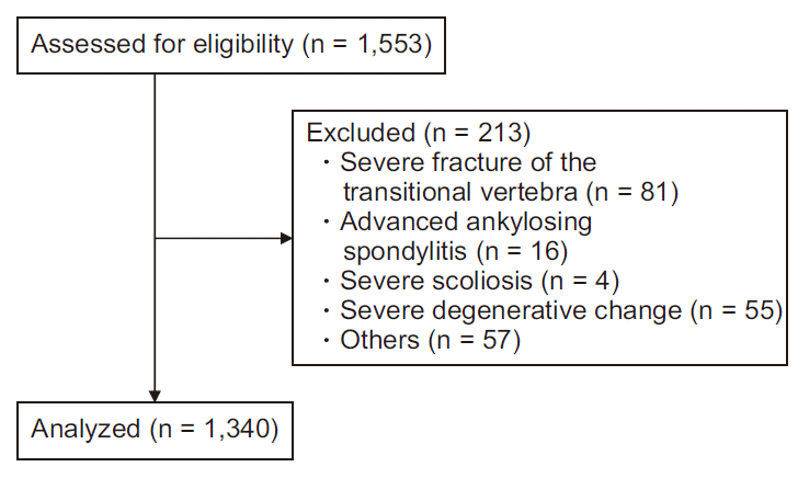 | Fig. 1
|
Table 1
Patient Demographics and Spinal Morphological Features between TLTV and No TLTV Group
|
Variable |
TLTV (n = 150) |
No TLTV (n = 1,190) |
P value |
|
Age (yr) |
55.0 (39.0, 66.0) |
58.0 (45.0, 69.0) |
0.014*
|
|
Sex |
|
M/F |
91/59 |
766/424 |
0.386 |
|
% of male |
60.7 |
64.4 |
|
Abnormal rib count |
96 (64.0) |
16 (1.3) |
< 0.001†
|
|
Rib count (in detail) |
|
11 |
96 |
11 |
|
|
12 |
54 |
1,174 |
|
13 |
0 |
5 |
|
LSTV |
51 (34.0) |
60 (5.0) |
< 0.001†
|
|
With 11 ribs |
28 |
2 |
|
With 12 ribs |
23 |
55 |
|
With 13 ribs |
0 |
3 |

The normal twelve ribs were observed in 1,228 of the 1,340 cases (91.6%). A case of thirteenth ribs (lumbar ribs) and lumbarization (LSTV) is described in
Fig. 2. If the lowest rib is interpreted as the 12th ribs (white arrows), the lumbar spinal configuration might be identified as sacralization (
Fig. 2A). However, by counting inferiorly from C1 using whole spine CT images, the lowest rib is confirmed as thirteenth ribs and the lowest incomplete-fused vertebra as S1 (lumbarization) (
Fig. 2B). Meanwhile, a case of thirteenth ribs without LSTV is also described in
Fig. 3. In this case, L1 was articulated at the facet joint with the thirteenth normal rib on axial CT image of L1 (black arrows) (
Fig. 3B). Of the 96 patients with 11 ribs and TLTV of the 19th vertebra, sacralization was observed in 28 patients (28/96) (
Table 1 and
Fig. 4), meanwhile, of the 54 patients with 12 ribs and TLTV of the 20th vertebra, lumbarization was observed in 23 patients (23/54). The images of our cases showing the different types of TLTV with or without LSTV are described in
Fig. 5.
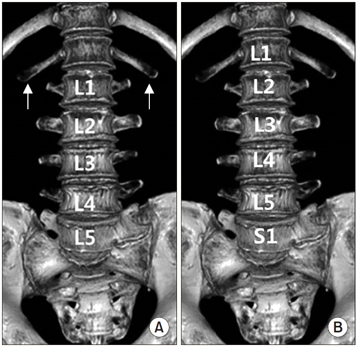 | Fig. 2Volume rendering reconstruction images of the anterior aspect of the lumbar vertebrae in case of thirteenth ribs (lumbar ribs) and lumbarization of S1. (A) If the lowest ribs are interpreted as 12th ribs (white arrows), the lumbar spinal configuration might be identified as sacralization. (B) By counting inferiorly from C1, the lowest ribs are confirmed as thirteenth ribs and the lowest incomplete-fused vertebra as S1 (lumbarization). 
|
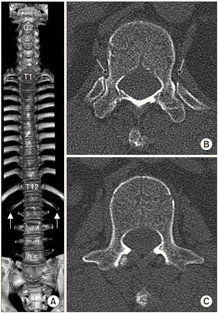 | Fig. 3Whole spine spiral three-dimensional computerized tomographic (CT) images of patient with thirteenth ribs (lumbar ribs) without lumbosacral transitional vertebrae. (A) Volume rendering reconstruction image of the thoracolumbar spine and ribs with the sternum removed. Thirteenth ribs are clearly shown (white arrows). (B) Axial CT image of L1. Articulations between L1 and the thirteenth ribs are well demonstrated (black arrows). (C) Axial CT image of L2. L2 has transverse processes typically. 
|
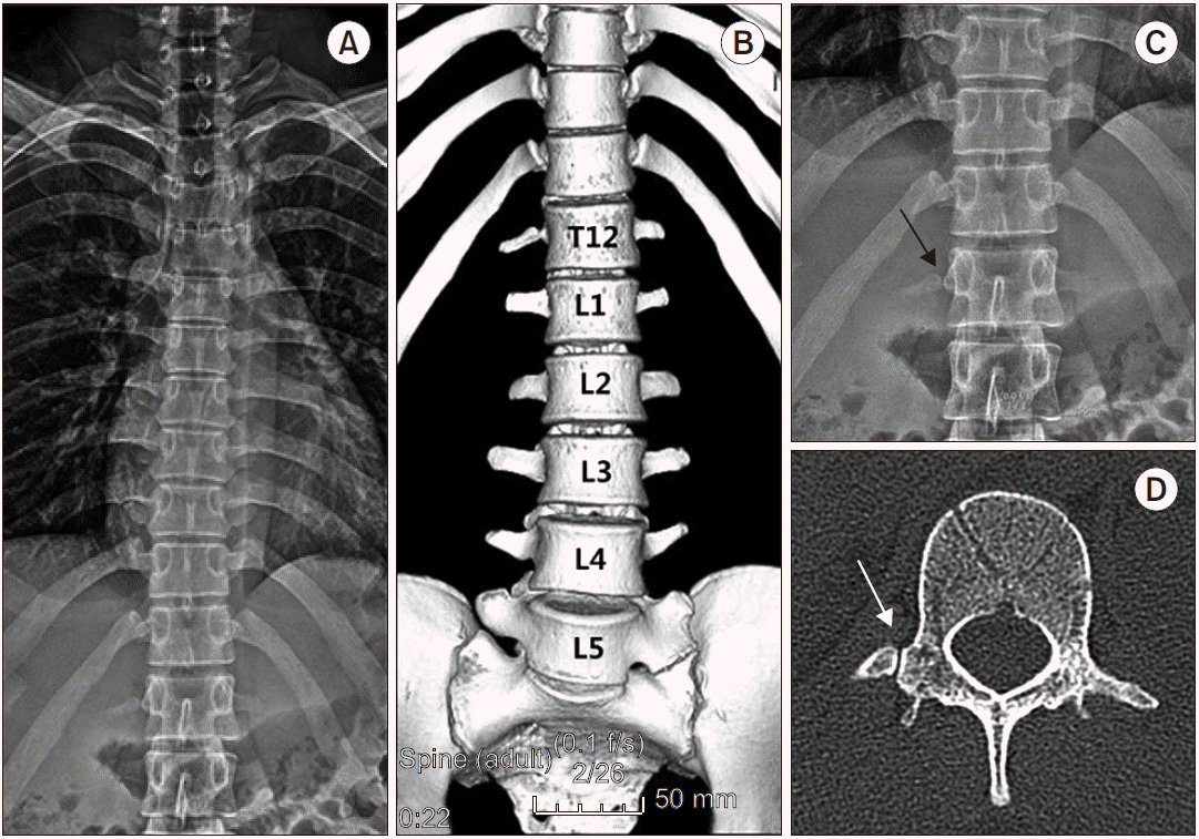 | Fig. 4Simple rib anterioposterior (AP) image and volume rendering reconstruction images of the anterior aspect of the lumbar vertebrae. This case has thoracolumbar transitional vertebra (TLTV) of 12th thoracic vertebra (type IV) and sacralization. (A) There are 11 normal ribs. (B) If the TLTV (12th vertebra) is interpreted as L1, the lumbar spinal configuration might be identified as lumbarization. (C) The facet joint (black arrow) is shown on the right side of 12th TLTV on the simple rib AP image. (D) An accessory ossification center with a facet joint (white arrow) on the right side on an axial computerized tomographic image. 
|
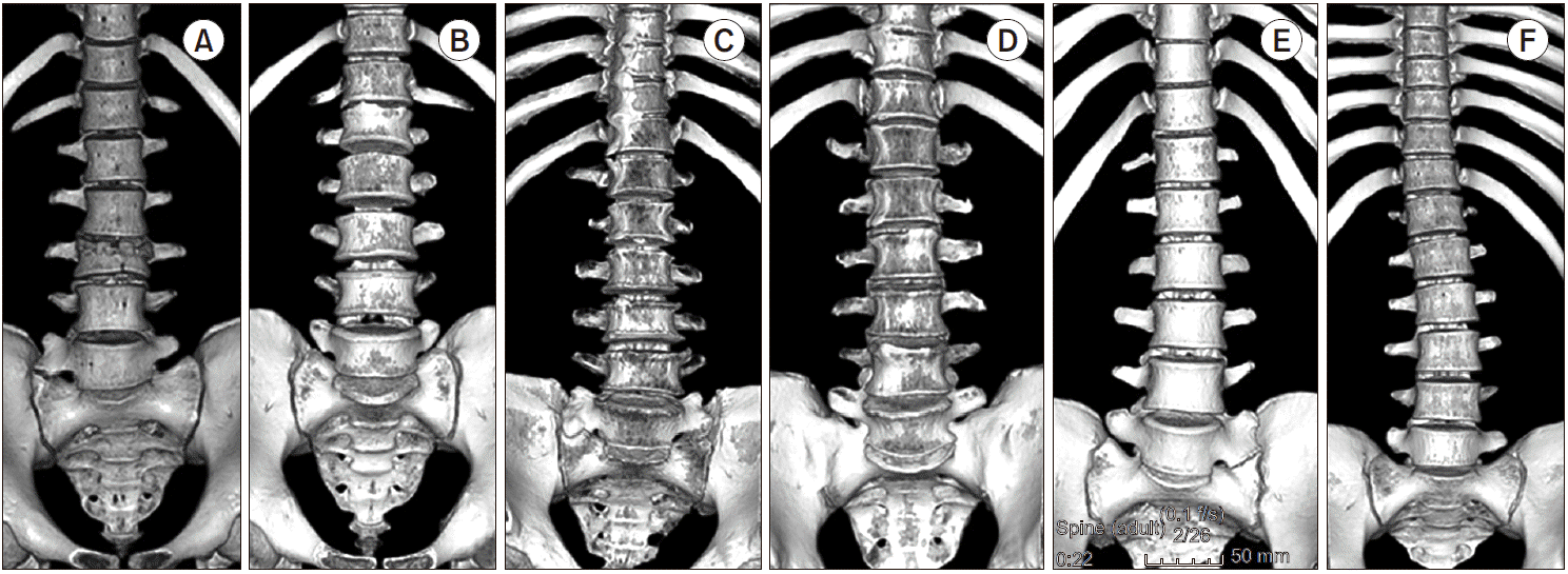 | Fig. 5Volume rendering reconstruction images of the anterior aspect of the lumbar vertebrae. This images showing different type of thoracolumbar transitional vertebra (TLTV) with or without lumbosacral transitional vertebra (LSTV). (A) TLTV type I with LSTV type IIa. (B) TLTV type I without LSTV. (C) TLTV type IIb with LSTV type IIb. (D) TLTV type IIb. (E) TLTV type IV with LSTV type IIa. (F) TLTV type IV. 
|
The prevalence of TLTV was 11.2% (150 of 1,340 patients) and that of LSTV was 8.3% (111 of 1,340 cases). In detail, sacralization was observed in 68 of 1,340 cases (5.1%) and lumbarization in 43 of 1,340 cases (3.2%). Patients with TLTV have a higher rate of coexistent abnormal rib count (11 or 13) when compared with patients without TLTV (P < 0.001) (
Table 1). The proportions of the patients with coexistent LSTV were 34.0% and 5.0% in the TLTV and no-TLTV group, respectively (P < 0.001). In patients with 11 ribs in the no-TLTV group, LSTV was observed in two of the patients (2/11), and all of them had sacralization. Of the 1,174 patients with 12 ribs and with no-TLTV, LSTV was observed in 55 patients (55/1,174), with 39 cases of sacralization, and 16 cases of lumbarization. In the no-TLTV patients, LSTV was observed in three of the patients with 13 ribs (3/5), all of which had lumbarization. There is a statistically significant probability that there is accompanying LSTV in subjects in the TLTV group (P < 0.001).
In multiple logistic regression analysis, there was a significant relationship between the existence of TLTV and the number of ribs and LSTV (P < 0.001) (
Table 2). Abnormal rib count and existence of LSTV had respective ORs of 117.26 (95% CI: 60.77–226.27) and 7.38 (95% CI: 3.99–13.63). Age and sex had no statistically significant associations on the existence of TLTV.
Table 2
The Relationships between the Existence of TLTV and Other Variables by Multiple Logistic Regression Analysis
|
Parameters |
Categories |
Odds ratio |
95% Confidence interval |
P value |
|
Abnormal rib count |
12 |
1 |
|
|
Not 12 |
117.26 |
60.77–226.27 |
< 0.001*
|
|
Existence of LSTV |
No |
1 |
|
Yes |
7.38 |
3.99–13.63 |
< 0.001*
|
|
Age (yr) |
< 65 |
1 |
|
≥ 65 |
0.56 |
0.29–1.09 |
0.089 |
|
Sex |
Female |
1 |
|
Male |
1.22 |
0.67–2.21 |
0.516 |

Go to :

DISCUSSION
The current study using whole-spine spiral 3D-CT images revealed a strong association between the existence of TLTV and the number of ribs and the LSTV. If TLTV was present, it is highly likely that there is a coexistent abnormality in the number of ribs and LSTV. Therefore, if a LSTV is present in fluoroscopic images during interventional procedures of the lumbar spine, pain physicians should concern that there is a strong possibility of the presence of TLTV. And in patients with TLTV at thoracolumbar junction, there is a higher probability of an abnormal rib count compared to the patients without TLTV. In these cases, whole spine imaging should be used to identify the accurate vertebral structure. To the best of our knowledge, this is the first investigation that demonstrates these associations.
Even though the cause of vertebral abnormality has not been clearly determined, genetic factors are thought to play a certain role in the segmental development of the spine [
14]. The number of cervical vertebrae is extremely stable at seven. However, the number of thoracic vertebrae and lumbar vertebrae may range from 11 to 13 and from 4 to 6, respectively [
8]. Variations in the thoracolumbar junction can have the potential to promote morphological shifts in the lumbosacral junction because the thoracic, lumbar and sacral spine develop craniocaudally in early fetal life [
15]. If TLTV forms on T12 (19th vertebrae), then L5 (24th vertebra) might fuse with S1, resulting in sacralization. Meanwhile, if TLTV forms on L1 (20th vertebrae), then S1 (25th vertebra) might separate from S2, resulting in lumbarization. For this reason, if TLTV is present, it is highly likely that there is a coexistent abnormality such as LSTV, as shown in the current study.
Many pain physicians use fluoroscopy to identify the segment of the lumbar vertebrae. If there is an anatomical deformity in the lumbar spine, such as LSTV (lumbarization or sacralization), the pain physician is more likely to map the lumbar segment by counting down from the T12 vertebra. Meanwhile, considering the high frequency of back pain in patients with LSTV, the incidence of LSTV may be higher in patients who visit pain clinics than others [
16,
17]. The lumbar spinal level is easily defined on radiographs by counting inferiorly from T12, which is defined as the lowest vertebra with ribs. As our study results demonstrated in
Fig. 2, this method is prone to errors because there may be a significant variation in the number and morphology of the most caudal ribs and the possibility of the presence of TLTV. Instead of counting inferiorly from T12, if possible, whole spine imaging should be used to identify the accurate vertebral structure. In addition, the facet gap can be used as a meaningful marker to judge the vertebra as TLTV as described in
Fig. 4. In 143 cases of the 150 patients with TLTV (95.3%) in the current study, the facet gap was observed even in simple images (chest anteroposterior [AP], T-spine AP, and rib AP).
We used the criteria to distinguish the deformation of the most caudal ribs [
9]. The criteria include measuring the length of separated bones, and determining the presence of joints with separated bones. In the current study, we also used this method to determine whether vertebrae segments in the thoracolumbar junction were TLTV or not, and the frequency of TLTV (11.2%) was similar to that observed by Park’s et al. [
6]. Meanwhile, Wigh [
8] suggested that segments with short ribs on thoracolumbar junction can be considered thoracic-type TLTV, while segments with accessory ossification centers can be considered lumber-type TLTV. They also suggested that showing that Type 1 vertebrae is thoracic TLTV and Type 4 vertebrae is lumbar TLTV. The consensus may be obtained through the expert discussion or the further studies.
The study by Nakajima et al. [
2], in which counting was done from C1 or C2 inferiorly on a whole-spine image, revealed that the incidence of lumbarization and of sacralization was respectively 6.2% and 2.7%. However, in the present study, the incidence of lumbarization was 3.2% and that of sacralization was 5.1%. Widely variable incidences of LSTV have been reported in the literature, ranging from 4% to 35.9% [
1,
9,
11,
14,
18,
19]. This variation may be explained by differences in diagnostic criteria, imaging techniques and confounding factors among the investigated populations.
Hughes and Saifuddin [
19] reported another technique to identify LSTV by locating the iliolumbar ligaments. Sacralization was determined by the lack of the iliolumbar ligament at the level above the sacrum. When an iliolumbar ligament was identified above the LSTV, the vertebra could be considered L5 and the LSTV was termed lumbarization. However, this technique assumes that there are always 7 cervical, 12 thoracic and 5 lumbar vertebrae. They suggested that the most ideal scenario would be to acquire whole spine images in every case to avoid any potential mistake in vertebral counting. Therefore, caution in numbering the lumbosacral vertebrae in patients with LSTV is of the utmost importance in spinal interventional procedures and surgeries because the presence of LSTV and rib anomalies can lead to inaccurate identification of vertebral levels.
Our study had a few limitations. First, we could not rule out the possibility of selection bias because most of the patients included in our study were those who were treated for trauma or disease. The second limitation is that rib length measurements are prone to errors. Although we used 3D-CT images to reduce such errors, inaccuracies in rib length measurements still remain unavoidable.
In conclusion, our results suggested that patients with TLTV were more likely to have abnormal rib counts or LSTV. If TLTV or LSTV was present on the fluoroscopic images, the order of the lumbar segment needed to be well understood. However, there is no proper way to identify accurately the number of the transitional segment without imaging of the entire spine. Only a whole spine image allows correct numbering of the lumbar vertebra in all cases and remains, therefore, the gold standard. Another alternative method, such as the thoracolumbar scout MR images, should be used and counting done caudally from T1.
Go to :


 PDF
PDF Citation
 Print
Print


 XML Download