This article has been
cited by other articles in ScienceCentral.
Abstract
A 58-year-old male patient with situs inversus totalis, a rare congenital malformation characterized by all asymmetric organs being formed as the mirror images of their normal morphologies, underwent mitral valve repair due to mitral valve prolapse. This case was reported to suggest that anesthesiologists should thoroughly understand the anatomy of these types of patients before providing cardiac anesthesia that often requires advanced monitoring and rely on their accurate interpretation. Accordingly, a few key points will be discussed with emphasis on reversing lead placement during electrocardiogram monitoring, using the left internal jugular vein for pulmonary artery catheterization, and firmly comprehending mirror image heart morphology to better conduct transesophageal echocardiography.
Keywords: Anesthesia, Situs inversus totalis, Thoracic surgery
Situs inversus totalis is a rare congenital anomaly in which all internal organs are arranged in the mirror image of their normal morphologies. Patients with situs inversus totalis are asymptomatic and usually lead normal lives, but the recognition of this condition is important to interpret patient symptoms of other conditions accurately and to avoid clinical or surgical mistakes. In addition, accurate understanding of the situs inversus totalis anatomy would be a prerequisite to an optimal anesthetic management for these patients, especially when advanced hemodynamic monitoring devices and their accurate interpretation are required as in cardiac surgery. This case was reported to share this medical team’s experience of a 58-year-old male patient with situs inversus totalis who underwent mitral valve repair.
CASE REPORT
A 58-year-old male patient was scheduled to undergo elective mitral valve repair surgery to repair mitral valve prolapse (P3). The preoperative chest X-ray showed that his heart was directed to the right with mild cardiomegaly. The preoperative electrocardiogram (ECG) was conducted with chest leads in their normal positions and showed a negative P-wave in the I and aVL leads and a positive P-wave in the aVR lead, a typical right-axis deviation pattern. Another ECG was then conducted with reversed lead placement positions, which produced normal results (
Fig. 1). In his history, the patient stated that he had been diagnosed with situs inversus totalis 50 years ago and had lived without any symptoms of this condition for the duration of his life. The preoperative physical examination and evaluations did not produce any other significant findings except for mild dyspnea.
Fig. 1
(A) Echocardiogram with normal lead placement showing right axis deviation. (B) Normal echocardiogram with reverse lead placement. ECG: electrocardiogram.

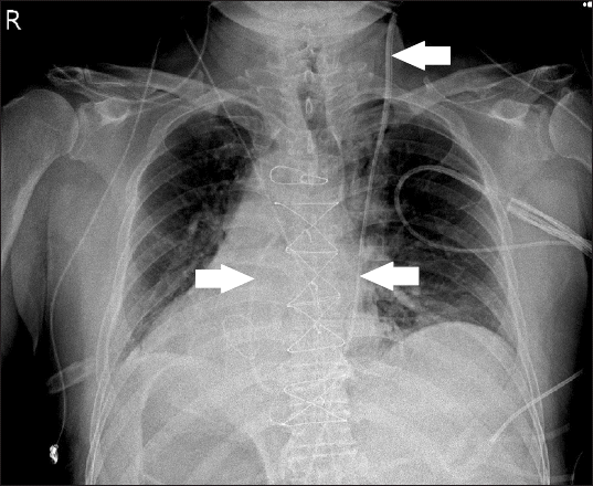
Upon the patient’s arrival in the operating room, ECG leads were applied in reverse to reduce confusions over the ECG findings and unnecessary treatments to correct abnormal ECG results produced by improperly placed leads. Other standard institutional monitoring methods were applied, including left radial arterial cannulation. Anesthetic induction was performed according to institutional guidelines and pulmonary artery catheterization was performed via the left internal jugular vein with MAC™ Multi Access Catheter (ARROW international Inc., USA) to guarantee a direct approach to the right atrium without crossing the midline. The medical team attempted to maintain the right-curved shape of the pulmonary artery catheter when inserting it into the left internal jugular vein to facilitate the advancement of the catheter though the morphologic right heart. Catheterization was successfully performed uneventfully on the first attempt. The postoperative chest X-ray revealed that the right-curved shape of the pulmonary artery catheter was maintained as the mirror image of its normal shape (
Fig. 2).
Fig. 2
Anteroposterior chest X-ray on postoperative day 1. Arrows show the right curved shape of pulmonary artery inserted directly via left internal jugular vein without crossing the midline.

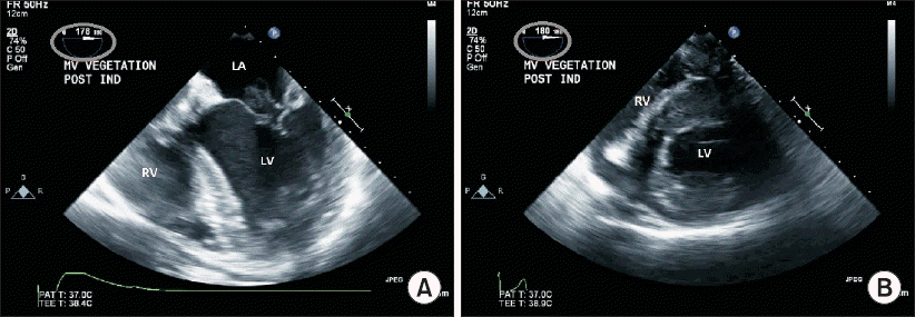
Intraoperative transesophageal echocardiography (TEE) was challenging. In this patient, a different angle was needed to acquire the desired views because of his atypical anatomy. Unlike the TEE images of normal hearts, the midesophageal four chamber and transgastric short axis views obtained near 0° in normal hearts were obtained with the left ventricle on the left side of the TEE screen, which is the inverse of normal TEE views, so these views were obtained at a 180° angle to reduce confusion (
Fig. 3). The bicaval view, which is normally obtained at 110°, was obtained at 70° while rotating the probe to the left in the direction of the right atrium (
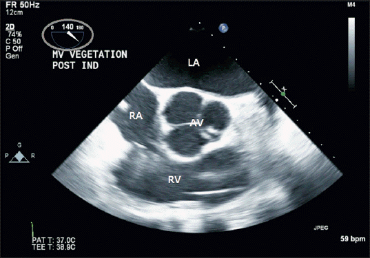
Fig. 4). The aortic valve short-axis view, normally found at 30°–40°, was obtained at 140°–150° (
Fig. 5). These views were best obtained at an omniplane angle which was the difference of 180° and the typical omniplane angle.
Fig. 3
(A) Transesophageal echocardiographic midesophageal four chamber view. (B) Transesophageal echocardiographic transgastric short axis view. These views were obtained at angle of 180° (circle). LA: left atrium, LV: left ventricle, RV: right ventricle.

Fig. 4
Transesophageal echocardiographic bicaval view obtained at 70° (circle). LA: left atrium, RA: right atrium, SVC: superior vena cava.

Fig. 5
Transesophageal echocardiographic aortic valve short-axis view obtained at 140° (circle). AV: aortic valve, LA: left atrium, RA: right atrium, RV: right ventricle.

The operation was performed without incident. The patient was stable during and after the operation. The patient was transferred to the general ward after spending two days in the intensive care unit and discharged on the seventh day after the surgery without any complications.
DISCUSSION
Situs inversus totalis is a rare congenital condition in which all internal organs are the mirror images of their normal anatomies, occurring in 0.01–0.02% of people [
1]. Situs inversus totalis is mainly an autosomal recessive disorder, but is sometimes related to X-chromosomes. Human anatomical changes occur during embryogenesis. Although most patients with situs inversus totalis have normal life expectancies without any significant related morbidities, they should be meticulously evaluated because the condition may coexist with other congenital comorbidities in the cardiovascular, respiratory, and digestive systems [
2]. When situs inversus totalis is combined with recurrent respiratory infections such as sinusitis and bronchiectasis in a patient, it is diagnosed as Kartagener’s syndrome or primary ciliary dyskinesia. Repeated respiratory infections result from impaired mucociliary function [
3]. In this case, the supplementation of humid oxygen, adequate patient’s hydration and analgesia is mandatory to reduce any inspissation of secretion in the airway and prevent the compromised respiratory mechanics [
4].
The recognition of anatomical changes and comorbidities by careful preoperative evaluation is necessary to prevent clinical mistakes and ensure that such patients receive appropriate treatment. It has been reported that thorough preoperative evaluations confirmed acute appendicitis in patients with situs inversus totalis suffering from left lower quadrant abdominal pain [
2,
5]. Moreover, accurate obtainment and interpretation of data from various monitoring devices would be challenging to the attending anesthesiologists, especially in case of cardiac surgery that requires additional monitors including the TEE.
As mentioned in the case report, the preoperative ECG with conventional lead placement showed right-axis deviation because of the anatomical variations resulting from situs inversus totalis. However, the ECG with reversed lead placement did not show any abnormalities, so this lead placement was maintained during and after surgery to prevent attending physicians from misinterpreting ECG results and giving unnecessary treatments. Likewise, physicians should remember that defibrillation pads should be applied to situs inversus totalis patients in reverse. Central venous catheterization via the left internal jugular vein has been recommended for patients with situs inversus totalis [
3]. Accordingly, we chose the left internal jugular vein for pulmonary artery catheterization to avoid thoracic duct injury, which would be on the patient’s right side, and guarantee a direct approach to the right atrium. The postoperative chest X-ray showed that the pulmonary artery catheter cannulated through the left internal jugular vein followed a direct course to the superior vena cava instead of crossing the midline (
Fig. 2).
In spite of a few recommandation in TEE practice in patients with situs inversus totalis [
6], it may be difficult for anesthesiologists to conduct TEE monitoring of situs inversus totalis patients during surgery, so they must be vigilant when examining cardiovascular structures such as the ventricles, atria, and aorta. Views obtained near 0°, including the mid-esophageal four chamber and trans-gastric short axis views are mirror images of the normal views (
Fig. 3). Other standard views can be obtained at an omniplane angle of 180° minus the typical omniplane angle. For instance, the bicaval view, which is normally obtained at 110°, can be obtained at 70° while rotating the probe to the left because the right atrium is located to the left (
Fig. 4). The aortic valve short-axis view, normally found at 30°–40°, can be obtained at 140°–150° (
Fig. 5). In order to reduce confusion regarding the omniplane angle adjustment, clinicians may prefer to acquire these views via right-left invert of the obtained images through the TEE machine. Still, the left-right maneuvering directions of the TEE probe need to be opposite, which can be quiet challenging to the attending physician.
Patients with situs inversus totalis are difficult to anesthetize and monitor while under anesthesia. However, these patients can be managed more safely with careful preoperative planning and increased vigilance. This case was reported to describe a few key considerations for the proper anesthetic management of a situs inversus totalis patient during cardiac surgery with special emphasis to monitoring devices including the TEE.
CONFLICTS OF INTEREST
No potential conflict of interest relevant to this article was reported.


