Progressive multifocal leukoencephalopathy (PML) is a demyelinating disease of the central nervous system (CNS) caused by the John Cunningham (JC) virus. This virus usually affects immunocompromised individuals, especially those with HIV infection. According to a study from the United States, HIV-infected patients account for 82% of all cases of PML [1]. The neurological signs of PML include muscle spasticity, cognitive dysfunction, speech alternation, aphasia, and lack of coordination [1,2]. All of these symptoms can lead to challenges with preoperative evaluation and intraoperative management of the airway. A previous case report described difficult intubation due to limited mouth opening in a patient with Rett syndrome, a rare neurological disorder accompanied by intellectual disability and spasticity [3]. The present article describes another unusual case of difficult intubation because of limited mouth opening, this time in a man with PML. The patient’s legal guardian provided informed consent for publication of anonymized case details.
CASE REPORT
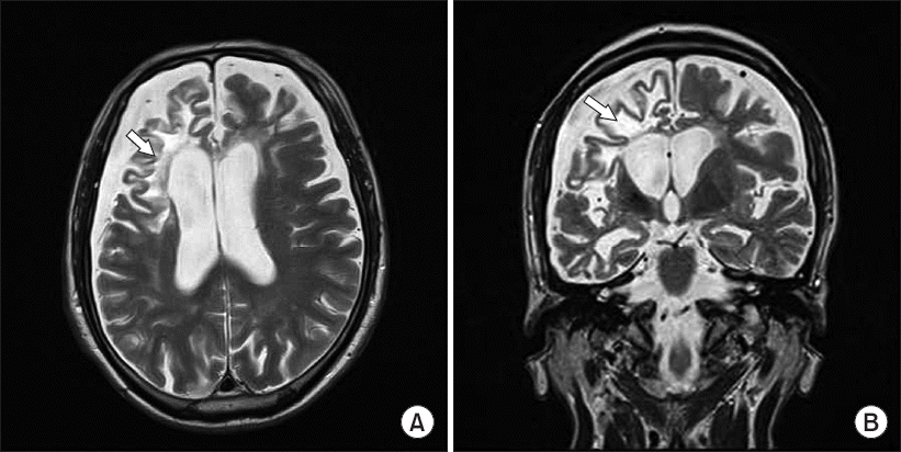
A 62-year-old man with PML was scheduled for video-assisted thoracoscopic (VATS) bullectomy. He had been diagnosed as HIV positive 6 years earlier, at which time no neurological lesions were observed. One year after his HIV diagnosis, he was admitted in a confused state via the emergency room, and PML was diagnosed on the basis of findings revealed on brain magnetic resonance imaging (Fig. 1) and JC virus positivity on polymerase chain reaction (PCR) analysis of the cerebrospinal fluid (CSF). As the disease progressed, he developed difficulty with ambulating because of spasticity in the lower limbs, and began visiting the neurology outpatient clinic.
Fig. 1
(A) Axial T2-weighted magnetic resonance imaging of the brain. (B) Coronal T2-weighted magnetic resonance imaging of the brain. The arrow indicates the lesion showing diffuse high intense areas and atrophy of the bilateral cerebral white matter, including the corticobulbar tract.

Six months before the planned VATS surgery, the patient was admitted to the intensive care unit with aspiration pneumonia and septic shock. He was in a bedridden state with spasticity in the upper and lower limbs. Two months earlier, a chest radiograph revealed a spontaneous pneumothorax in the left lung that was drained using a chest tube. The patient became delirious while hospitalized, and communication was difficult. Cognitive impairment and mild dementia were diagnosed by the neurologist. At that time, chest tube drainage revealed continuous air leak and oscillations; consequently, a VATS bullectomy was scheduled.
Blood tests, chest X-ray, and electrocardiogram were unremarkable. The patient’s medical record revealed that he had been undergoing high-dose antiretroviral therapy. His height and weight were 165 cm and 47 kg, respectively. Preoperative arterial blood gas analysis was normal on administration of oxygen (5 L/min) via nasal cannula. Chest computed tomography (CT) revealed partial expansion of the left lung after insertion of a chest drainage catheter. His medical record described dysarthria, a confused state, spastic hemiparesis, and cognitive dysfunction. Communication was not possible when the anesthesiologist attempted to take a history and assess the patient’s airway. Furthermore, a preoperative airway evaluation, including Mallampati classification and ability to extend the neck, could not be performed because of lack of cooperation from the patient.
However, general physical examinations, including an evaluation of mouth opening and assessment of dental status by opening the patient’s mouth, were not performed. Due to insufficient information regarding the patient’s airway, a video laryngoscope, which is often used when difficult intubation is expected, was prepared (GlideScope, Verathon Medical Inc., Canada).
The patient was brought to the operating room without premedication. Intraoperative monitoring was started using electrocardiography, pulse oximetry, noninvasive recording of blood pressure, and measurement of the bispectral index. Oxygen saturation was 99% on oxygen (5 L/min) via nasal cannula, and blood pressure was 115/78 mmHg. After pre-oxygenation, midazolam (2 mg) was administered intravenously. Anesthesia was then induced by continuous intravenous infusion of propofol (200 μg/kg/min) and remifentanil (0.5 μg/kg/min). Manual ventilation was performed using oxygen (8 L/min) and rocuronium (40 mg) was administered intravenously. When the bispectral index value decreased to below 60 and the anesthesiologist believed that muscle relaxation was satisfactory, laryngoscopy was attempted using a Macintosh blade (no. 4). However, the blade could not be inserted because the patient’s mouth opening was limited to the width of one finger, in addition to limited neck extension. An additional 10 mg of rocuronium was administered intravenously to enable further muscle relaxation; however, this did facilitate an increase in the width of mouth opening.
Manual mask ventilation was well maintained, even after the second failure in mouth opening. However, a more experienced anesthetist was called to perform the intubation due to inadequate mouth opening. Additionally, the third attempt at intubation was made using the GlideScope, at which the patient’s mouth opened by an additional half-finger width. The blade of the GlideScope (size 4) was barely inserted into the oral cavity, and endotracheal intubation was achieved using a single-lumen endotracheal tube (internal diameter 7.5 mm) with a stylet. The outer diameter of the double-lumen endotracheal tube (39 Fr) prepared for the surgery was larger than the opening of the mouth and, therefore, could not be used.
Following a discussion with the surgeon, the surgeon suggested that the operation could be performed under two-lung ventilation and, therefore, was performed accordingly. Anesthesia was maintained with oxygen at 2 L/min, medical air at 2 L/min, and a continuous infusion of propofol 100 μg/kg/min and remifentanil 0.25–0.5 μg/kg/min. The tidal volume, respiratory rate, peak inspiratory pressure, and end-tidal CO2 pressure were set at 300 ml, 14 breaths/min, 10–12 mmHg and 34–36 mmHg, respectively. The total operating time was 2 hours 45 minutes. Sugammadex (200 mg) was administered intravenously for reversal of muscle relaxation at the conclusion of surgery. The patient was extubated uneventfully in the operating theater and moved to the recovery room. He was transferred to the general ward on recovery.
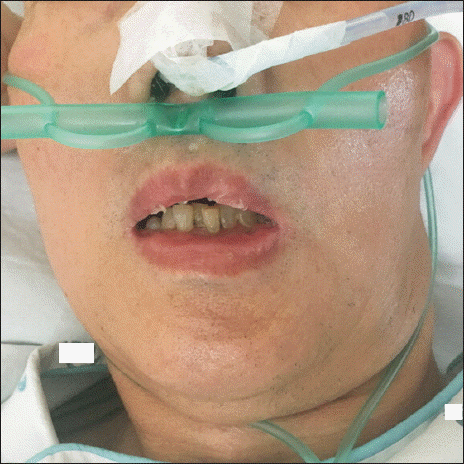
The risks due to difficult intubation were explained to the patient’s legal guardian and, after explaining the need for exploring the reason behind limited mouth opening, consent was obtained for additional tests. Three-dimensional CT of the face was performed to investigate the patient’s restricted mouth opening and revealed no abnormalities at the temporomandibular joint. A neurological consultation suggested that the limited mouth opening was a result of severe muscle spasticity related to PML. After surgery, the patient’s mouth opening remained limited because of muscle spasticity (Fig. 2).
DISCUSSION
PML is a brain disease resulting from symptomatic JC virus infection. Reactivation of the JC virus occurs when the infected individual becomes immunosuppressed by HIV infection. Upon reactivation, the virus migrates to the brain, where lytic infection in the host cells leads to PML. The diagnosis of PML is confirmed by immunohistochemistry of a brain biopsy specimen or by detection of JC virus DNA in the CSF on PCR analysis. Additional diagnostic criteria for PML include specific imaging findings and clinical features. The brain lesions in patients with PML are usually found in the white matter on magnetic resonance imaging [1]. High-intensity lesions with a scallop-shaped appearance are apparent on T2-weighted images [4]. Our patient had clinical features characteristic of PML, including diffuse high-intensity areas and atrophy of the cerebral white matter on magnetic resonance imaging, along with JC virus in the CSF confirmed by polymerase chain reaction positivity and, as such, was diagnosed with PML.
The clinical neurological symptoms of PML are diverse, and common manifestations include mental confusion, limb paresis, behavioral and cognitive abnormalities, speech or language disorders, and coordination and gait abnormalities [1,4]. Spastic hemiparesis also develops as the disease progresses [2]. In the present case, the patient’s neurological manifestations included an altered mental state, cognitive impairment, spastic hemiparesis and dysarthria, which contributed to his inability to communicate or cooperate with medical staff. Although performing a preoperative physical evaluation of the airway, including Mallampati classification, would have been difficult, we should have performed another physical examination of the airway, such as manually opening the patient’s mouth and examining the airway. Moreover, there are various additional physical evaluations that can be performed by anesthetists on patients with impaired mental status or loss of consciousness [5]. Generally, physical evaluation of the airway includes: inspection of the nasal cavity; verifying that it is possible to insert at least two finger widths between the upper and lower incisors; that there are no deformities of the mouth and/or palate; and assessing the patient’s jaw protrusion and movement of the temporomandibular joint. Measuring the submental space, and verifying the anomalies of the face and head, neck and spine are also helpful in predicting difficult airway. Performing these physical assessments of the airway would avoid unexpected encounters with problems associated with limited mouth opening. This case was meaningful in that it raises awareness to physicians to prepare for unanticipated difficult airway by performing various physical assessments of the airway, even in patients with poor cooperation.
Restricted mouth opening is also known as trismus. This condition has many causes including ankylosis, infection (odontogenic origins, tetanus, parotid abscess), trauma (e.g., fracture of mandible and zygomatic arch), temporomandibular joint disorder (myofascial muscle spasm, disc displacement, and other conditions), oral tumor, drugs, radiotherapy, and chemotherapy. Temporomandibular joint disorder is a common cause of trismus [6]. Normal temporomandibular joint function depends on coordinated contraction of the masticatory muscles acting on an intact condyle disc complex. Therefore, patients with a disorder of the masticatory muscles or disc condyle experience difficulty with mouth opening [7]. We initially attributed the patient’s limited mouth opening to dysfunction of the articular disc, but no abnormality was found on CT imaging of the face. A masticatory muscle problem was also considered; however, after seeking the opinion of a neurologist, we concluded that the patient’s restricted mouth opening was caused by a PML-related brain lesion.
PML is a demyelinating disease that affects the subcortical white matter of the CNS [4]. Spasticity occurs in patients with CNS disorders affecting the upper motor neurons [8]. In our patient, the PML lesions, to some extent, involved the corticobulbar tract and resulted in intractable spasticity of the masticatory muscles, which caused limited mouth opening. Stroke is also an example of an upper motor neuron disorder in which physicians can encounter muscle spasticity in the operating room [8]. In stroke, as in PML patients, there may be trismus due to brain lesion(s) [9]; therefore, extra precision during airway assessment must be exercised if the patient has a history of stroke. A previous case report described spasticity causing limited mouth opening that led to difficulty in tracheal intubation in a patient with Rett syndrome who exhibited trismus and spasticity [3]. The medical staff predicted limited mouth opening with adequate physical assessment and comprehensive understanding of disease, and hoped to be successful with fiberoptic intubation [3].
As with the previous case, fiberoptic intubation can be attempted when anticipating trismus [7]. Retrograde guidewire-assisted intubation also can be used in patients with restricted mouth opening [10]. If fiberoptic intubation fails, a light wand may also be a suitable alternative given that successful cases of intubation using light wand have been reported [11]. Awake fiberoptic intubation is another method that can be attempted when difficult intubation is expected due to limited information about the patient’s airway [12]. In this case, awake intubation would have been difficult due to the lack of patient’s cooperation, leading to biting of the fiberoptic scope and unstable posture. As mentioned, there are many reports describing intubation techniques for patients with limited mouth opening. Additionally, if endotracheal intubation is successfully achieved with a single-lumen tube through such airway management, we can substitute the double-lumen endotracheal tube that was prepared by using an endobronchial blocker to proceed with the operation [13]. As with this case, waking the patient as per the difficult airway management guidelines is also an option if trismus is not anticipated and mask ventilation is possible. If mask ventilation fails, immediate emergency cricothyroidostomy must be performed [14]. Fortunately, mask ventilation was maintained in this case; therefore, we could have postponed the surgery to conduct further, more thorough preparations by accurately assessing the airways. Despite the availability of such various options for airway management, the authors fell short by only preparing the GlideScope in this case. We also made a mistake in preparing the Macintosh no. 4 blade, which is commonly used in adults, even though the airway was not properly assessed. Fortunately, the patient was intubated successfully using the GlideScope and the operation was completed uneventfully without the need for one-lung ventilation. However, our limitation in this case was that we did not prepare various airway management strategies. Importantly, this case highlights the fact that inadequate airway assessment can lead to poor airway management preparation, thereby posing harm to the patient. Through this case, we are reminded the need to learn and prepare various airway assessment and management strategies under all circumstances.
In conclusion, spasticity in patients with PML could be a predictor of limited mouth opening. Patients with PML exhibit many neurological manifestations that could make preoperative airway evaluation difficult and endotracheal intubation challenging. The anesthesiologist should have a thorough knowledge of pre-operative assessments and perform active physical assessment of the airway to plan appropriate airway management in patients with this disease.


 PDF
PDF Citation
 Print
Print


 XML Download