This article has been
cited by other articles in ScienceCentral.
Abstract
A 6-month-old boy was scheduled for a laryngeal mass excision and tracheal bougienage for secondary subglottic stenosis. Following successful excision of the laryngeal mass, a tracheal tube was temporarily extubated for tracheal bougination. However, tracheal re-intubation using a direct laryngoscope with the Miller blade failed because of mucosal swelling and bloody secretions. Following multiple intubation attempts, the patient’s peripheral oxygen saturation had decreased to 52%. Immediately, a video laryngoscope was requested, and, by using the C-MAC® video laryngoscope, the patient was successfully re-intubated. Because pediatric patients are more vulnerable to desaturation, extreme caution should be used in securing airways even during a short apneic period. Using a video laryngoscope at the first intubation attempt would be useful for successful tracheal intubation.
Keywords: Difficult intubation, Pediatrics, Video laryngoscope
INTRODUCTION
Airway management in pediatric patients can be especially difficult because of their anatomy and an increased risk of hypoxia. A video laryngoscope can provide a better image of laryngeal anatomical structures than can a conventional direct laryngoscope, thereby improving patient safety. Even in an emergency situation which requires immediate intubation, a video laryngoscope would be more successful than a conventional direct laryngoscope [
1]. In this case report, we describe successful tracheal re-intubation using a C-MAC
® video laryngoscope after multiple intubation failures under direct laryngoscopy in a temporarily extubated pediatric patient.
CASE REPORT
A 6-month-old boy (weight 4.2 kg) was scheduled for laryngomicroscopic surgery (LMS) and bougienage for subglottic stenosis. He was born at 24 weeks’ gestation, weighing 800 g at the time of birth. His Apgar scores at birth were 3 at 1 minute and 7 at 5 minutes. He received surgical ligation of the patent ductus arteriosus 3 months prior to the current procedure. Chest radiography showed mild bronchopulmonary dysplasia in the right upper lobe. Computerized tomography of the trachea revealed secondary subglottic stenosis due to tracheal buckling, the largest diameter of which was nearly 3.5 mm. Preoperative electrocardiogram and laboratory findings were all within normal ranges. Two months earlier, he was admitted to the neonate intensive care unit (NICU) and treated for an upper respiratory infection. Although he generally recovered well, inspiratory stridor was still heard and chest wall retraction was observed. In the NICU, the patient’s peripheral oxygen saturation (SpO2) was well maintained at more than 98% with an oxygen hood. However, a day before the scheduled operation date, on the suspicion of aspiration, a transient desaturation occurred and emergency tracheal intubation was performed using a cuffless tracheal tube with an inner diameter of 3.5 mm in the NICU.
On arrival at operating room, the patient was connected to a ventilator system and general anesthesia was induced with sevoflurane. Rocuronium bromide 2.0 mg was administered and a tracheal tube was fixed at a depth of 9.5 cm in the left side angle of the mouth. Breath sounds were bilaterally audible on chest auscultation. Anesthesia was maintained with sevoflurane in 50% oxygen/air. After laryngeal exploration via suspension laryngoscope without tracheal tube change, a 0.4 cm false vocal cord mass was successfully excised with a CO2 laser; LMS duration was 30 minutes. Following vocal cord mass excision, the surgeon temporarily removed the tracheal tube to expose the subglottic area for bougienage. Ventilating bronchoscopy was not considered because it was too long and large for subglottic stenosis. After successful tracheal bougienage, the surgeon tried re-intubation with a 3.5 mm cuffless tracheal tube by direct laryngoscopy with a #0 Miller blade but failed because of a poor laryngeal view and bloody secrtions. Despite multiple attempts at direct laryngoscopy by the surgeon and attending pediatric intensivist, the vocal cord could not be adequately observed because of soft tissue swelling and mucosal bleeding. The patient’s SpO2 decreased from 100% to 52% and manual ventilation with a facial mask was immediately begun. However, the patient’s spontaneous breathing movement re-appeared, inhibiting appropriate ventilatory control. Therefore, we decided to use a C-MAC® video laryngoscope (Karl-Storz, Tuttlingen, Deutschland). After additional administration of rocuronium 2.0 mg and gentle oropharyngeal suction under C-MAC® laryngoscopy, the patient’s vocal cord was adequately exposed. The patient was successfully re-intubated with a 3.5 mm cuffless tracheal tube under C-MAC® video laryngoscopy with a #0 Miller blade, and tracheal intubation was confirmed by the presence of end-tidal carbon dioxide curve and bilateral breath sounds. After surgery, the patient was delivered to the NICU and extubated six hours later without any complications.
DISCUSSION
In pediatric patients, anatomic features such as a relatively large tongue, a longer epiglottis, and an anteriorly-located larynx make for greater difficulty in airway management than is the case in adults [
2]. Also, pediatric patients are at increased risk for hypoxia during tracheal intubation because of limited oxygen reserves and a higher rate of oxygen consumption. Thus, particularly in pediatric patients, fast and accurate tracheal intubation is necessary. As video laryngoscopy provides a better glottic view than conventional direct laryngoscopy, it can be effective in pediatric tracheal intubation [
3].
C-MAC
®, a video laryngoscope which has features that are similar to a direct laryngoscope, can be useful for successful tracheal intubation on the first attempt [
1,
4]. The C-MAC
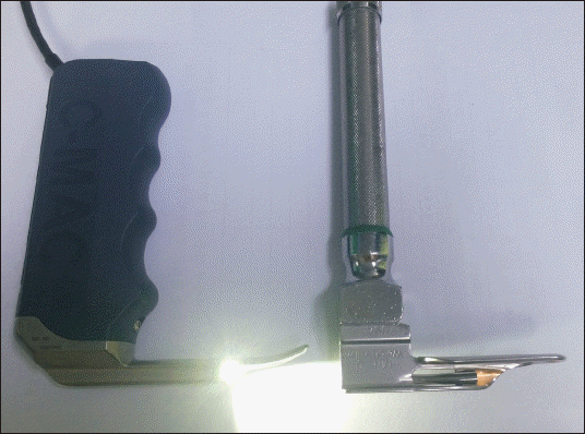
® video laryngoscope has an external light source and a camera on the distal third of the blade, thereby providing an indirect, magnified view on the display monitor. The pediatric-sized blade used with C-MAC
® (#0 Miller blade) is longer and at a slightly different angle than the conventional Miller blade of the same size (
Fig. 1) and can be effectively used for tracheal intubation in pediatric patients [
2,
5]. Compared with direct laryngoscopy, C-MAC
® is effective in patients with unexpected difficult airway [
6,
7], or as a rescue device in failed intubations [
8]. Compared with other video laryngoscopes, such as Trueview and Glidescope, C-MAC
® provides a better laryngeal view [
9,
10].
Fig. 1
Comparison of two intubation devices. The C-MAC® video laryngoscope with a #0 Miller blade (left) has a longer blade with a slightly angular tip and a brighter light source than does the conventional direct laryngoscope with a #0 Miller blade (right).

There are several specific ventilation techniques associated with LMS, such as transtracheal jet ventilation, high frequency oscillatory ventilation, and intermittent ventilation with a microlaryngoscopy tube [
11,
12]. However, in the present case, a jet ventilator or microlaryngoscopy tube was not available and the tracheal tube needed to be removed for tracheal bougienage. After tracheal bougienage, prompt re-intubation using a direct laryngoscope was not successful because of mucosal bleeding and edema. Following multiple intubation attempts under a poor laryngoscopic view, soft tissue injury could cause a further airway edema and may aggravate the difficulty of intubation. The patient was successfully intubated using a C-MAC
® video laryngoscope within a few minutes; therefore, it would have been more appropriate if we had used a video laryngoscope as a first choice. One cause of the failed re-intubation was that the anesthesia was wearing off and muscle relaxation was partially reversed at the time of temporary extubation. A partially re-appeared spontaneous breathing movement also contributes to difficult intubation due to poor alignment in the pharyngeal-laryngeal axis and unsynchronized respiratory movement. Delayed intubation time and repeated laryngeal stimuli could have also caused vigorous movement. However, we think that bloody secretions, mucosal swelling, and multiple intubation attempts under poor laryngoscopic view were the major causes of failed intubation in this situation, and the patient’s unsynchronized respiratory movement due to partially reversed neuromuscular blockade only slightly aggravated the situation. Thus, using C-MAC
® for visualizing the glottis may contribute to successful intubation more than would administration of additional neuromuscular blocking agents. Even during a very short period, temporary tracheal extubation can cause a serious clinical problem and, if necessary, a video laryngoscope should be used as a first choice for postoperative re-intubation. Additionally, a supraglottic airway device such as i-gel™ should be prepared as a rescue device, which is suggested in a published guideline for difficult airway management in pediatric patients [
5,
13].

