INTRODUCTION
There are 10,000–20,000 newly diagnosed osteonecrosis of the femoral head (ONFH) patients annually in the United States [1]. Proposed risk factors of ONFH include corticosteroid use, alcohol consumption, trauma, and coagulation abnormality [2]. Because ONFH is a progressive disease, early diagnosis and proper treatment are important [3]. If untreated, ONFH is known to result in the collapse of the femoral head in 20% to 75 % of patients within 1 to 3 years. About 50% of patients need surgical treatment within 3 years after its diagnosis [1]. If the depression of the femoral head is severe or if there are degenerative changes, total hip replacement (THR) is required [4,5]. To make appropriate decision on hip joint replacement, physician should consider the severity of pain and the degree of radiographic progression [5]. It is important to prevent depression of the femoral head and to reduce pain in old patients. In this study, patients (Association Research Circulation Osseus classification stage III) underwent Botulinum toxin type A (BoNT-A) injection into the psoas major muscle. Here we report five cases of ONFH managed by BoNT-A injection to the psoas major muscle.
Go to : 
CASE REPORT
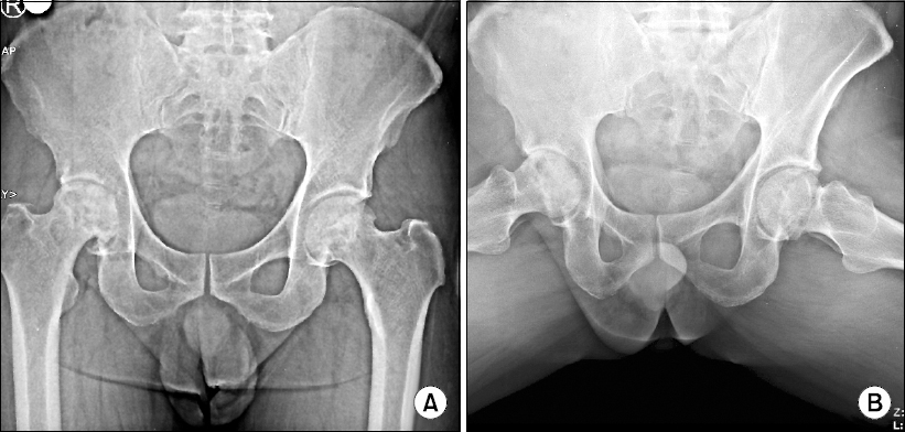
A 73-year-old male patient with height of 168 cm and weight of 65 kg visited our hospital with right hip pain. He had been receiving medications including tramadol 200 mg/day and mefenamic acid 1,000 mg/day at another hospital for two years due to pain in his hip joint under suspected ONFH. At the time of his hospital visit, the patient presented pain in the right hip joint area with radiating pain in the right thigh. Pain was aggravated on walking. Cramping pain radiated to the anterior thigh. His visual analogue scale (VAS) for pain (0/100 mm means no pain, 100/100 mm means worst pain imaginable) was 90/100 mm. In physical examination, his right hip joints had positive responses to Patrick test and Leg rolling test. When he visited our hospital, stage III (Association Research Circulation Osseus classification) osteonecrosis of the right femoral head was diagnosed based on hip simple radiography by a radiologist (Figs. 1A and 1B).
We conducted a non-operative treatment of his ONFH through restricted weight-bearing and extracorporeal shock wave therapy (ESWT). During ESWT, the patient had severe pain. The patient had poor compliance in restricted weight bearing due to his poor quality of life. After 6 months of this treatment, the patient was not satisfied with the degree of pain relief.
Previously performed diagnostic block of psoas major muscle with 20 ml of 0.5% lidocaine in this patient showed a duration of pain relief for less than 8 hours. Therefore, we performed fluoroscopy guided intra-muscular injection of BoNT-A into the psoas major muscle in order to release contracted psoas major muscle between L3 and L4 vertebrae.
The patient was prepared in prone position with a pillow under the abdomen in order to widen the gap between lumbar spines. The shadow of the psoas major muscle was first checked from the anterior-posterior view of fluoroscopy. The shadow of psoas muscle between transverse processes of L3 and L4 was confirmed as the needle entry point. After anesthetizing the entry site with 1 ml of 1% lidocaine, a 23-gauze, 10-cm Spinal needle was inserted vertically with gun-barrel technique.
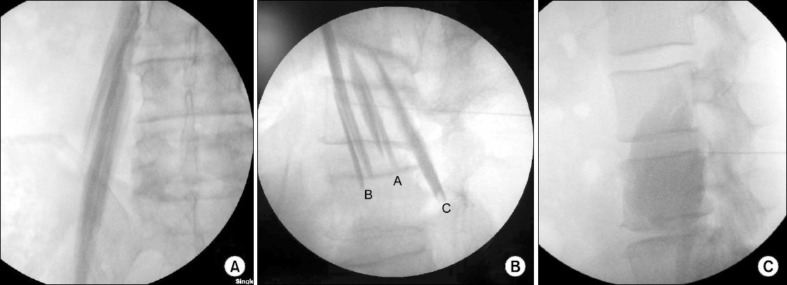
At 3–4 cm depth from the skin, needle entry into the psoas major muscle was confirmed on both AP view and lateral view. The psoas compartment plexus was then confirmed by loss of resistance (LOR) technique. After LOR, radiating pain down to the lower limb was developed. Needle was then advanced about 2 cm more from this point, reaching the deep part of the psoas major muscle. This was checked with a contrast agent (Fig. 2A). The depth of the deep part of the psoas major muscle from the skin was 8.5 cm, while the psoas compartment plexus was approximately 6.5 cm (Fig. 2B). After we injected dye solution into the deep part of the psoas major muscle (Fig. 2B), we confirmed that the dye could spread over the psoas major muscle without blood aspiration. Subsequently, we injected 20 ml of 0.5% lidocaine, 1 ml of contrast agent (Iopromide), and BoNT-A supplied in vials of 100 units of mixed powder including 0.5 milligrams of human albumin and 0.9 milligrams of sodium chloride (Botulex®, Hugel, Korea) (Fig. 2C). Due to the large volume of the solution (20 ml of 0.5% lidocaine, 1 ml of contrast, 100 Botox Units were reconstituted in 1 ml of 0.9% normal saline), after we injected it to the deep part of the psoas major muscle, solution was spread into psoas compartment. After we confirmed that the solution was spread into the entire muscle (Fig. 2C), numbness of the lower limb occurred due to psoas compartment block.
 | Fig. 2(A) Anteroposterior image of Psoas major muscle after fluoroscopic guided muscle injection with contrast media. (B) Lateral image of psoas major muscle after fluoroscopic guided injection with contrast media. A: psoas compartment plexus, B: Deep part of the psoas major muscle, C: superficial part of the psoas major muscle. (C) Lateral image of psoas major muscle after fluoroscopic guided injection with 20 ml of 0.5% lidocaine, 1 ml of contrast agent (Iopromide), and 100 unit of botulinum toxin type A (BoNT-A) were reconstituted in 1 ml of 0.9% normal saline. |
After performing the procedure, the patient’s oxygen saturation, blood pressure, and pulse were checked in the recovery room for 30 minutes in 5-minute intervals. The patient complained of post-injection back pain and sensory decrease at the frontal side of the femoral region. The duration of the sensory decrease was 30 minutes. The patient’s hip pain was decreased from 90/100 mm to 50/100 mm in visual analogue scale (VAS) at one day after the procedure. It was decreased further to 20/100 mm in VAS at two weeks after the procedure. The VAS score of pain in the right hip of the patient was reduced to below 20/100 mm in VAS between two weeks and six weeks after the procedure. Medication was decreased to tramadol 100 mg/day at one week after the procedure. Medication was no longer required between two weeks and six weeks after the procedure. However, at 6 weeks after the procedure, the pain in the right hip during walking was increased to VAS score of 50/100 mm. The patient resumed medication to extent dosage before a procedure. The pain level was increased to VAS score of 90/100 mm at 8 weeks after the procedure. We repeated the same procedure. BoNT-A injection to the psoas major muscle was conducted a total of three times in 6 months with 2-month intervals. There was no radiographic progression change during follow-up of 6 months.
For another four patients with ONFH (Association Research Circulation Osseus classification stage III) who were not responsive to drug therapy (non-steroidal anti-inflammatory drugs) for two years, we performed the same conservative treatments (ESWT, restricted weight-bearing) for 6 months as described above. However, these patients were not satisfied with the degree of pain relief. Alternatively, we performed psoas major muscle injection with BoNT-A for pain control.
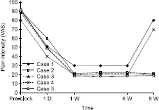
The amount of BoNT-A used was 2 U per kg of body weight. In addition, 20 ml of 0.5% lidocaine and 1 ml of contrast agent (Iopromide) were injected along with BoNT-A. In the five patients, the mean hip pain before procedure was 88 ± 0.4/100 mm in VAS. At one day after the procedure, the pain was decreased to 50 ± 0.6/100 mm in VAS. At two weeks after the procedure, the pain was significantly reduced to 22 ± 0.4/100 mm in VAS. The period of pain relief was a minimum of 6 weeks and a maximum of 8 weeks (Fig. 3). The amount of medication needed was reduced after the procedure (Table 1).
 | Fig. 3Change of VAS after psoas major muscle relaxation with botulinum toxin type A in the five ONFH patients. Duration: 1 day–8 weeks, D: day, W: week, ONFH: Osteonecrosis of the femoral head. VAS: visual analog scale, Ranging from 0 = no pain to 10 = worst pain imaginable. |
Table 1
Patient Demographics and Clinical Changes
Go to : 
DISCUSSION
There are several conservative measures proven to be effective for ONFH [6], we performed conservative treatments for these patients as described above case. However, patients were not satisfied with the degree of pain relief. In our study, patients (Association Research Circulation Osseus classification stage III) underwent BoNT-A injection into the psoas major muscle. This procedure was performed as an alternative management in old patients when there was no effect from other conservative treatments.
Conservative treatments in early stage of ONFH are as effective as surgical treatments [6]. Approximately 10% of patients diagnosed with ONFH will undergo THR [1]. However, THR is not a permanent cure for ONFH. It should be used as a last resort [5]. There are non-operative treatments for ONFH, including restricted weight-bearing, bisphosphonates, anticoagulants, statins, extracorporeal shock wave therapy, pulsed electromagnetic therapy, and hyperbaric oxygen [6]. The goal of non-operative treatment of ONFH in early stage is to reduce pain, prevent depression in the femoral head, and improve function of the hip joint [3]. Long term effect of conservative treatments for ONFH has not been confirmed yet [4]. After collapse of the femoral head, THR is the main treatment option for ONFH [1]. In spite of collapse of the femoral head, ONFH patients without severe pain can use conservative treatments [4]. It is important to prevent depression of femoral head and control pain in old patients.
This procedure was performed as alternative management in old patients who were not responsive to other conservative treatments. Psoas major muscle originates from the vertebral body of T12 to L4. It is combined with iliacus muscle. Its tendon is inserted into the lesser trochanter of the femur. Tendon of iliopsoas is located immediately adjacent to the femur joint capsule. Therefore, pathologic contracture of the psoas major muscle can cause excessive flexion of the hip joint, thus compressing the femoral head into the acetabulum [7]. As a result, when hip joint pressure increases, femoral head perfusion is reduced [7], which can lead to osteonecrosis of the femoral head. Psoas major muscle is separated into a superficial part and deep part. Psoas compartment exists between the superficial part and the deep part, including lumbar plexus consisted of L1–L4 nerve [8]. Sensory innervation of the hip joint area is composed of obturator nerve and femoral nerve from the lumbar plexus [9].
In other studies, muscles around hip joint including adductor muscle, tensor fascia lata muscle, and rectus femoris muscle can also affect its pressure [10]. However, some studies have reported that injection into the adductor muscle has a risk of femoral nerve injury and hematoma [8]. Although psoas major muscle plays an important role as a stabilizer of the hip joint [7], several other muscles and ligaments (iliofemoral ligament, pubofemoral ligament, ischiofemoral ligament) around the hip joint contribute to the stabilization and motion of the hip joint [10]. Therefore, psoas major muscle relaxation only with BoNT-A injection might be insufficient to reduce the pressure of the hip joint.
In this study, we suggested an alternative pain management of ONFH by injecting BoNT-A into the psoas major muscle by inducing psoas muscle relaxation and blocking lumbar plexus. For muscle relaxation, we used BoNT-A together with 0.5% lidocaine. It has been reported that BoNT-A can decrease muscular stiffness, mitigate dystonia, and extend the range of joint movement upon intramuscular injection [11]. Therefore, we used BoNT-A for muscle relaxation to decrease stiffness and dynamic contracture in the psoas major muscle, which could ultimately decrease hip joint pressure. It has been reported that BoNT-A is safe when used at less than 15 U per kg of the body weight [12]. Therefore, we used BoNT-A for muscle relaxation at the amount of 2 U per kg of body weight. Higher dose of BoNT-A can decrease muscular activity in a greater degree and for a longer time [13]. Depending on the response of patient, the amount of BoNT-A might be adjusted to achieve the best effect.
When we performed psoas compartment block without BoNT-A, the duration of pain relief was less than 8 hours [14]. We had performed epidural block and L2–L4 nerve root block with 4 ml of 0.5% mepivacaine to other patients of ONFH. The duration of pain relief was less than 4 hours. According to other paper, lumbar plexus block with BoNT-A has reduced diabetic leg pain for 4 months [15].
In this report, for the total of five patients, their hip pain before treatment was 88 ± 0.4/100 mm in VAS. At one day after the procedure, their pain was decreased to 50 ± 0.6/100 mm in VAS. At two weeks after the procedure, their pain was significantly reduced to 22 ± 0.4/100 mm in VAS. The period of pain reduction was a minimum of 6 weeks and a maximum of 8 weeks. Our results in the duration of pain relief were similar to results on the duration of electrical stimulation change with BoNT/A in a previous report [13]. It has been reported that BoNT-A can decrease compound muscle activity starting from the day after intramuscular injection and reaching a maximum decrease in muscle activity at week 2. Starting from week 6 after the treatment, the muscle activity is then recovered up to the level at day 1 [13]. After procedure, duration of the numbness at the frontal side of the femoral region in patients was 30–120 minutes.
In some patients of ONFH, core decompression is effective in reducing the pressure of the hip joint, which might prevent ONFH from progressing to osteoarthritis [6]. However, core decompression must be conducted under anesthesia in an operating room. On the other hand, it is possible to perform psoas major muscle injection using BoNT-A without hospitalization or anesthesia.
The limitation of this study includes the relatively short term follow-up, the small number of patients (n = 5), and possible patient selection bias. In addition, this is a retrospective case series without comparison group. Therefore, more research studies including comparison of different drugs and injection sites are needed.
In summary, BoNT-A injection to the psoas muscle could be considered as an alternative procedure to control pain for ONFH caused by increased hip joint pressure due to excessive contraction of the psoas major muscle. More progressive degeneration of hip joint might be prevented by directly injecting BoNT-A into the deep part of the psoas major muscle. However, more studies are merited to confirm the efficacy and duration of this procedure in more patients with ONFH.
Go to : 


 PDF
PDF Citation
 Print
Print


 XML Download