This article has been
cited by other articles in ScienceCentral.
Abstract
Objectives
Lymphatic malformations of the orbit are rare lesions that constitute approximately 1% to 8% of all orbital masses. They are difficult to treat since they do not remain within anatomic boundaries and tend to penetrate into normal orbital structures. The aim was to analyze clinical courses and therapy options in patients with lymphatic malformations of the orbit.
Methods
Thirteen patients with orbital lymphatic malformations confirmed by magnetic resonance imaging between 1998 and 2009 were enrolled in this study. Patients' charts were retrospectively reviewed to analyze clinical courses and treatment options.
Results
Four patients suffered from isolated intraorbital lymphatic malformations without conjunctival involvement, in three of them the masses were completely resected, in one patient close controls were performed. Three patients had isolated intraorbital lymphatic malformations with conjunctival involvement. Surgical volume reduction of the exterior parts of the lymphatic malformation were performed without any complications and satisfying outcome in these cases. Six patients suffered from intra- and periorbital lymphatic malformations. In 3 patients a watch-and-wait strategy was initiated. In the other 3 patients a surgical therapy was performed, one patient additionally received sclerotherapy with OK-432; however, these 3 patients suffered from residual lymphatic malformations.
Conclusion
The presented cases underline the inconsistencies in the malformations behavior and underscore the inability to make specific recommendations regarding treatment. The treatment decision should be based on the size and location of the lymphatic malformation. The untreated patient must be watched for signs of visual detoriation, which may signal the need for therapeutic intervention.
Go to :

Keywords: Lymphatic malformation, Orbit, Orbitotomy, OK-432
INTRODUCTION
Lymphatic malformations are benign malformations of the lymphatic system which represent between 1% and 8% of all orbital masses [
1]. Although different theories have been suggested, the aetiology of lymphatic malformations remains unclear [
2]. Lymphatic malformations in general have no predilection for sex, however, lymphatic malformations of the orbit seem to be slightly more common in females than in males, with gender ratio of 1.4:1 [
3]. Lymphatic malformations of the orbit characteristically involve the subconjunctival and periocular tissues. Those lesions that involve the superficial or anterior orbital structures are diagnosed earlier [
4]. Vesicles may be noted in the conjunctiva. Patients typically present with a variety of symptoms like swelling, intraorbital hemorrhage, ocular proptosis, blepharoptosis and cellulitis.
Complete surgical resection still remains the best treatment for lymphatic malformations; since incomplete resection may lead to recurrence [
5]. Other treatment options, such as sclerotherapy have been proposed as an alternative to reduce the impact and complications of surgery. Various products, such as doxycycline, bleomycin, ethibloc, and OK-432, have been used as sclerotherapeutic agents [
6]. Apart from OK-432, the other agents were reported to cause perilesional fibrosis and thus to complicate eventual surgical excision. Treating orbital lymphatic malformations with sclerosants the risk of orbital compartment syndrome leading to a loss of vision due to an expansion of the lesion after injection of the sclerosant has always to be kept in mind. Therefore the treatment of lymphatic malformations of the orbit can be challenging.
The aim of the present study was to evaluate the disease-related impairments and the clinical courses of patients with lymphatic malformations of the orbit and to discuss the treatment opportunities.
Go to :

MATERIALS AND METHODS
All patients who presented with orbital and periorbital lymphatic malformations to the Department of Otolaryngology, Head and Neck Surgery, Philipps University of Marburg between 1998 and 2009 were included in this analysis. The diagnosis was made when magnetic resonance (MR) imaging and clinical features were both consistent with the diagnosis. In some patients the diagnosis was confirmed by histopathologic examination. Treatment response was assessed by clinical aspects, recurrences were evaluated by clinical features and MR imaging. Retrospective data were collected from clinical charts, radiology and pathology reports.
In case of surgery the surgical approach was determined by the location and extent of the malformation and intraoperative antibiotics and corticosteroids (dexamethasone 250 mg) were administered. Lateral orbitotomy was performed in cases of periorbital and intraorbital tumors, located dorsal, basal and lateral to the optic nerve. Medial orbitotomy was performed via a transnasal endoscopic approach. It is especially suitable for masses of the medial and inferomedial orbit. The lamina papyracea was exposed and partly removed to expose the periorbita. The periorbit was incised to allow access to the orbital content.
Sclerotherapy with OK-432 was performed under general anaesthesia according to the method of Ogita et al. [
7]. The aspiration of cyst fluid was performed with ultrasound guidance to localize the cysts. A 20-gauge sleeve needle was advanced under ultrasound guidance until the respective area was reached, 1.5 mL fluid was aspirated and replaced by 1.5 mL of 0.01 mg/mL dilution of OK-432 in physiological serum.
Go to :

RESULTS
Thirteen patients with lymphatic malformations of the orbit were investigated: 5 male and 8 female patients (
Table 1). The mean age at presentation was 16 years, ranging from 3 months to 39 years. Their ages at diagnosis ranged from birth to 23 years. Seven patients had isolated orbital lymphatic malformations. An adjacent periorbital involvement was seen in 6 patients. The lymphatic malformation was unilateral in all cases. The left orbit was affected in 10 cases and the right orbit in 3 cases.
Table 1


Isolated intraorbital lymphatic malformation without conjunctival involvement
Four patients (2 males and 2 females) suffered from isolated intraorbital lymphatic malformations without conjunctival involvement. In two cases (case 1 and 4) the clinical manifestation was a progressive proptosis without restriction of eye movements or loss of vision. The mass was completely resected via medial orbitotomy in one patient, in the other case a wait-and-see strategy was indicated since the optical nerve lies within the retrobulbar lymphatic malformation. The other two patients (case 2 and 3) presented with lymphatic malformations resembling symptoms of orbital complications of a rhinosinusitis with loss of vision. In both cases the masses were completely resected via lateral orbitotomy; histologic examination confirmed the diagnosis of lymphatic malformation. The vision improved to 1.0 postoperatively in both cases.
Isolated intraorbital lymphatic malformations with conjunctival involvement
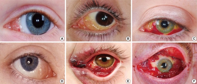
Three patients (case 5-7) presented with intraorbital lymphatic malformations with conjunctival involvement which were apparent since birth and had been treated by surgery at other hospitals before (
Fig. 1A-C). Due to the aesthetic impairment and the potential risk of hemorrhage a surgical volume reduction of the exterior parts of the lymphatic malformation was recommended in all cases. The operative procedures were performed without any complications and satisfying outcome in all cases (
Fig. 2).
 | Fig. 1Typical aspects of conjunctival involvement in lymphatic malformations of the orbit. Frosh egg like vesicles in the medial area of the conjunctiva in cases 6 (A), 7 (B), and 8 (D). Subconjunctival hemorrhage and small vesicles covering the conjunctiva 5 (C). Multiple lymph-filled cysts with purple color due to occasional rupture of capillaries in cases 9 (E) and 13 (F). 
|
 | Fig. 2Lymphatic malformation of the orbit with conjunctival involvement before (A) and after (B) partial resection (case 7). 
|
Patients with intra- and periorbital lymphatic malformations
Six patients (3 males and 3 females; case 8-13) suffered from lymphatic malformations of the orbital and periorbital region (
Figs. 1D-F,
3). Five of them were treated at other hospitals before. In a 2
1/2-month-old girl who was referred with a left-sided peri- and intraorbital mass since birth a surgical volume reduction was performed. 3
1/2 years later the vision of the left eye was 0.7. In one patient with an exophthalmia, ptosis and visual impairment of the left eye due to a lymphatic malformation with venous components an incision of the periorbit after lateral orbitotomy in combination with Nd:YAG laser therapy of venous components of the malformation was performed (
Fig. 1F). A 4-year-old girl was treated by ultrasound guided intralesional injection of OK-432. After OK-432 therapy a significant improvement of the eye opening has been noted (
Fig. 1C). Due to recurrent swelling attacks of the lymphatic malformation with exophthalmia 2 years later an incision of the periorbit via lateral orbitotomy was performed. In two cases revision surgery was recommended to reduce the malformations. The patients decided to comply with the advice in case of further symptoms. In another girl a complete resection of the malformation seemed not to be possible without violating the function, therefore the wait-and-see strategy was indicated.
 | Fig. 3Effect of OK-432 therapy. (A) Four-year-old girl with orbital lymphatic malformation before OK-432 therapy. (B) One day after OK-432 injection the typical inflammatory reaction and swelling after OK-432 sclerotherapy is seen. (C) Result 7 months after OK-432 sclerotherapy: a significant improvement of the eye opening has been noted. 
|
Go to :

DISCUSSION
Lymphatic malformations of the orbit must be differentiated from haemangiomas, venous malformations, rhabdomyosarcomas and lymphomas. The diagnosis usually depends on clinical examination and different imaging modalities. MR imaging precisely delineates these lesions. However, there is a heterogenous pattern. The lymph-containing spaces tend to be hypointense. After haemorrhage into the cysts there will be hyperintense parts on T1-weighted sequences because of the paramagnetic properies of blood breakdown products [
8]. Due to typical morphologic appearance histological confirmation of the diagnosis is only rarely necessary. In this series, lymphatic malformations of the orbit were more common in females, consistent with data reported in the literature [
3]. To date numerous theories have been proposed regarding the embryonic aetiology of lymphatic malformations, however, the precise pathogenesis is still unknown [
2]. As the orbit is free of lymphatic vessels orbital lymphatic malformations originate either from displaced foetal cells of the lymphatic channels [
9] or from lymphatic malformations extending from the lid and conjunctiva.
Lymphatic malformations of the orbit have the tendency to insituate within the intra- and extraconal space thereby violating normal anatomic boundaries [
10]. They often have visible superficial components in the conjunctiva or eyelid, deeper lesions within the orbit especially without conjunctival involvement may are quiescent for many years and often became symptomatic in case of infection and sudden swelling. This was also true for the cases presented in this series. In the presented series 8 patients had a visible component of the lymphatic malformation at birth, in the other cases the lymphatic malformation was diagnosed later in life; the oldest patient was 23 years old.
Clinical findings of lymphatic malformations of the orbit like swelling of the eyelid and exophthalmia can resemble orbital complications of rhinosinusitis [
11]. In the presented series two patients presented with symptoms resembling an orbital complication of rhinosinusitis since sinusitis led to expansion of the occult lymphatic malformation. Lymphatic malformations typically fluctuate in size with upper respiratory tract infections and expand suddenly due to intralesional hemorrhage and infection. Moreover there is always the risk for ocular emergencies like optic neuropathies and permanent visual disturbances due to spontaneous intracystic haemorrhage. However, in comparison to the extension of the lymphatic malformation most of the patients in the present analysis only have few complaints.
Various treatments methods have been described for lymphatic malformations of the orbit. Surgical removal still appears to be the best choice of treatment of lymphatic malformations. The problem is that important structures often run within the septa of the lymphatic malformation. Especially in extensive lymphatic malformations of the orbit it is difficult to perform a complete resection but preserve the function. Many orbital lymphatic malformations are diffusely infiltrative which makes their complete excision even more difficult. A complete resection is mainly possible in cases of well-delineated extraconal lymphatic malformations without involvement of the conjunctiva. If complete removal does not seem to be possible, the indications for surgery were varied. In some cases preservation of function or restoration of aesthetic appearance were the goals of surgery. Exploration of an unidentified orbital mass may also be the reason for surgical exploration. Visually threatening or aesthetically disfiguring disease should be treated. However, in many cases also a partial resection leads to an improvement of symptoms. A useful alternative treatment method in patients with proptosis or recurrent swelling episodes due to massive lymphatic malformation of the orbit which cannot be totally resected or in cases where a postoperative visual impairment would be inevitable after total resection is a bony orbital decompression [
12] or an incision of the periorbit. In the presented series, in 3 cases an orbital decompression was performed via medial or orbital orbitotomy. After a medial orbitotomy, there is a possible risk of extension of the lymphatic malformation to the sinuses with obstruction and sinus dysfunction. However, this was not seen in the presented cases.
Non-surgical procedures should be particularly proposed in unresectable and recurrent tumors. Especially sclerotherapy with OK-432 has been proposed as an alternative to surgery and has shown promising results. Recent studies indicate that the shrinkage of the cysts seems to be related to an immunomodulatory effect [
13,
14]. The efficiency of OK-432 is especially proven for macroscopic lesions. A regression of head and neck lymphatic malformations after OK-432 therapy is reported in up to 96% of the patients [
15]. The main advantage of OK-432 with respect to other sclerosing agents is the absence of perilesional fibrosis. OK-432 therapy has also been described to be effective in orbital lymphatic malformations [
16,
17]. One patient of the present series received OK-432 therapy with good outcome. The main disadvantage of this sclerosant is the local inflammatory reaction and swelling that occurs after injection and bears the risk of orbital compartment syndrome. Therefore OK-432 has to be used with caution in orbital lymphatic malformation and close controls after injection are indispensable. Moreover the patients must be informed about the off-label use.
A wait-and-see policy can be adopted for lymphatic malformations of the orbit before deciding for surgical removal especially if symptoms are slight and steady, if a complete removal is not possible without violating the function or if the patients refuse to treatment. In case of acute swelling episodes due to infection pharmacological intervention, predominantely with steroids and antibiotics, is considerate appropriate. Especially in small children a close control is necessary as it is known that occlusion of the pupil at an early age results in a poor visual prognosis.
In conclusion, there is no treatment modality that is ideal for all cases of lymphatic malformations of the orbit, since each treatment modality has its own risks, side effects and benefits. The treatment decision should be based on the size and location of the lymphatic malformation. The untreated patient must be watched for signs of visual detoriation, which may signal the need for therapeutic intervention.
Go to :


 PDF
PDF Citation
 Print
Print


 XML Download