Abstract
Objectives
Laryngomicrosurgery (LMS) is used to manage most vocal fold lesions. However, the functional voice outcome of the LMS might be diverse due to the influence of various factors. We intend to evaluate the incidence and etiologic factors of persistent dysphonia after LMS for benign vocal fold disease (BVFD).
Methods
We performed a retrospective review of 755 patients who underwent LMS for BVFD. We analyzed the clinical characteristics, preoperative and postoperative two onths voice studies. Postsurgical dysphonia was defined as grade 1 or above in GRBAS (grade, roughness, breathiness, asthenia, and strain) scale. Thirty nine patients (5.2%; 25 males and 14 females; average, 42.9 years; range, 21 to 70 years) were diagnosed with postsurgical dysphonia.
Results
There was no correlation between the diagnosis, coexistence with laryngopharyngeal reflux disease, habit of smoking, or occupational voice abuse and voice outcome. The patients with a worse preoperative acoustic parameter had aworse voice outcome. Stroboscopic findings showed excessive scarring or bowing in 21 cases, presence of lesion remnant in eight cases, prolonged laryngeal edema in five and no abnormal findings in three.
Go to : 
Benign vocal fold disease (BVFD), such as vocal polyp, vocal nodule, intracordal cyst or Reinke's edema is one of the most common causes which deteriorate the voice [1]. These laryngeal mucosal diseases are known to be caused by voice abuse; so-called 'phonotrauma'. Repeated collision forces and shear stress lead to microvascular injury and trauma to the epithelial basement zone [2,3]. Treatment of BVFD includes medical therapy, voice therapy by speech language pathologists or surgical therapy by otolaryngologists [1]. In patients whose voice has not improved by medical or voice therapy, laryngomicrosurgery (LMS) might be needed [4]. LMS is used to manage most cases of BVFD for the restoration of voice quality or diagnosis of the disease. LMS aims to improve the vibratory characteristics of the layered microvascular structure of vocal fold [5-7].
A patient who has persistent or recurrent dysphonia after LMS presents therapeutic challenges to the clinician, because the functional voice outcome of LMS might be diverse due to the influence of various factors. For example, in case of laryngopharyngeal reflux disease (LPRD), acid and pepsin give rise to the inflammation of laryngeal mucosa, which can render the, vocal folds susceptible to phonotrauma [8]. In addition, smoking, alcohol consumption, or coexistence of pulmonary disease also has been reported to influence the functional outcome of LMS [1,9,10]. However, there have been only a few studies about the incidence or etiologic factors of persistent dysphonia after the treatment of BVFD. Therefore, we aimed to evaluate the incidence and etiologic factors of persistent dysphonia after LMS for benign vocal fold lesions.
Go to : 
The charts of 1,088 patients who underwent LMS at Ajou University Hospital from March 2003 to April 2011 were retrospectively reviewed. A single operating surgeon (CHK) performed the LMS with a standardized microflap technique mainly with cold instruments. Each patient was set in supine the position under general anesthesia with the neck flexed and head extended. The largest laryngoscope possible was used. In vocal polyp or nodule, mucosal incision was made at the junction of the lesion and the superior surface of the vocal cord, and then the lesion was carefully dissected and removed. The microflap was redraped over the operative site. In the case of an intracordal cyst, the mucosal incision was made alongside the cyst which under the vocal fold mucosa and the cyst was carefully dissected with a cotton ball and cup forceps. Every effort was made to remove the cyst intact and the overlying mucosa was preserved as much as possible and draped over the area where the cyst was removed. In Reinke's edema, the mucosal incision was made on the lesion with CO2 laser or sickle knife and swollen Reinke's layer was sucked out. The redundant mucosa was resected with microscissors and the mucosal flap was re-draped. Depending on the 2-month postoperative voice analysis results, postoperative voice therapy was added as needed.
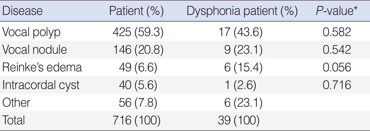
We excluded patients who underwent LMS due to malignant lesion, and who had prior history of LMS, other head and neck surgery or irradiation. Of these patients, 755 patients whose preoperative and 2-month postoperative voice analyses were completed were included. The mean age of the patients was 45.9 years (range, 18 to 70 years) and the diagnosis of the patients is described in Table 1. The patients were identified as having coexistent LPRD if they had ever been diagnosed with LPRD or prescribed a proton pump inhibitor. The habit of smoking or occupational voice abuse such as teachers, sales persons, or singers was also reviewed.
Preoperative and postoperative voice was analyzed perceptually with the GRBAS (grade, roughness, breathiness, asthenia, and strain) scale, acoustically using the multi-dimensional voice program (MDVP; KayPENTAX, Lincoln Park, NJ, USA), and morphologically with videostroboscopy (model 9200c, KayPENTAX). Patients were instructed to read a paragraph, which is used for voice analysis in Korea, at a comfortable loudness level and rate. A GRBAS score was given at the end of the evaluation session by one experienced speech pathologist and one otolaryngologist with the consensus on its score. The voice was scored using the five GRBAS parameters. As described by Hirano, the "G" represents grade or overall quality, "R" for roughness, "B" for breathiness, "A" for asthenia, and "S" for strain. Each parameter is rated on a 4-point scale: 0 means that there is no deficit in this parameter, 1 is for a mild deficit, 2 for a moderate deficit, and 3 for a severe deficit. Patients were instructed to pronounce the vowel "a" at a comfortable loudness level and a constant pitch for at least 3 seconds Each vowel pronunciation was recorded with a constant mouth to-microphone (SM48, Shure, Niles, IL, USA) distance of 5 cm using Computerized Speech Lab (model 4500, KayPENTAX). All digital recordings were made in a quiet room. On stroboscopy, the same speech pathologist and an otolaryngologist examined and categorized the dysphonia patients into four groups: 1) excessive scarring or bowing, 2) presence of lesion remnant, 3) prolonged laryngeal edema, or 4) no abnormal finding.
Persistent dysphonia after LMS was defined as grade 1 or above according to the GRBAS scale rated by one professional speech therapist and one otolaryngologist. We compared the clinical parameters and voice outcome between the postoperative dysphonia and normophonia group.
Comparisons were performed with the paired t-test, Fisher's exact test, and multivariate logistic regression analysis. Statistical significance was defined as P<0.05.
Go to : 
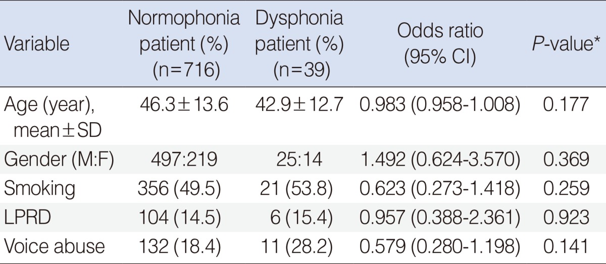
Of the 755 enrolled patients, 39 patients (5.2%) were classified into the persistent dysphonia group and the rest were classified into the normophonia group. Twenty-five patients were male and 14 were female, and the mean age was 42.9 years (range, 21 to 70 years). The age and gender distribution of the dysphonia group were not different from those of the normophonia group (Table 2).
Regarding the original disease, there was no correlation between the diagnosis and voice outcome (Table 1). Patients with Reinke's edema showed a tendency of worse outcome, but it was not statistically significant (P=0.056). Co-existence of LPRD, habit of smoking or occupational voice abuse was not correlated with the worse voice outcome (Table 2).
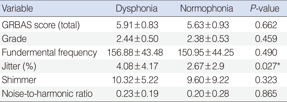
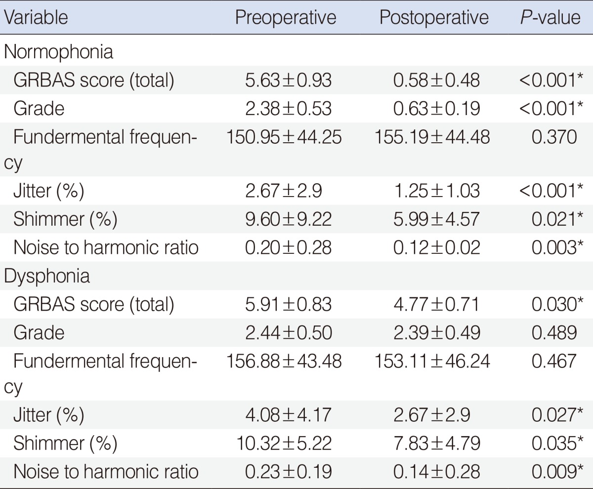
Preoperative GRBAS scales did not show a significant difference in both groups (Table 3). As we defined the dysphonia with GRBAS score, postoperative total GRBAS score and the grade was significantly poorer than the normophonia group (Table 4). The GRBAS scores significantly improved after LMS in both groups, but the grade did not improved in the dysphonia groups (Table 4).
Jitter, shimmer, and noise-to-harmonic ratio (NHR) significantly improved after LMS in both groups. Postoperative jitter and NHR of the dysphonia group was worse than those of the normophonia group, but shimmer was not different (Table 4). Preoperative acoustic analysis showed that preoperative jitter of the dysphonia group was significantly worse than that of the normophonia group (Table 3).
We reviewed the videostroboscopic findings of the patients classified in the dysphonia group. Excessive scarring or bowing was found in 21 patients, lesion remnants in eight, and prolonged laryngeal edema in five. In the remaining five patients, no abnormal findings on the vocal cord were found (Table 5). These stroboscopic findings were not correlated with the disease entities.
Go to : 
During the past two decades, there has been a lot of effort to introduce a variety of techniques for objectively measuring voice quality. Among the measurements currently used, perturbations of fundamental frequency (jitter) and amplitude (shimmer) is the most representative method [11,12]. Measurements of these parameters and comparison of the treatment outcomes have been performed in several studies for the objective assessment of preoperative and postoperative voice status [11,13].
However, it is difficult to diagnose BVFD or to identify the cause of dysphonia only with acoustic analysis, because the value of normal individuals also shows diversity to some degree [11,14]. In social life, voice dysfunction is entirely recognized by perceptual judgment rather than any acoustic measurement and voice which is assessed by other people might be vital above everything else. Hence, we chose a perceptual rating scale, "grade" of the GRBAS scale rated by inspectors to define persistent dysphonia. The GRBAS scale has been reported to be a reliable and relevant assessment of voice, and the scale is easy to use [15]. We classified the patients into the dysphonia group and the normophonia group, according to the GRBAS scale.
Total GRBAS scores, jitter, shimmer, and NHR were significantly improved after LMS in this series. These acoustic parameters might be sensitive markers that indicate the improvement after LMS for BVFD. These findings coincided with previous reports, such as Uloza's study where these objective measures (the MDVP scales; jitter and shimmer) were strongly correlated with subjective assessment (the GRBAS scale) and serial measurement of jitter and shimmer score represented the treatment outcomes [11]. In the dysphonia group, preoperative jitter was significantly worse than that of the normophonia group. Jitter and shimmer focus on the vocal stability, and jitter is thought to assess the stability of vocal fold vibration, while shimmer is believed to reflect the regularity of glottal closure [16,17]. Therefore, jitter might better represent the size of a vocal fold lesion than other acoustic parameters. This finding coincided with a study in which that the size of the vocal polyp significantly correlated with the preoperative voice quality and postoperative outcome [18]. However, in this study, we did not investigate the correlation between the size of vocal fold lesion and voice outcome. Further studies might be needed to determine the exact correlation of voice outcome, size of the vocal fold lesion and acoustic parameters. Even though acoustic parameters do not reflect the severity of BVFD exactly, surgeons should pay great attention when treating patients with worse preoperative acoustic parameters.
In this series, regarding the etiologic factors, no single clinical factor could successfully predict postoperative dysphonia. Because the percentage of patients who had habit of smoking in both groups was higher than that of the normal Korean population (49.9% vs. 20.2%), smoking might contribute to the development of BVFD, but was not correlated with persistent dysphonia after LMS. LPRD and occupational voice abuse also were not significantly connected with a worse voice outcome. However, even though we could not analyze and compare properly due to the lack of medical records, there seemed to be more patients who could not take sufficient voice rest after LMS because of occupational reasons or patient's compliance in the dysphonia group. Because this study was a retrospective study, we could not properly evaluate the influence of clinical factors such as smoking cessation, LPRD medication, or voice rest after surgery. A well-designed prospective controlled study might be needed to accurately validate the effect of these clinical factors.
Videostroboscopic analysis of the dysphonia group showed findings primarily representing inattentive surgical procedure. Excessive bowing and scarring, which might be caused by unnecessary injury to normal surrounding mucosa or inordinate deep removal of the lesion, was seen in more than half of the dysphonia patients. A primary phonomicrosurgical principle in vocal surgery is to maximally preserve the vocal folds' layered microstructure [7,19]. In addition, lesion remnants, which are also caused by inappropriate surgery, resulted in worse voice outcome. Therefore, with few exceptions, postoperative dysphonia could be avoided by use of a precise surgical technique. In terms of surgical equipments, the use of the CO2 laser did not deteriorate the voice outcome, which was discordant with a previous study [19]. The CO2 laser was only used in selected cases of Reinke's edema. Even though the difference was not statistically significant, as patients with Reinke's edema generally had a worse outcome than patients with other types of BVFD, might be attributed to the use of the laser or the disease in itself. A large scale prospective study regarding Reinke's edema might be needed to determine the surgical voice outcome of Reinke's edema.
In this series, LMS is a relatively effective procedure for benign vocal fold lesions. In patients with worse preoperative jitter, surgeons should pay particular attention to their procedure. The worse voice outcome might be mainly attributed to the inattentive surgery. Hence, surgeons should take care to avoid any injury to mucosa, and minimize the risk of scarring of the remaining and regenerated mucosa to the underlying vocal ligament.
Go to : 
ACKNOWLEDGMENTS
The authors thank Seol Ah Kang for her contribution as a speech pathologist. This research was supported by a grant of the Ministry of Health and Welfare, Republic of Korea (A110688).
Go to : 
References
1. Zeitels SM, Healy GB. Laryngology and phonosurgery. N Engl J Med. 2003; 8. 349(9):882–892. PMID: 12944575.

2. Remacle M, Degols JC, Delos M. Exudative lesions of Reinke's space: an anatomopathological correlation. Acta Otorhinolaryngol Belg. 1996; 4. 50(4):253–264. PMID: 9001635.
3. Gray SD, Hammond E, Hanson DF. Benign pathologic responses of the larynx. Ann Otol Rhinol Laryngol. 1995; 1. 104(1):13–18. PMID: 7832537.

4. Scalco AN, Shipman WF, Tabb HG. Microscopic suspension laryngoscopy. Ann Otol Rhinol Laryngol. 1960; 12. 69:1134–1138. PMID: 13747029.
5. Courey MS, Gardner GM, Stone RE, Ossoff RH. Endoscopic vocal fold microflap: a three-year experience. Ann Otol Rhinol Laryngol. 1995; 4. 104(4 Pt 1):267–273. PMID: 7717615.

6. Sataloff RT, Spiegel JR, Heuer RJ, Baroody MM, Emerich KA, Hawkshaw MJ, et al. Laryngeal mini-microflap: a new technique and reassessment of the microflap saga. J Voice. 1995; 6. 9(2):198–204. PMID: 7620542.

7. Ford CN. Advances and refinements in phonosurgery. Laryngoscope. 1999; 12. 109(12):1891–1900. PMID: 10591344.

8. Koufman JA. The otolaryngologic manifestations of gastroesophageal reflux disease (GERD): a clinical investigation of 225 patients using ambulatory 24-hour pH monitoring and an experimental investigation of the role of acid and pepsin in the development of laryngeal injury. Laryngoscope. 1991; 4. 101(4 Pt 2 Suppl 53):1–78. PMID: 1895864.
9. Hantzakos A, Remacle M, Dikkers FG, Degols JC, Delos M, Friedrich G, et al. Exudative lesions of Reinke's space: a terminology proposal. Eur Arch Otorhinolaryngol. 2009; 6. 266(6):869–878. PMID: 19023584.

10. Kambic V, Radsel Z, Zargi M, Acko M. Vocal cord polyps: incidence, histology and pathogenesis. J Laryngol Otol. 1981; 6. 95(6):609–618. PMID: 7252338.
11. Uloza V, Saferis V, Uloziene I. Perceptual and acoustic assessment of voice pathology and the efficacy of endolaryngeal phonomicrosurgery. J Voice. 2005; 3. 19(1):138–145. PMID: 15766859.

12. Orlikoff RF. Vocal stability and vocal tract configuration: an acoustic and electroglottographic investigation. J Voice. 1995; 6. 9(2):173–181. PMID: 7620540.

13. Petrović-Lazić M, Babac S, Vukovic M, Kosanovic R, Ivankovic Z. Acoustic voice analysis of patients with vocal fold polyp. J Voice. 2011; 1. 25(1):94–97. PMID: 20083380.

14. Bough ID Jr, Heuer RJ, Sataloff RT, Hills JR, Cater JR. Intrasubject variability of objective voice measures. J Voice. 1996; 6. 10(2):166–174. PMID: 8734392.

15. Webb AL, Carding PN, Deary IJ, MacKenzie K, Steen N, Wilson JA. The reliability of three perceptual evaluation scales for dysphonia. Eur Arch Otorhinolaryngol. 2004; 9. 261(8):429–434. PMID: 14615893.

16. Jones TM, Trabold M, Plante F, Cheetham BM, Earis JE. Objective assessment of hoarseness by measuring jitter. Clin Otolaryngol Allied Sci. 2001; 2. 26(1):29–32. PMID: 11298163.

17. Vieira MN, McInnes FR, Jack MA. On the influence of laryngeal pathologies on acoustic and electroglottographic jitter measures. J Acoust Soc Am. 2002; 2. 111(2):1045–1055. PMID: 11863161.

18. Cho KJ, Nam IC, Hwang YS, Shim MR, Park JO, Cho JH, et al. Analysis of factors influencing voice quality and therapeutic approaches in vocal polyp patients. Eur Arch Otorhinolaryngol. 2011; 9. 268(9):1321–1327. PMID: 21547388.

19. Zeitels SM. Laser versus cold instruments for microlaryngoscopic surgery. Laryngoscope. 1996; 5. 106(5 Pt 1):545–552. PMID: 8628079.

Go to : 


 PDF
PDF Citation
 Print
Print


 XML Download