Journal List > Clin Exp Otorhinolaryngol > v.12(2) > 1157732

Bae, Kang, and Chung: Comparison of the Clinical Results of Attic Cholesteatoma Treatment: Endoscopic Versus Microscopic Ear Surgery

Abstract

Objectives

We aimed to compare clinical outcomes including hearing improvement and cholesteatoma recurrence between endoscopic and conventional microscopic surgeries in patients with attic cholesteatoma.

Methods

We collected data from patients with attic cholesteatoma who were treated using endoscopic (10 patients) and microscopic (10 patients) approaches by a single surgeon. The data were retrospectively reviewed for patient characteristics, intraoperative findings, hearing levels, and follow-up clinical status. Recurrence of the cholesteatoma, improvement of hearing, and operation time were evaluated.

Results

Ossiculoplasty was performed in four patients in the endoscopic group and two patients in the microscopic group. Lempert endaural incision II was used in all the patients in the microscopic group, whereas Lempert I incision was used in all the patients in the endoscopic approach group. There were no significant differences between the two groups regarding hearing improvement and operating time. And, there were no recurrences during the follow-up period in both groups.

Conclusion

The endoscopic approach for the management of attic cholesteatoma is as useful as the microscopic approach.

INTRODUCTION

Cholesteatoma arising from the attic area tends to invade medially with bone erosion [1]. Classification of attic cholesteatoma into five categories as well as its surgical excision is determined according to the extent of the cholesteatoma [2].
Traditionally, an endaural or postauricular approach using a microscope is used. The microscopic approach comprises removal of the cholesteatoma and reconstruction or obliteration of the attic area with or without ossiculoplasty. Since the reporting of successful endoscopic removal of retraction pocket cholesteatoma in 36 patients, there have been many reports on clinical results of endoscopic ear surgery (EES) [3].
EES has many advantages because the endoscope allows for a broader and closer view of the surgical field than the microscope. Furthermore, for educational purposes, EES may provide better visualization of the middle ear. However, using an endoscope does not allow proper manipulation of surgical instruments because one hand is engaged with holding the endoscope. Although new instruments that can allow two-hand surgery during EES have been recently introduced [4], their use is not widespread yet. Along with other disadvantages such as heat damage and involuntary movement, one-hand surgery is regarded as a significant disadvantage of EES.
Although some studies have compared the microscopic and endoscopic approaches, the comparison is not exact because most of the microscopic approaches have been used by performing mastoidectomy [5]. In this study, we collected data from patients whose attic cholesteatomas were removed using either a microscopic or endoscopic approach and analyzed the intraoperative and postoperative results to evaluate the efficacy of EES. Mastoidectomy was not needed in these patients because the cholesteatoma extended only within the antrum area (stages Ib and II).

MATERIALS AND METHODS

We retrospectively reviewed the medical records of 68 patients who had been diagnosed with attic cholesteatoma and had undergone surgery between 2015 and 2016 at the Asan Medical Center, a tertiary referral hospital. Patients aged <19 years and those who had undergone mastoidectomy were excluded. Medical records of the enrolled 20 patients were collected and reviewed for their clinical features, intraoperative findings, and postoperative results. Among these 20 patients, 10 were treated using the endoscopic approach and the remaining 10 were treated using the conventional microscopic approach.
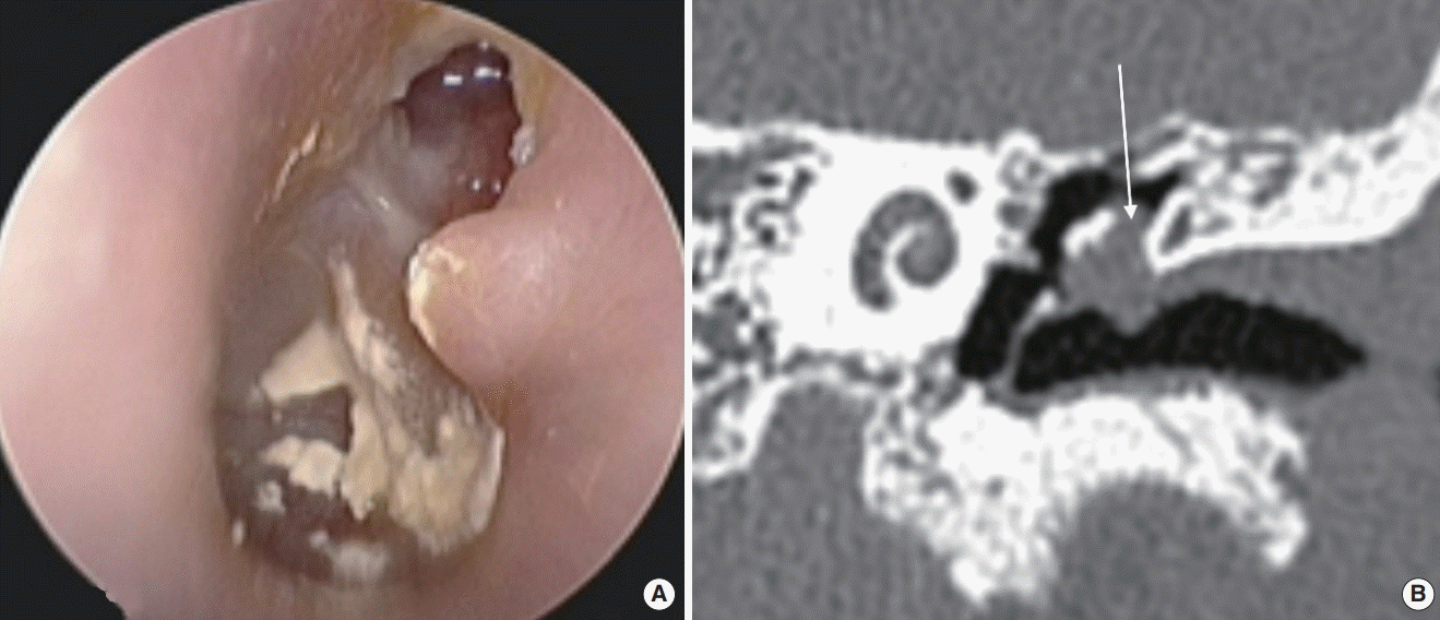
The diagnosis was made by performing otomicroscopic inspection and temporal bone computed tomography (CT) (Fig. 1). Based on the temporal bone CT, patients with cholesteatoma extension limited to the attic area were selected for atticotomy and attic reconstruction. Hearing levels were determined using the four-tone pure tone average, and postoperative hearing improvement was analyzed according to the reporting guidelines of American Academy of Otolaryngology-Head and Neck Surgery [6]. All patients were followed up for at least 1 year to evaluate their hearing and to observe them for cholesteatoma recurrence. The protocol of this retrospective study was approved by the Institutional Review Board of the Asan Medical Center (IRB No. 2017-1224). Informed consent was waived.
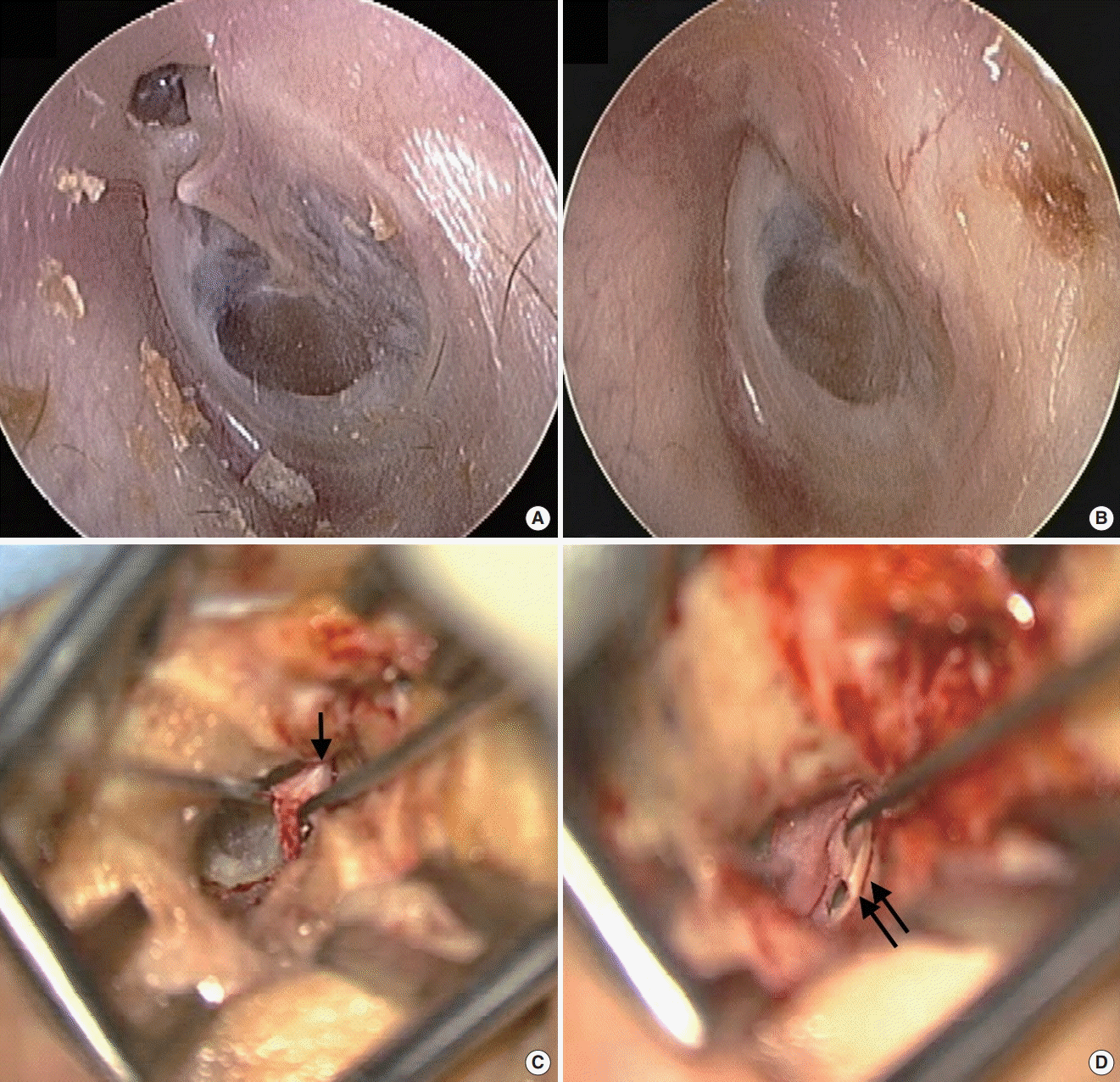
In all the patients in the microscopic approach group, a Lempert endaural incision II was made to visualize the tympanic membrane and middle ear. A tympanomeatal flap was raised with skin incision along the tympanic annulus at 6 and 12 o’clock positions (Lempert I incision) and an anterior extension to expose the attic area. Two-prong retractors were used to keep the wide opening of the external auditory canal. Atticotomy was performed using a drill, and the entire extent of the cholesteatoma was visualized. After removal of the cholesteatoma, the attic area was inspected for any remnant cholesteatoma matrix under direct microscopic view with or without a mirror. The attic was reconstructed using cartilage that was harvested from tragus, and the tympanomeatal flap was repositioned supported by perichondrium (Fig. 2). In cases of ossicular destruction, ossiculoplasty was performed using a prosthesis; mastoidectomy was not performed.
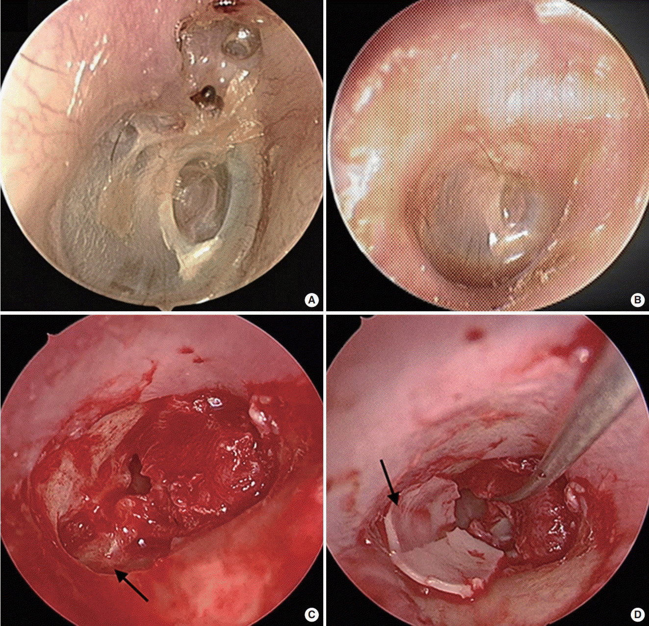
The procedure used in the endoscopic approach group was similar to that used in the microscopic approach group but without a Lempert endaural incision II. With the endoscope held in the left hand, a skin incision was made along the tympanic annulus at 6 and 12 o’clock positions; bleeding was controlled using a cotton pledget. A curette or a gouge and mallet were used for atticotomy. After removing the cholesteatoma, the attic area was inspected using 0º and 30º endoscopes. The attic was reconstructed using cartilage and repositioning the tympanomeatal flap (Fig. 3).

RESULTS

Patients’ data are summarized in Table 1. The mean patient age in the endoscopic and microscopic groups was 43.5 and 49.8 years, respectively. Surgery was performed under general anesthesia in six of 10 patients in both the groups. Ossicular continuity was found to be normal in seven patients in the microscopic and six patients in the endoscopic group. Four patients in the endoscopic group and two patients in the microscopic group underwent ossiculoplasty. Tympanoplasty without ossiculoplasty (tympanization) was performed in one patient of microscopic group.
The preoperative and postoperative air conduction threshold averages were 36.13±5.8 and 25.56±4.9 dB in the endoscopic group and 36.63±4.0 and 31.25±3.4 dB in the microscopic group (Table 2). The preoperative air-bone gap (ABG) was 17.25 dB in the endoscopic group and 13.63 dB in the microscopic group. The postoperative ABG was 8.75 dB in the endoscopic group and 11.88 dB in the microscopic group. Gap closure (preoperative ABG−postoperative ABG) was 6.67 dB in the endoscopic group and 1.75 dB in the microscopic group. There was no difference in hearing improvement between the groups (P=0.13) (Fig. 4). One of the endoscopic groups did not have postoperative hearing tests.
The mean follow-up period was 19.75 months in the endoscopic group and 41.05 months in the microscopic group. The mean operation time in the endoscopic group was 1.65 hours; this was a little less than the 1.79 hours in the microscopic group, but the difference was not significant (P=0.63). There were no postoperative complications in both groups.
Regarding the cholesteatoma stage [2], 10 patients (seven in the endoscopic group and three in the microscopic group) were categorized into stage Ib and the remaining 10 into stage II.

DISCUSSION

Management of attic cholesteatoma

According to the results of this study, the endoscopic approach seems to be as useful as the microscopic approach in the management of attic cholesteatoma. The mean operation time and postoperative hearing improvement were similar in both groups. Lempert endaural incision II was not necessary while using the endoscopic approach but it was necessary in nine of 10 patients treated using the microscopic approach.
Cholesteatoma is clinically classified as congenital cholesteatoma, acquired cholesteatoma [2], and recidivistic cholesteatoma depending on its origin, with acquired cholesteatoma being the most common form. Acquired cholesteatoma is divided into epitympanum (attic) cholesteatoma and mesotympanum cholesteatoma depending on its initial location. Attic cholesteatoma grows from the Prussak’s space through the epitympanum and the aditus ad antrum to the mastoids. In cases with attic cholesteatoma extending up to the antrum, an endoscope with curette can provide an enough surgical view [7]. If the cholesteatoma extends into a mastoid cavity, mastoidectomy is usually needed [8]. While performing surgery for attic cholesteatoma, it is essential to completely remove the lesion and secure a ventilation way to prevent the recurrence of the disease [9].

Endoscopic approach: advantages and disadvantages

Recently, endoscopic surgery has been actively performed in the areas of otology and neurotology due to the rapid development of endoscopic surgical instruments with high interest in minimally invasive surgery [5,10]. Compared with the use of a surgical microscope, the use of a surgical endoscope makes it easier to expose the hidden lesions in the middle ear and provides a better close-up view of the surgical field [10,11]. With these advantages, results of middle ear surgery using endoscopy have been reported since the 1990s. Marchioni et al. [12] and Tarabichi [10] have chosen the transcanal approach as the primary surgical approach for middle ear cholesteatoma and have reported that preservation of the ossicles with complete removal of the cholesteatoma is easier using the endoscopic approach. Ayache et al. [13] reported that there was no difference between endoscopic and microscopic surgeries regarding the rate of cholesteatoma recurrence but the range of surgery was significantly reduced in endoscopic surgery; furthermore, endoscopic findings were helpful in determining the need for revision surgery. Postoperative pain was lower and the healing process was faster with the endoscopic approach than with the microscopic approach [5].
Because the mastoid cavity is not affected by the cholesteatoma in stage I and II cases, a conventional mastoidectomy is not necessary to remove the cholesteatoma. In stage III cases, mastoidectomy may be needed to remove the cholesteatoma in the antrum and mastoid cavity. Kakehata et al. [7] reported that an endoscopic approach in addition to retrograde mastoidectomy using an ultrasonic bone curette could be used for cholesteatomas extending into the antrum. Visualization of the antrum and the posterior part of the lateral semicircular canal is possible with retrograde mastoidectomy and a 30º or 45º endoscope. With these, the authors could remove the attic cholesteatoma as one large piece or sometimes as small number of large pieces.
In our study, using an endoscope, we could easily observe the internal anatomical structures that are difficult to observe using a microscope for removal of residual lesions. Furthermore, there was no visible postoperative scar in the endoscopic group because the Lampert endaural incision II was not used in this group. However, because endoscopic surgery is difficult to perform with both hands and bleeding may interfere with the surgical field, it is advisable for beginners to use the microscope and endoscope in combination.
In this study, there were no residual lesions or recurrences as well as no postoperative complications and excellent results were obtained in hearing improvement. However, the follow-up period was short and the number of cases was small. Taken together, we can confirm that endoscopic removal of attic cholesteatoma was efficient for the eradication of the disease.

Combined approach

Although the benefits of the endoscope can be achieved in transcanal middle ear cholesteatoma surgery, an endoscopic approach offers significant benefits when used with the conventional microscope to treat larger cholesteatomas using modified combined approaches [8].
Nogueira and Cohen [14] reported treating 112 patients using a combined approach. This approach has been advantageous, especially in cases with more extensive lesions, offering residual and recurrence rates as low as those reported with traditional canal wall-down surgery for these invasive cholesteatomas.
The microscopic portion of the combined approach is performed in the same fashion as a traditional microscopic surgery with the addition of endoscopic visualization of the mastoid cavity to confirm and facilitate the removal of affected mastoid part. If the precise extent of the attic cholesteatoma is known using preoperative imaging workup, the surgeon may be able to choose the appropriate approaches in advance [15].
An endoscopic approach for the management of attic cholesteatoma is as useful as a microscopic approach. For patients with attic cholesteatoma extending into a mastoid cavity, a combined endoscopic and microscopic approach may be used with mastoidectomy.

HIGHLIGHTS

• Transcanal endoscopic ear surgery is useful for removing the attic cholesteatoma.
• Hearing improvement and operation time is similar in microscopic and endoscopic surgeries.
• Lempert II endaural incision is not needed in the endoscopic surgery.

Notes

No potential conflict of interest relevant to this article was reported.

REFERENCES

1. Kobayashi T, Toshima M, Yaginuma Y, Ishidoya M, Suetake M, Takasaka T. Pathogenesis of attic retraction pocket and cholesteatoma as studied by computed tomography. Am J Otol. 1994; Sep. 15(5):658–62.
2. Tono T, Sakagami M, Kojima H, Yamamoto Y, Matsuda K, Komori M, et al. Staging and classification criteria for middle ear cholesteatoma proposed by the Japan Otological Society. Auris Nasus Larynx. 2017; Apr. 44(2):135–40.
crossref
3. Tarabichi M. Endoscopic management of acquired cholesteatoma. Am J Otol. 1997; Sep. 18(5):544–9.
4. Khan MM, Parab SR. Endoscopic cartilage tympanoplasty: a two-handed technique using an endoscope holder. Laryngoscope. 2016; Aug. 126(8):1893–8.
crossref
5. Magliulo G, Iannella G. Endoscopic versus microscopic approach in attic cholesteatoma surgery. Am J Otolaryngol. 2018; Jan-Feb. 39(1):25–30.
crossref
6. Committee on Hearing and Equilibrium. Committee on Hearing and Equilibrium guidelines for the diagnosis and evaluation of therapy in Meniere’s disease. Otolaryngol Head Neck Surg. 1995; Sep. 113(3):181–5.
7. Kakehata S, Watanabe T, Ito T, Kubota T, Furukawa T. Extension of indications for transcanal endoscopic ear surgery using an ultrasonic bone curette for cholesteatomas. Otol Neurotol. 2014; Jan. 35(1):101–7.
crossref
8. Marchioni D, Villari D, Mattioli F, Alicandri-Ciufelli M, Piccinini A, Presutti L. Endoscopic management of attic cholesteatoma: a single-institution experience. Otolaryngol Clin North Am. 2013; Apr. 46(2):201–9.
9. Choi JE, Kim HJ, Kim BK, Moon IJ. Minimally invasive transcanal removal of attic cholesteatoma. Korean J Otorhinolaryngol Head Neck Surg. 2017; Apr. 60(4):158–63.
crossref
10. Tarabichi M. Transcanal endoscopic management of cholesteatoma. Otol Neurotol. 2010; Jun. 31(4):580–8.
crossref
11. Alicandri-Ciufelli M, Marchioni D, Kakehata S, Presutti L, Villari D. Endoscopic management of attic cholesteatoma: long-term results. Otolaryngol Clin North Am. 2016; Oct. 49(5):1265–70.
12. Marchioni D, Mattioli F, Alicandri-Ciufelli M, Presutti L. Transcanal endoscopic approach to the sinus tympani: a clinical report. Otol Neurotol. 2009; Sep. 30(6):758–65.
13. Ayache S, Tramier B, Strunski V. Otoendoscopy in cholesteatoma surgery of the middle ear: what benefits can be expected. Otol Neurotol. 2008; Dec. 29(8):1085–90.
14. Nogueira JF, Cohen MS. Combined endoscopic and microscopic approaches to cholesteatoma. Oper Tech Otolayngol Head Neck Surg. 2017; Mar. 28(1):36–8.
crossref
15. Edfeldt L, Stromback K, Danckwardt-Lilliestrom N, Rask-Andersen H, Abdsaleh S, Wikstrom J. Non-echo planar diffusion-weighted MRI increases follow-up accuracy after one-step step canal wall-down obliteration surgery for cholesteatoma. Acta Otolaryngol. 2013; Jun. 133(6):574–83.
crossref

Fig. 1.
Preoperative findings. (A) Attic destruction was observed on otoscopic examination. (B) Coronal computed tomography scan of the left temporal bone shows erosion of the scutum and soft tissue density (arrow) in the left Prussak’s space.
ceo-2018-00507f1.tif
Fig. 2.
Microscopic approach for attic cholesteatoma in patient who suffered from intermittent otorrhea and otalgia (case 16 in Table 1). (A) Attic destruction was observed on preoperative examination of the left tympanic membrane. (B) Postoperative examination shows the tympanic membrane with clear attic area. Intraoperative findings show the elevated tympanomeatal flap (arrow) after Lempert endaural incision II and Lempert I incision (C), and inserted cartilage for reconstruction of the attic area (double arrows, D).
ceo-2018-00507f2.tif
Fig. 3.
Endoscopic approach for attic cholesteatoma in patient who suffered from hearing loss and intermittent otorrhea (case 10 in Table 1). (A) Attic destruction was observed on preoperative examination of the right tympanic membrane. (B) Postoperative examination shows a clear attic. (C) Endoscopically, the head of the malleus is shown (arrow) after elevation of the tympanomeatal flap. (D) After removal of the cholesteatoma, the attic was reconstructed with cartilage (arrow).
ceo-2018-00507f3.tif
Fig. 4.
Preoperative and postoperative air conduction hearing levels (HL) of the patients. HL was improved postoperatively in both groups (asterisk). There was no statistically significant difference of improvement between endoscopic and microscopic approaches (P=0.13).
ceo-2018-00507f4.tif
Table 1.
Summary of patients’ data
Case Use of endoscope Age/sex Stagea) Operation time (hr) Anesthesia Ossiculoplasty Lempert incision Preop AC (dB) Postop AC (dB) Preop ABG (dB) Postop ABG (dB) ABG closure (dB) Recurrence Follow-up month
1 Yes 47/M 2 1.3 G No I 27.5 23.75 17.5 10 7.5 No 13.9
2 Yes 60/F 1b 2.1 G Yes I 67.5 32.5 28.75 7.5 21.25 No 19.6
3 Yes 26/F 1b 1.1 L No I 18.75 7.5 15 0 15 No 19.9
4 Yes 23/F 1b 2 L No I 8.75 3.75 7.5 0 7.5 No 23.0
5 Yes 61/F 1b 1.2 L No I 51.25 43.75 17.5 12.5 5 No 26.0
6 Yes 52/M 1b 1.4 L No I 26.25 30 8.75 16.25 –7.5 No 41.7
7 Yes 28/M 2 2.1 G Yes I 47.5 23.75 28.75 15 13.75 No 10.8
8 Yes 38/M 1b 1.7 G Yes I 48.75 - 33.75 - - No 5.6
9 Yes 65/M 2 2.3 G Yes I 43.75 47.5 0 12.5 –12.5 No 9.0
10 Yes 35/F 1b 1.3 G No I 21.25 17.5 15 5 10 No 28.0
11 No 39/M 2 1.5 L No II 43.75 37.5 33.75 27.5 6.25 No 30.2
12 No 69/F 2 1.3 G No II 46.25 42.5 10 6.25 3.75 No 27.8
13 No 44/M 2 2 G Yes II 36.25 22.5 17.5 12.5 5 No 30.7
14 No 62/F 2 1.5 G No II 42.5 38.75 18.75 13.75 5 No 30.7
15 No 47/F 2 3 G No II 28.75 27.5 1.25 8.75 –7.5 No 36.1
16 No 21/M 1b 1.3 G No II 20 28.75 5 15 –10 No 37.9
17 No 58/F 2 2 G Yes II 45 22.5 28.75 12.5 16.25 No 38.9
18 No 43/M 1b 2 L No II 16.25 16.25 0 3.75 –3.75 No 61.0
19 No 65/F 2 1.8 L No II 31.25 25 15 15 0 No 53.2
20 No 50/M 1b 1.5 L No II 56.25 51.25 6.25 3.75 2.5 No 64.0

Preop, preoperative; AC, air conduction; Postop, postoperative; ABG, air-bone gap; G, general; L, local.

a) Stage according to the classification of Tono et al. [2].
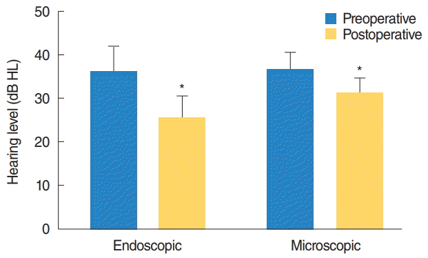
Table 2.
Comparison between endoscopic and microscopic approach
Variable Endoscopic surgery (n=10) Microscopic surgery (n=10)
Mean age (yr) 43.5 49.8
Sex (male:female) 5:5 5:5
Anesthesia (general:local) 6:4 6:4
Stage (1b:2)a) 7:3 3:7
Air conduction PTA (dB HL), mean±SE
 Preoperative 36.13±5.8 36.63±4.0
 Postoperative 25.56±4.9 31.25±3.4
Air-bone gap (dB HL), mean±SE
 Preoperative 17.25±3.4 13.63±3.6
 Postoperative 8.75±2.1 11.88±2.2
Tympanoplasty type I:ossiculoplasty 6:4 7:2b)
Operation time (hr), mean±SD 1.65±0.4 1.79±0.5
Recurrent:residual disease 0:0 0:0
Mean follow-up period (mo) 19.75 41.05

PTA, pure tone average; HL, hearing level; SE, standard error; SD, standard deviation.

a) Stage according to the classification of Tono et al. [2].

b) One patient received tympanoplasty without ossiculoplasty (tympanization).

TOOLS
Similar articles