

 PDF
PDF Citation
 Print
Print


INTRODUCTION
The obstructive sleep apnea syndrome (OSAS) is an apnea and hypopnea disorder caused by the collapse of the upper airway during sleep, which is characterized by nocturnal sleep snoring, apnea, and superficial breathing. This disorder may result in nocturnal hypoxemia, hypercapnia, and sleep disorders. Some cases of severe OSAS were reported to cause functional damages in critical organs which gave rise to a high risk of disability [1-4]. A number of symptoms occur in OSAS patients, for example, daytime sleepiness, apathetic, memory loss and cognitive disorders, which may suggest functional damages in the nervous system [5]. In general, the functional impairment is reversible in the early hypoxemia due to OSAS, but it can be irreversible for chronic hypoxemia [6]. Since the energy consumption occurred in the process of signal transmission on the various levels of central nucleus and cochlea transduction, such energy consumption is highly dependent on blood oxygen. Thus, the persistent hypoxemia may lead to auditory impairments in OSAS patients. It has been found that some severe OSAS patients showed abnormalities in terms of conventional audiometry testing, such as pure-tone audiogram (PTA), distortion product otoacoustic emissions (DPOAEs), and auditory brainstem response (ABR) suggesting functional impairments in the auditory system [7]. However, it still lacks reverent research to investigate the auditory function in the case of less severe OSAS.
In clinic, the integrity of auditory neural pathway is generally examined by ABR elicited by a series of impulse sounds via airor bone-conduction ways [8]. The ABR reflects the synaptic activity of auditory brainstem neurons, and makes it a useful tool in studying the functional integrity for OSAS. Some studies reported prolonged peak latencies and latencies in wave intervals such as I–V and III–V of the click-evoked auditory brainstem response (click-ABR) in OSAS patients [7,9,10]. The pathophysiologic mechanism may be accounted for by the neurotransmitter synthesis disorders and the abnormal conduction of the nerve impulse in the auditory pathway, which is due to hyperlipidemia caused by respiratory disturbances [10]. However, other studies claimed that no abnormal click-ABR found in the OSAS patients, suggesting no acute or chronic injury in the auditory brainstem caused by repeated nocturnal hypoxemia [11-13]. These conflicting findings inspire the need of new methods and more sophisticated studies in this regard.
Speech-evoked auditory brainstem response (speech-ABR) is a new technique of auditory evoked brainstem response in recent years. A commonly used stimulus is a 40-ms synthesized syllable /da/ [14]. The speech-ABR elicited by /da/ contains seven feature peaks termed waves V-A-C-D-E-F-O, which are commonly analyzed in both time and frequency domains. The time domain indexes include the latencies of these feature peaks and slopes of VA complex; the frequency domain indexes include the amplitude of the fundamental frequency (F0), the first formant (F1) and high formant (HF) [15]. For a speech-ABR, the transient components defined by waves V, A, C and O, as well as F1 and HF were considered to reflect the filter information (temporal and formant related information) which was processed by the “where” pathway; while the periodic components defined by peaks D, E, and F, as well as F0 were considered to reflect the sound source information (pitch-related information) which was processed by “what” pathway [15,16]. Actually, “what/where” pathways are functional pathway represented by independent neural networks with different synaptic connections [17].
It was reported that the transient components of speech-ABR showed significant abnormalities for some patients, such as learning problem children (LPC) [18-21] and persistent developmental stuttering (PDS) population [22], while these patients are with normal click-ABR. These findings suggest that speech-ABR may be a more effective method for detecting auditory function than click-ABR. Therefore, speech-ABR may play a role in the study of neurological status of auditory system, and may be a useful tool to detect the early impairments of neurological functions in patients with OSAS. Based on this hypothesis, we conducted an auditory system testing on the selected group of OSAS patients of less severe condition and the control group of normal hearing participants. Since the hemispheric dominance is related to handedness, the right ears for righthanded participants were recorded and analyzed.
Go to : 
MATERIALS AND METHODS
Participants
Participants were recruited from 116 snoring patients undergoing overnight polysomnogram monitoring from January 2016 to May 2017 at the Sleep Center of Guangdong Second Provincial General Hospital. OSAS was diagnosed according to clinic criteria, and degree of severity was categorized as mild, moderate and severe according to the apnea hypopnea index (AHI) and the lowest oxygen saturation (SaO2) [23-25]. Specifically, OSAS was diagnosed for a patient with AHI ≥5, where 5≤ AHI ≤15 was classified as mild degree, 15< AHI ≤30 moderate degree, and AHI >30 severe degree, meanwhile the degree of hypoxemia is required to be attached as well. The hypoxemia criterion is 85% ≤ lowest SaO2 ≤90% for mild degree, 65%≤ lowest SaO2 <85% for moderate degree, and lowest SaO2 <65% for severe degree [24,25]. For example, AHI of 35 times/hr and lowest SaO2 of 86% are reported as severe OSAS with mild hypoxemia.
The OSAS group contains mild to moderate OSAS patients with mild hypoxemia. Participants without OSAS and hypoxemia can be in control group. Both groups also required to be right-handed (Persian version of Edinburgh handedness questionnaire), fluent in Chinese Mandarin, and free from any history of cardiovascular, pulmonary, otological, neurological or mental disorders. Participants with any risk factors which may affect hearing such as noise exposure, smoking, diabetes, or hypercholesterolemia, were also excluded. In addition, they had normal otology examination and were asked to complete auditory tests. They were asked to participate in tests voluntarily, and to sign informed consent forms. The study was approved by the Medical Ethics Committee of Guangdong Second Provincial General Hospital (No. 2015-KYLL-004).
According to the selection criteria, 52 patients (34 males and 18 females; age, 23 to 39 years; mean age, 32.2±4.0 years) were selected. Based on the value of AHI, the participants were divided in OSAS group (31 patients with 5≤ AHI ≤30 and 85%≤ lowest SaO2 ≤90%; 21 males and 10 females; mean age, 32.5±4.4 years) and control group (21 participants with AHI <5 and lowest SaO2 >90%; 13 males and 8 females; mean age, 31.7±3.5 years).
Sleep monitoring
Sleep monitoring was performed to all participants with an Embla N7000 System (Natus Medical, Pleasanton, CA, USA). The monitoring sleep time is more than 7 hours with simultaneous recording of AHIs and lowest SaO2. They were banned from taking hypnosis, caffeine, concentrated tea, sedatives, and antihypertensive drugs before the day of polysomnogram monitoring.
PTA tests
The pure-tone hearing threshold of all participants were tested with the GSI-61 (Grason-Stadler, Eden Prairie, MN, USA) diagnostic audiometer according to the international standard. The threshold from 250 Hz to 8,000 Hz for right ear was calculated.
DPOAE tests
DPOAE tests were performed by SmartOAE ver. 4.5 (Intelligent Hearing System, Miami, FL, USA). The frequency ratio f2/f1 of two original stimuli is 1.22, the intensity of f2 is 55dB sound pressure level (SPL), intensity of f1 is 65dB SPL, and fdp=2F1–f2. The geometric mean of the two initial pure tone frequencies were from 500 Hz to 8,000 Hz for a total of nine frequencies, which are 500, 700, 1,000, 1,400, 2,000, 3,000, 4,000, 6,000, and 8,000 Hz, respectively. The signal-to-noise ratio in right ear was recorded.
ABR tests
ABR tests were accomplished using a SmartEP System (Intelligent Hearing System) in an acoustically attenuated and electromagnetically shielded room. The signal were recorded from three gold-cup electrodes placed at forehead (+), ipsilateral mastoid (–), and nasion (ground). Conductive electrode gel was applied to keep the contact impedances lower than 3 kΩ. Three blocks (1,024 sweeps each) of responses were collected. Stimulus clicks of alternating polarity were presented at 80 dB SPL through insert earphone (ER-3A Etymotic Research, Elk Grove Village, IL, USA). The analogue bandpass filter is 100–3,000 Hz, the response signals were digitalized at 800 Hz stored in a computer. Trials with signal voltage over 35 μV were rejected. Participants were required to be quiet and ignore the stimulus sounds during testing. For click-ABR, the stimulus rate was 19.3 S/s (stimulus/sec) and the recording window was 12.8 ms; for speech-ABR, the stimulus rate was 11.1 S/s and the recording window was 64 ms. The stimulus for speech-ABR was syllable /da/, of five formants, in which the F0 and F1 rise from 105–125 Hz and 455–720 Hz, respectively, while the second formant decreases from 1,700 to 1,222 Hz, the third formant reduces from 2,550 to 2,000 Hz. The last two formants remain fixed at 3,600 Hz and 4,600 Hz [14,15].
The temporal measurements of click-ABR, such as the latencies of featured peaks I, III and V, and the interpeak periods I–V, I–III and III–V, were identified by the trained experts and measured in the SmartEP system. The data of speech-ABR were transferred to a Brainstem toolbox [14-16] on MATLAB platform (MathWorks, Natick, MA, USA) which is increasingly used in the customized analysis of ABR research [26] for further analysis. The automated peak-picking algorithm in the toolbox was used to detect all peaks objectively for a higher confidence. The detailed information regarding the frequency encoding in the sustained segment of speech-ABR was obtained using fast Fourier transform, in which the spectral magnitudes of the frequency following response were measured over a 11.4–40.6 ms time window in three frequency ranges, i.e., F0, 103–121 Hz; F1, 454–719 Hz; and HF, 721–1,155 Hz, respectively. F2 and the higher formants of higher frequencies were not measured because of the upper limit of phase locking (1,500 Hz) [14,21] at the level of the rostral brainstem. A series of featured peaks V-AC-D-E-F-O, VA slope, and spectral formants F0-F1-HF were also identified and measured.
Statistical analysis
The statistical analysis was carried out with SPSS ver. 13.0 (SPSS Inc., Chicago, IL, USA). The level of significance was set at P<0.05 (two-tailed). Group differences were tested by independent sample t-test. Bivariate correlation analysis was applied to assess the relationship between AHI and speech ABRs.
Go to :
RESULTS
Sleep data
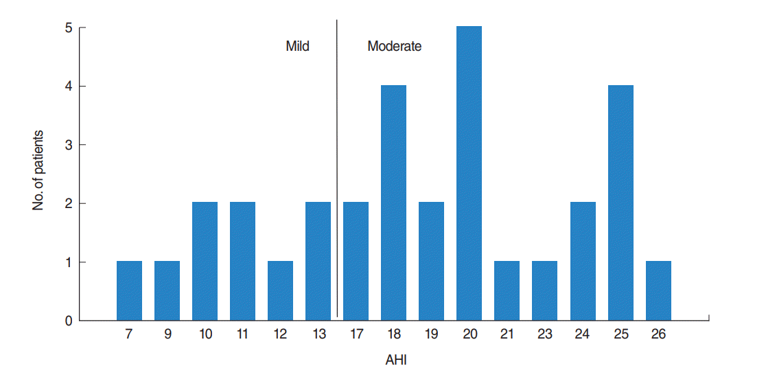
The AHI distribution of the 31 OSAS participants is shown in Fig. 1. It shows an uneven distribution of AHI from 7 to 26 (18.00±5.50), with a large number of participants of moderate degree. All of them are with mild hypoxemia (85%≤ lowest SaO2 ≤90%).
PTA data
The data of PTA showed no significant differences between the OSAS patients and control group (P>0.05), as shown in Table 1.
Table 1.
Auditory thresholds in OSAS and control groups
| Frequency (Hz) | OSAS group | Control group | P-value |
|---|---|---|---|
| 250 | 11.13±9.19 | 11.19±9.86 | 0.982 |
| 500 | 13.55±8.48 | 12.86±7.51 | 0.764 |
| 1,000 | 12.10±9.38 | 13.81±8.05 | 0.498 |
| 2,000 | 11.13±9.19 | 14.76±7.98 | 0.147 |
| 4,000 | 11.29±8.85 | 13.33±7.80 | 0.396 |
| 8,000 | 12.26±9.56 | 14.52±7.57 | 0.368 |
| Averagea) | 11.51±4.85 | 12.66±4.58 | 0.394 |
![]()
DPOAE data
No significant differences are found between OSAS group and control group (P>0.05) for DPOAE test, as shown in Table 2.
Table 2.
Averages and standard deviations of the DPOAE amplitudes
![]()
Click-ABR data
The results of click-ABR are shown in Table 3, no significant differences are found between OSAS group and control group in terms of the peak latencies (I–III–V) and the interpeak latencies (I–III, III–V, and I–V) (P>0.05).
Speech-ABR data
Speech-ABR waves for both groups are shown in Fig. 2. All the featured peaks are clearly elicited as labeled. By comparing the latency of these waves, we found that OSAS patients appeared to have longer latency values than the control group for the transient portion of the speech-ABR (except for peak A) as shown in Fig. 3.
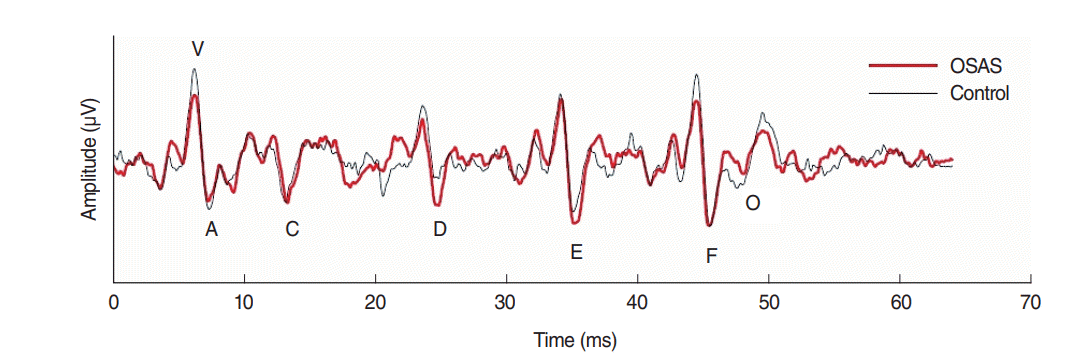
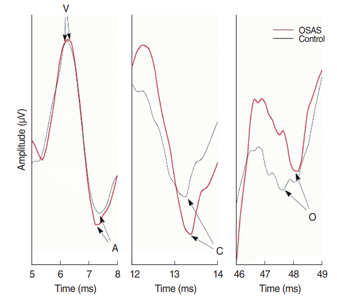
Table 4 presents all the statistics in time and frequency domains of the speech-ABR. Statistical analysis results show that there are significant latency differences between OSAS and control groups for peak V, peak C, and peak O (P<0.05). No significant latency differences are founded for peak A, peak D, peak E and peak F (P>0.05). Analysis of composite measure VA slope and the spectral amplitude of F0, F1, and HF revealed no significant group differences (P>0.05).
Table 4.
Statistical analysis for latency, composite and spectral measures of speech-ABR
![]()
Correlations between the peak latencies of speech-ABR and AHIs
To assess whether the brainstem timing of speech-ABR is related to the severity of OSAS, latencies of the waves were correlated with the AHIs using bivariate correlation analysis method. The results of Pearson correlation coefficients showed significant correlations for peak V (r=0.37, P=0.040), peak C (r=0.36, P=0.045), and peak O (r=0.55, P=0.001).
Go to :
DISCUSSION
Many studies have always been interested in the functional state of auditory nervous systems in patients with OSAS. In recent years, Casale et al. [7] found that severe OSAS patients may have lower DPOAE amplitudes, higher PTAs, and prolonged latencies as well as wave intervals in click-ABRs. It was considered that hypoxia of severe OSAS may be a risk-factor for auditory system dysfunction [7,9,10]. In our study, no significant differences are found for PTA, DPOAE, and click-ABR measurements as shown in Tables 1-3. The reasons may be ascribed to the small sample size, or, more likely, because it is a long persistent process for auditory impairment caused by hypoxemia [7], and the mild to moderate hypoxemia may not be sufficient to cause the above auditory impairments in patients. In this study, typical speech-ABR waveforms for both groups are morphologically similar (Fig. 2), while the latencies of peak V, C, and O are significantly longer in OSAS patients (Table 4, Fig. 3), which indicates the abnormality may occur in the transient components of speech-ABR in mild-moderate OSAS participants without observed changes appeared in the conventional click-ABR. This finding is similar to cases of LPC [18-21] and PDS [22] patients in speech-ABR researches.
Although both click-ABRs and speech-ABRs are the auditory evoked potentials reflecting the synchronized activity of neurons in the auditory brainstem, they may have different sensitivities to the same stimulation properties. A feasible explanation involves possible differences in neural populations recruited during the encoding click and speech syllable. The process of complex sounds in auditory brainstem may have an independent mechanism in contrast with simple sounds [27]. Some researchers advocated this notion in the case of the abnormal performance of speech-ABR in LPC populations [18]. Note that the acoustical structure of click and speech sounds are of great difference. A click sound is a burst of impulse with a plain frequency distribution over a large hearing range, while a speech syllable generally contains tones (mainly reflected in the sound of the F0), timbre (reflected by harmonics), timing (mainly reflected in the start, end of sound and phoneme transitions), and other acoustic characteristics related to the meaningful language. Therefore, the auditory brainstem may need to adequately mobilize more neurons or regulate their functions for the speech stimulus, so that the auditory mechanisms involved in speech syllable coding are more complicated than that of click acoustic stimuli. Furthermore, it is more challenging in coding speech sound in auditory system due to the backward masking effect [28]. This may lead to the separation of click-ABR and speech-ABR in the case of the LPC [18-21], the PDS [22], and the mild to moderate OSAS population in this experiment. Thus, the normality of click-ABR cannot completely rule out the auditory dysfunction in patients with OSAS though it is sensitive to the detection of auditory brainstem injury in severe OSAS patients [7,9]. The incompetence of click-ABR is further supported by an animal study, which claimed that one-sided inferior colliculus degeneration and medial geniculate degeneration cannot be detected by click-ABR [29]. This study implies that speech-ABR may have broader application prospects since it can provide additional information on the auditory brainstem center to better reflect the auditory function [27].
The abnormality in the transient components (Table 4) of OSAS patients is considered to reflect the filter information, which is related to temporal and formant, as coding with “where” pathway. No significant differences were found for periodic components of speech-ABR, which reflects sound source information related to sound of F0 or pitch as coding with “what” pathway. The latency delay in peak V, C, and O of the transient components may support that the auditory whatwhere functional pathways consist of different neural networks and have independent functions [17]. These abnormal performances of speech-ABR may be related to the interference of temporal information encoded in the auditory brainstem center [15,18-22]. It is presumed that peak V, C, and O occurred in response to transient stimulus events (in contrast to the periodic acoustic events in the speech stimulus), specifically, peak V may reflect a highly synchronized neural response to the onset of the stimulus; peak C is probably a response to the onset of the voicing that occurs after stimulus onset; peak O is probably a response to the cessation of sound, as it temporally corresponds to the offset of the stimulus [15,18,30].
These transient peaks of speech-ABR may originate from the consequence of activity of octopus cells in the brainstem nuclei representing the timing information of speech [31]. Burst responses produced by the auditory neurons require high energy consumptions, or equivalently more oxygen [7,31], these neurons may be more likely to be impaired by hypoxemia. The auditory neurons that generate transient components of speech-ABR are more susceptible to adverse effects than those that generate periodic components. This notion was supported by other studies under noisy environment [32] and in the old population [33]. Studies have shown that the higher the AHI, the lower the SaO2 of OSAS patients [1-3]. Therefore, the severe OSAS should be more likely to damage the involved neurons, or their temporal coding functions.
Although OSAS participants have mild hypoxemia, higher AHI means longer durations of apnea and hypopnea, or more frequent hypoxia, which leads to the damage of auditory functions due to the lack of blood supply in the brainstem [34]. The significantly positive correlations between the latency of peaks (V, C, and O) and AHI suggest that the damage may be aggravated with the severity of the OSAS. In this study, OSAS patients only have mild to moderate degree of AHI, which suggests that the damage occurred in the neurons may be responsible for speech-ABR transient elements, but it may not be adequate to cause observable changes in the periodic elements that required severe and accumulated effects from hypoxia.
In this study, the latency of peak O showed the strongest correlation coefficient (r=0.55, P=0.001), suggesting that the latency of peak O can be a sensitive indicator to the effects of OSAS severity on auditory system. Some researchers declared that the latency of peak O may reflect the ability of auditory brainstem to discriminate temporal information in speech syllable, which may have originated from neurons responsible for the termination and duration of acoustic signals [35]. These auditory neurons are present in the nucleus of the inferior colliculus [36], and can be used to distinguish temporal information from speech sound in environment noise [32]. It has been reported that the latency of peak O may be a strong predictor of weak temporal resolution abilities in some deficit populations. For example, the latency of peak O was used to measure the postmasking effects in LPC population [22,35]. In addition, it was also found that the latency of peak O was significantly delayed in the elderly population with impaired auditory function and speech cognition which related to the temporal information processing capacity in the auditory brainstem of hearing loss associated with age [33,37,38]. These findings prompt that further research of the relationship between characteristic of peak O and temporal processing of auditory brainstem may be particularly informative, and then the latency of peak O may be an important indicator to the auditory deficits in OSAS patients.
In summary, we reported that the transient components of speech-ABR were significantly and positively correlated with the AHI in mild and moderate OSAS patients, while the change in conventional click-ABR is not detectable. As these components reflect the coding of temporal information in speech syllable, this finding suggests that OSAS may be responsible for the deficits of temporal information processing which can be represented by the speech-ABR. Thus speech-ABR can be a potential biomarker in the diagnosis and evaluation at early stage of OSAS.
Go to :
HIGHLIGHTS
▪ Auditory dysfunction without abnormal click-evoked auditory brainstem response (click-ABR) might be found in obstructive sleep apnea syndrome (OSAS) patients.
▪ More speech-evoked ABR (speech-ABR) than click-ABR helps to detect auditory deficits in OSAS patients.
▪ Therefore, speech-ABR may be a potential biomarker for auditory dysfunction of OSAS patients.
Go to :
 XML Download