INTRODUCTION
MATERIALS AND METHODS
Subjects
Data acquisition
Postprocessing
Calculation
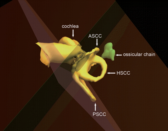
 | Fig. 2.Right inner ear and ossicular chain. The semicircular canal planes were represented by the planes passing through each canal. ASCC, anterior semicircular canal; PSCC, posterior semicircular canal; HSCC, horizontal semicircular canal. |
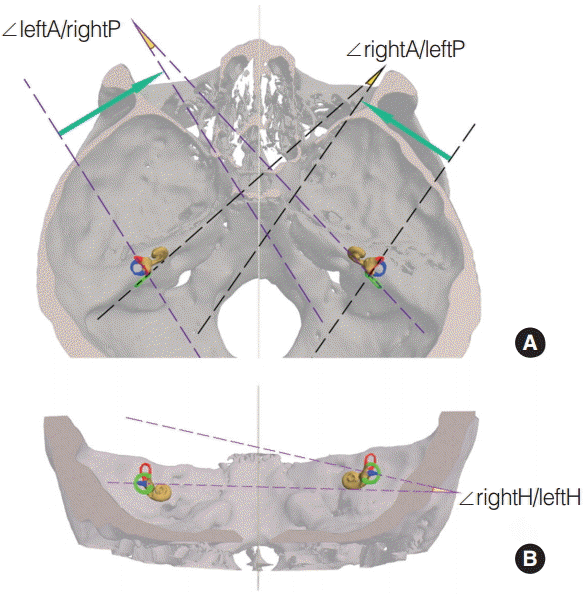
 | Fig. 3.The angles between contralateral synergistic canals. Panel A is apical view. Panel B is posterior view. The anterior, posterior, and horizontal semicircular canals are red, green and blue colored, respectively. ∠ indicate the angle between 2 planes; A, P, and H, indicate the anterior, posterior, and horizontal semicircular canal plane, respectively. |
Statistical analysis
RESULTS
Table 1.
Values are presented as mean±SD.
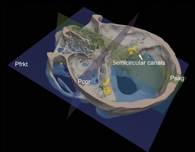
Psag, median sagittal plane; Pcor, coronal plane; Pfrkt, Frankfurt horizontal plane; L, left; R, right; NS, not significant.
Groups A–D indicate 1–6, 7–12, 13–18, and >18 years old group, respectively; Pab, Pac, Pad, Pbc, Pbd, Pcd indicate P-value for differences between groups A and B, between groups A and C, between groups A and D, between groups B and C, between groups B and D, and between groups C and D, respectively; ∠ indicate the angle between 2 planes; A, P, and H indicate the anterior, posterior, and horizontal semicircular canal plane, respectively.
Table 2.
| Side | ∠A/Psag | ∠A/Pcor | ∠A/Pfrkt | ∠P/Psag | ∠P/Pcor | ∠P/Pfrkt | ∠H/Psag | ∠H/Pcor | ∠H/Pfrkt | |
|---|---|---|---|---|---|---|---|---|---|---|
| JB–Psag | L | 0.06 | - | - | 0.09 | - | - | 0.04 | - | - |
| R | –0.16 | - | - | 0.16 | - | - | –0.42*** | - | - | |
| JB–Pcor | L | - | –0.29** | - | - | 0.20 | - | - | –0.10 | - |
| R | - | –0.26* | - | - | 0.19 | - | - | 0.06 | - | |
| JB–Pfrkt | L | - | - | –0.06 | - | - | 0.38*** | - | - | –0.12 |
| R | - | - | 0.03 | - | - | 0.11 | - | - | –0.14 |
JB, jugular bulb; Psag, median sagittal plane; Pcor, coronal plane; Pfrkt, Frankfurt horizontal plane; Pcor, coronal plane; L, left; R, right.
∠ indicate the angle between 2 planes; A, P, and H indicate the anterior, posterior, and horizontal semicircular canal plane, respectively; JB–Psag, JB–Pcor, and JB–Pfrkt: The JB position in the cranial base was defined as its distances from Psag, Pcor, and Pfrkt planes, respectively.
Table 3.
Values are presented as mean±SD.
ipsi, ipsilateral; L, left; R, right; NS, not significant.
Groups A–D indicate 1–6, 7–12, 13–18, and >18 years old group, respectively; Pab, Pac, Pad, Pbc, Pbd, Pcd indicate P-value for differences between groups A and B, between groups A and C, between groups A and D, between groups B and C, between groups B and D and between groups C and D, respectively; ∠ indicate the angle between 2 planes; A, H, and P indicate the anterior, horizontal, and posterior semicircular canal plane, respectively.
Table 4.
| Plane |
JB–Psag |
JB–Pcor |
JB–Pfrkt |
|||
|---|---|---|---|---|---|---|
| L | R | L | R | L | R | |
| ∠ipsiA/ipsiH | –0.22 | –0.19 | –0.08 | –0.07 | 0.06 | –0.01 |
| ∠ipsiP/ipsiH | –0.21 | –0.04 | 0.15 | –0.26* | 0.07 | 0.01 |
| ∠ipsiA/ipsiP | 0.10 | 0.01 | 0.03 | –0.14 | 0.16 | –0.14 |
JB, jugular bulb; Psag, median sagittal plane; Pcor, coronal plane; Pfrkt, Frankfurt horizontal plane; L, left; R, right; ipsi, ipsilateral.
∠ indicate angle between 2 planes; A, H, and P indicate the anterior, horizontal, and posterior semicircular canal plane, respectively; JB–Psag, JB–Pcor, and JB–Pfrkt: The JB position in the cranial base was defined as its distances from Psag, Pcor, and Pfrkt planes, respectively.
DISCUSSION
Table 5.
| Source | No. | ∠ipsiA/ipsiH | ∠ipsiA/ipsiP | ∠ipsiP/ipsiH | ∠rightH/leftH | ∠rightA/leftP | ∠leftA/rightP |
|---|---|---|---|---|---|---|---|
| Current | 76 | 88.3±5.1 | 110.3±8.8 | 87.6±4.8 | 8.6±4.9 | 19.0±7.2 | 19.0±7.8 |
| El Khoury et al. [10] | 137 | 74.2±4.4 | 111.2±6.4 | 88.2±6.2 | 19.7±8.9 | 10.6±5.4 | 11.4±6.4 |
| Aoki et al. [11] | 11 | 91.5±6.7 | 94.9±3.8 | 91.0±4.9 | 9.8±5.1 | 17.2±9.5 | 19.0±8.8 |
| Blanks et al. [2] | 12 | 111.8±7.6 | 86.2±4.7 | 95.8±4.7 | 19.8±14.9 | 23.7±6.7 | 24.6±7.2 |


 PDF
PDF Citation
 Print
Print


 XML Download