Abstract
Heterotopic gastric mucosa tissue is also called gastric choristoma, and this type of lesion can be found anywhere in the alimentary tract. However, gastric choristoma in the pharynx is very rare; only 10 cases of pharyngeal gastric choristoma have been reported in the English medical literature. A 32-yr-old woman was referred to our institution for the evaluation of a large mass that originated from the posterior wall of the oropharynx. The mass did not cause any symptoms except for the occasional sensation of a foreign body. Gadolinium-enhanced T1 weighted imaging showed a 5 cm-sized mass with central enhancement and hypointense portions, yet the radiological diagnosis was not clear. Transoral mass excision was performed with using electrocautery for making the diagnosis and for treating the mass. The microscopic analysis revealed gastric choristoma.
Choristoma is defined as microscopically normal cells or tissues change to "which" are present in an abnormal location (1). Gastric choristoma is also referred to as a gastric heterotopia, ectopic gastric mucosa, foregut duplication cyst and enterocystoma, and this can be found in any part of the alimentary tract. The most common site for heterotopic gastric mucosa is the upper esophagus (2). The finding of gastric choristoma in the pharynx is very rare. Since the first report by Stout and Lattes in 1957, only 10 cases of pharyngeal gastric choristoma have been reported in the English medical literature (3-6). Here we report one more case of gastric choristoma in the oropharynx and we review the relevant medical literature.
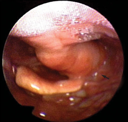
A 32-yr-old woman was referred to our institution for the evaluation of an oropharyngeal mass. She reported that the mass was found incidentally at the age of three. However, because it was asymptomatic there was no further diagnostic work up or treatment. The patient had no symptoms such as swallowing difficulties or upper airway obstruction; however, she did report an occasional foreign body sensation. Flexible laryngoscopy revealed a pedunculated mass attached to the left posterior oropharyngeal wall (Fig. 1). A Gadolinium-enhanced T1 weighted MRI showed a 5×3×1.5 cm tumor that originated from the posterior pharyngeal wall with central enhancement and hypointense portions (Fig. 2). The mass had not invaded the adjoining structures. The peduncle of the mass was located at the level of C3 and the inferior portion of the mass filled the left pyriform sinus. These radiologic findings suggested a rare benign pharyngeal tumor such as a fibrous polyp and rhabdomyoma, but the radiologic diagnosis was not clear.
Although the mass didn't cause any symptoms at that time and it showed completely benign features according to the history and the MRI finding, we recommended a simple excision since the mass had the potential risk of upper airway obstruction due to its location just above the larynx and its huge size. A preoperative procedure for making the histological diagnosis was not performed because of the potential risk of blood aspiration and airway obstruction caused by swelling that would occur after the biopsy at the out-patient department.
Under general anesthesia, a pedunculated 5×3×1.5 cm sized mass was exposed with using a McIvor mouth retractor. The mass was completely excised from the posterior wall of the oropharynx with using monopolar electrocautery. The prevertebral fascia was exposed after the resection; the mass had not infiltrated to the adjacent structures. Frozen biopsy was not performed since the mass had benign features and an adequate resection margin was acquired. The patient was discharged on the same day of the operation. At three months follow up, the wound had healed completely and there was no evidence of recurrence or a residual mass.
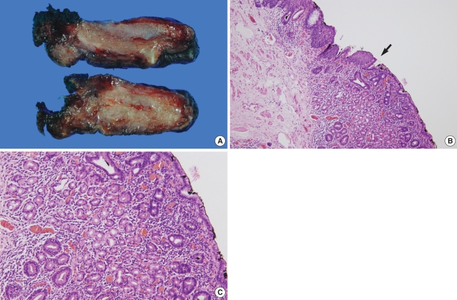
Gross examination of the specimen showed a firm mass with a homogeneous cut surface. Histopathological examination of the specimen revealed that some foci of the lesion were lined by heterotopic gastric foveolar epithelium with gastric glands beneath the surface. In addition, there were glandular structures that were characterized by parietal and chief cells. Moreover, there was focal intestinal metaplasia that contained goblet cells in the glands. The muscularis propria was found beneath the submucosal layer. These findings were consistent with gastric choristoma (Fig. 3).
Gastric choristoma is one of the foregut duplication cysts or masses and it is diagnosed when the following criteria are satisfied: 1) it is covered by a smooth muscle coat, 2) it is lined by epithelial cells derived from the foregut, and 3) it is attached to some portion of the foregut (7). In this case, the mass was covered by gastric mucosa and it contained the muscularis propria beneath the submucosal. The mass was found in the oropharynx, which is a part of the alimentary tract. Based on these findings, the present case was diagnosed as gastric choristoma.
A total of 11 cases of pharyngeal gastric choristoma have been reported in the medical literature, including the present case. Among the 11 patients, four patients were diagnosed as adults and they did not have symptoms except for an occasional foreign body sensation; their lesions were found incidentally. However, in another seven cases, the diagnosis was made during infancy because of the presence of symptoms related to the mass; six out of seven had upper airway obstructive symptoms such as inspiratory stridor and cyanosis. One patient had no airway symptoms, but the patient had had dysphasia and emesis (1, 3-6, 8).
Symptoms of heterotopic gastric tissue in the cervical esophagus are known to be related to acid production, which causes ulcer formation, tracheoesophageal fistula, bleeding, scarring and stricture formation (5). Adenocarcinoma can develop from heterotopic gastric tissue in the esophagus (9). A case with an esophageal gastric choristoma and fatal aspiration due to cricopharyngeal spasm, which was caused by acid production, has previously been reported (10). Unlike esophageal lesions, the symptoms of a pharyngeal gastric choristoma are associated with the presence of the mass. Symptoms associated with acid production have not been reported.
The treatment of esophageal gastric choristoma depends on the symptoms. Asymptomatic patients do not require any treatment, and medical therapy such as a H2 blocker or proton pump inhibitor is the primary mode of treatment for patients with dysphagia, odynophagia or globus pharyngeus. Surgical intervention might be a treatment option for patients with complications such as a web, stricture or stenosis (11). However, the treatment for a pharyngeal gastric choristoma has not been well established. It is questionable whether or not an asymptomatic pharyngeal gastric choristoma needs to be treated. Surgical resection, as in the present case, is indicated for making the diagnosis and for treatment even in some asymptomatic patients. Although choristoma of the pharynx is rare, a pharyngeal gastric choristoma can cause severe airway symptoms, and especially in infants; surgical excision is curative in such cases. Excisions of the mass are performed using electrocautery or a carbon dioxide laser. In one previously reported case, laser ablation was successfully used to treat small remnant gastric tissue (5). To the best of our knowledge, there has been no report of recurrence in cases with completely excised pharyngeal choristoma. After partial excision, the symptom could recur after a certain period of time (6).
References
1. Johnston C, Benjamin B, Harrison H, Kan A. Gastric heterotopia causing airway obstruction. Int J Pediatr Otorhinolaryngol. 1989; 9. 18(1):67–72. PMID: 2807756.

2. Borhan-Manesh F, Farnum JB. Incidence of heterotopic gastric mucosa in the upper oesophagus. Gut. 1991; 9. 32(9):968–972. PMID: 1916499.

3. Edwards J, Pearson S, Zalzal G. Foregut duplication cyst of the hypopharynx. Arch Otolaryngol Head Neck Surg. 2005; 12. 131(12):1112–1115. PMID: 16365227.

4. Fraser L, Howatson AG, MacGregor FB. Foregut duplication cyst of the pharynx. J Laryngol Otol. 2008; 7. 122(7):754–756. PMID: 18086334.

5. Hsu JM, Mortelliti AJ. Gastric choristoma of the hypopharynx presenting in an infant: a case report and review of the literature. Int J Pediatr Otorhinolaryngol. 2000; 11. 30. 56(1):53–58. PMID: 11074116.

6. Kim JK, Park KK. Foregut duplication cyst of the hypopharynx: a rare cause of upper airway obstruction. J Pediatr Surg. 2007; 6. 42(6):E5–E7. PMID: 17560193.

7. Azzie G, Beasley S. Diagnosis and treatment of foregut duplications. Semin Pediatr Surg. 2003; 2. 12(1):46–54. PMID: 12520472.

8. Wolff M, Rankow RM. Heterotopic gastric epithelium in the head and neck region. Ann Plast Surg. 1980; 1. 4(1):53–64. PMID: 7377677.

9. von Rahden BH, Stein HJ, Becker K, Siewert RJ. Esophageal adenocarcinomas in heterotopic gastric mucosa: review and report of a case with complete response to neoadjuvant radiochemotherapy. Dig Surg. 2005; 22(1-2):107–112. PMID: 15849472.

10. Libcke JH. Heterotopic gastric mucosa in the cervical esophagus a possible cause of fatal aspiration. Pediatrics. 1969; 9. 44(3):447–448. PMID: 5809902.

11. von Rahden BH, Stein HJ, Becker K, Liebermann-Meffert D, Siewert JR. Heterotopic gastric mucosa of the esophagus: literature-review and proposal of a clinicopathologic classification. Am J Gastroenterol. 2004; 3. 99(3):543–551. PMID: 15056100.

Fig. 1
Laryngoscopic examination. A huge mobile pedunculated mass (arrow) originated from the posterior oropharyngeal wall. It partially occluded the laryngeal inlet during respiration.

Fig. 2
MRI findings of the mass. The gadolinium-enhanced T1 weighted images showed a posterior pharyngeal wall mass with central enhancement and hypointense portions (arrows).

Fig. 3
Pathologic findings of the mass. (A) Gross examination shows a firm mass with a homogeneous cut surface. (B) Microscopic examination shows the junction (black arrow) between the heterotopic gastric mucosa with foveolar epithelium and the oropharyngeal squamous mucosa (hematoxylin-eosin [H & E], ×100). (C) The gastric tissue contains chief cells and parietal cells (H & E, ×200).


 PDF
PDF Citation
 Print
Print


 XML Download