

 PDF
PDF Citation
 Print
Print


Introduction
Endoscopic ear surgery is an emerging technique that is playing an increasing role in the field of otologic surgery. Despite some shortcomings, such as one-handed surgery, poor bleeding control, and loss of depth perception, the endoscopic transcanal approach has the advantage of bypassing the narrow segment of the ear canal and providing a wide angle view, which allows for minimally invasive procedures [1]. Many surgical procedures, such as attic cholesteatoma and cochlear implant, are also performed using an endoscopic approach, but the most commonly performed and reported surgery is tympanoplasty. Previous studies that have compared a microscopic approach with endoscopic transcanal tympanoplasty have reported similar or better graft success rates, complication rates, and degrees of hearing improvement for the two techniques [2-7]. Moreover, the endoscopic approach provides effective access to the middle ear with no requirement for a postauricular incision or canaloplasty, thus reducing operating time, with cosmetic advantages [8].
In addition to the conventional underlay and overlay graft techniques, several modified approaches have been developed to increase the graft success rate of tympanoplasty, including sandwich graft tympanoplasty [9], over-under tympanoplasty [10], mediolateral graft tympanoplasty [11], window shade tympanoplasty [12], and loop overlay tympanoplasty [13]. Among these, the circumferential subannular graft technique produces good results [14,15]. This procedure is a modification of the conventional underlay technique. Instead of inserting the anterior edge of the graft blindly under the bony annulus, it is positioned between the fibrous annulus and its bony annular seat and extended onto the anterior bony canal wall. Although this approach maintains stability by securing the supportive area of the anterior edge of the graft, there are some limitations when performing the procedure. In addition, as a clear view of the anterior margin is important for this technique, endoscopy is more suitable than microscopy. In this study, we explored the shortcomings of previous techniques and performed modified circumferential subannular tympanoplasty (MCST) via an endoscopic approach. The results were compared to those of the underlay technique and the results of previous studies to assist in the selection of the appropriate graft technique.
Go to : 
Subjects and Methods
Patients
The medical records of patients who underwent endoscopic transcanal type 1 tympanoplasty at our hospital between June 2016 and May 2019 were reviewed retrospectively. Patients without otorrhea and no or minimal mastoid lesions were included in the study, and those with cholesteatoma, ossicular chain disorders, combinations of other procedures, such as mastoidectomy or ossiculoplasty, and revision cases were excluded. All patients included in our study underwent audiological evaluation at least 3 months after surgery, and had a follow-up period of at least 6 months through tympanoscopy. The total number of patients was 31, of which 11 were treated with MCST and 20 were treated via underlay tympanoplasty. Most perforations were caused by chronic otitis media, and one case was caused by trauma. Demographic data, size and location of the perforation, pre- and postoperative hearing, operating time, complication rate, and graft success rate were evaluated in each group. Anterior blunting and lateralization of tympanic membrane (TM) were evaluated by observing the shape of the TM through tympanoscopy at 3 months after surgery. Patients who fulfilled the inclusion criteria provided verbal consent at the time of recruitment. This study was approved by the Institutional Review Board of Gyeongsang National University Changwon Hospital, Changwon, Korea (2021-01-038).
Surgical procedure
The endoscopic tympanoplasty type I procedure was performed under general anesthesia, using an endoscopic system and a 20-cm-long, 2.7-mm-diameter, 0° rigid endoscope (Karl Storz, Tuttlingen, Germany). The transcanal approach was performed in all patients, and surgeries were performed by a single experienced surgeon. After injecting local anesthetic and irrigating the canal, the margins of the perforation were circumferentially freshened with an angled pick. A meatal incision was made 5-6 mm lateral to the tympanic annulus, between the 7 o’clock and 12 o’clock positions in the underlay group, and between the 11 o’clock and 1 o’clock positions in the MCST group. The tympanomeatal flap was slowly elevated with an Austin duckbill elevator, and the fibrous annulus was separated from the tympanic sulcus; the chorda tympani nerve was preserved. As the anterior fibrous annulus is tightly attached to the mucosa of the middle ear cavity, an incision was made at the border between the fibrous annulus and the mucosa with the Austin duckbill elevator and then the fibrous annulus was elevated by pulling out from the incision site. Then the middle-ear structures were assessed, including the integrity and mobility of the ossicular chain. Grafts were taken from the perichondrial layer of the tragus. In the underlay group, the perichondrial graft was placed medial to the fibrous annulus and the handle of the malleus. In the MCST group, the tympanomeatal flap was extended to expose the anterior meatal recess, and the fibrous annulus was circumferentially elevated. The lateral process and handle of the malleus were denuded, if necessary. The graft was placed over the bony annulus (subannular), and medial to the handle of the malleus. If the perforation was large and the malleus was exposed, a slit or hole was created in the graft and inserted into the handle of the malleus to stabilize the graft. The middle ear was packed with Gelfoam® (Pfizer Inc., New York, NY, USA) to support the graft, and the tympanomeatal flap was returned to its original position. The ear canal was packed with a nylon sheet and cottonoids (Fig. 1). Operating time was defined as the duration from the beginning of injecting local anesthetic to completion of wound dressing.
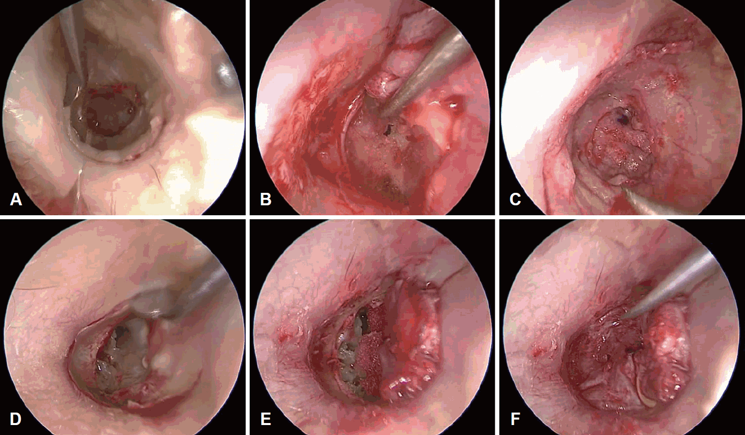 | Fig. 1.Surgical process of endoscopic tympanoplasty with underlay graft (A-C) and modified circumferential subannular graft (D-F). A: A meatal incision was made from the 7 o’clock position to the 12 o’clock position for the underlay graft technique. B and C: After inserting the graft medially to the fibrous annulus (B), the tympanomeatal flap was repositioned to its original position (C). D: A meatal incision in the modified circumferential subannular graft was made from the 11 o’clock position to the 1 o’clock position. E and F: After the fibrous annulus was elevated circumferentially (E), the graft was inserted into the lateral side of the bony annulus, and the medial side of the handle of the malleus (F).
|
Audiological analysis
Audiological evaluations including pure-tone audiometry (PTA) were performed in all patients preoperatively and postoperatively. A trained examiner performed the audiometry examination and obtained air and bone conduction thresholds in a double-walled audio booth. Pure-tone air conduction (AC) thresholds were measured at 0.5, 1, 2, 3, 4, 6, and 8 kHz over an intensity range of -10 to 120 dB. The average hearing threshold (in dB) values at 0.5, 1, 2, and 4 kHz were calculated using PTA. PTA was conducted again at least 3 months after the surgery, and the changes in hearing threshold and air-bone (AB) gap were compared to those before surgery.
Statistical analysis
The statistical analyses were performed using SPSS software for PC, version 21 (IBM Corp., Armonk, NY, USA). The Wilcoxon signed-rank test was used to compare postoperative improvements within groups, and significant differences between groups were determined using the Mann-Whitney test, the chi-square test, or analysis of variance. In all analyses, p-value<0.05 was taken to indicate statistical significance.
Go to :
Results
Of the 31 tympanoplasty patients, 11 were in the MCST group and 20 were in the underlay group. The male-to-female ratios were 4:7 and 7:13 and the mean ages of the patients were 51.8±13.2 years and 54.1±9.8 years, respectively. The perforation locations were divided into anterior, central, and posterior categories, being 6:2:3 in the MCST group and 5:6:9 in the underlay group, respectively. The average perforation sizes were 31.4±14.3% and 25.0±18.1% and the mean preoperative AC thresholds were 31.1±12.2 dB HL and 36.3±16.8 dB HL, respectively. No significant differences in age, location, or the size of the perforation, preoperative hearing threshold, or AB gap were observed between the two groups (Table 1). There was no need for canaloplasty during surgery in the MCST group, and a sufficient anterior field of view was obtained in all cases using an endoscopic approach. In addition, no other difficulties were encountered with the procedure. The average operating times were 68.6±16.5 min and 64.9±9.3 min in the MCST and underlay groups, respectively. The procedure took about 4 minutes longer on average in the MCST group, but the difference was not statistically significant (p=0.403). The postoperative hearing improvement was 12.1±5.0 dB in the MCST group and 9.0±6.3 dB in the underlay group (p=0.297). The respective postoperative AB gap values improved significantly to 4.1±3.2 dB and 4.9±3.1 dB, compared to the preoperative AB gap values (p=0.011 and 0.001, respectively), and no significant differences were detected between the two groups (p=0.570). No specific complications were observed in any patient, and no other problems, such as blunting or lateralization of the TM, were encountered during the follow-up period. The graft success rate was 90.9% in the MCST group and 85.0% in the underlay group. All recurrences were micro perforations, and no otorrhea or other complications were detected. As the graft was well maintained immediately after surgery, factors such as eustachian tube dysfunction could have been affected (Table 2). Eleven patients had anterior perforations before surgery. Among these, no recurrence occurred in the six patients in the MCST group, whereas graft failure occurred in two of the five patients in the underlay group. Of the four patients with graft failure, the average size of the perforation before surgery was 23.8±16.3%. As this result was not significantly different from the average size of perforation and showed wide-ranging variation, we did not find any significant correlation between the size of the perforation and graft failure.
Table 1.
Demographic findings between the MCST group and the underlay group in patients undergoing endoscopic tympanoplasty
![]()
Table 2.
Comparison of surgical outcomes according to the graft technique in patients undergoing endoscopic tympanoplasty
![]()
Go to :
Discussion
The reported graft success rates of tympanoplasty vary between studies, but a meta-analysis by Pap, et al. [16] showed that the graft uptake rate of endoscopic tympanoplasty was 86.9% (159/183) in randomized controlled trials and that the overall average success rate was 90.5% (503/556). A microscopic tympanoplasty success rate of 89.0% (163/183) was achieved in randomized controlled trials, and of 88.3% (550/623) in overall cases. The graft success rate of the present study was 90.9% (10/11) in the MCST group, 85.0% (17/20) in the underlay group, and 87.1% (27/31) overall. Our results are not significantly different from reported success rates in endoscopic tympanoplasty, and those reported for microscopic surgery. Therefore, the endoscopic approach is considered a useful surgical method that can replace conventional microscopic tympanoplasty. The wide-angle view of endoscopic transcanal tympanoplasty enables smooth manipulation of the anterior and anteroinferior parts of the TM that are difficult to access from a microscope, thus reducing the level of difficulty of the surgery and shortening the operating time. The wide and clear field of view also helps to stabilize attachment of the graft to the handle of the malleus, by making a hole or slit in the graft and inserting it into the handle of the malleus or covering the lateral side of the neck of the malleus. A wide supportive area is obtained using these procedures, and the graft does not fall or become lateralized (Fig. 2).
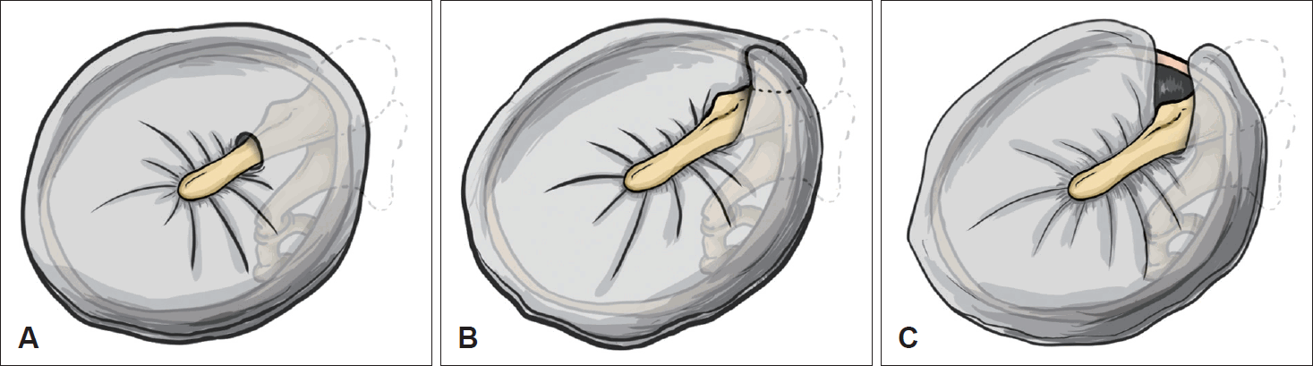 | Fig. 2.Graft positioning technique. Placing the graft on the lateral side of the malleus can cause detachment from the handle of the malleus, which causes blunting of the umbo angle and decreased hearing. In contrast, the graft can fall off when placed on the medial side of the malleus. To minimize this risk, a supportive area was made with a slit or hole in the graft and it was inserted into the handle of the malleus (A), or by placing both sides (B) or one side (C) of the graft on the lateral side of the neck of the malleus.
|
Two conventional graft techniques that have been developed are the underlay and overlay procedures. The difference between these two procedures is whether the graft is located laterally or medially to the fibrous annulus. In the underlay technique, the graft is placed entirely medial to the fibrous annulus, and the anterior side is placed freely under the bony annulus (Fig. 3A) [17]. Therefore, the middle-ear space is reduced, and the risk of the graft failing to adhere to the anterior remnant of the TM increases because there is no structure to support the medial side of the graft. This is the major reason why the graft success rate is lower for anterior perforations. However, the procedure is easy and fast, blunting or lateralization of the graft is avoided, and this procedure can be applied when transcanal visibility is limited. In the overlay technique, the graft is placed lateral to the fibrous layer and annulus after the squamous layer has been carefully removed (Fig. 3B) [18]. As the anterior edge of the graft is firmly fixed using this technique, a more stable postoperative result can be expected for an anterior perforation or subtotal perforation. Particularly, the use of the overlay technique is considered in cases of an anterior perforation when there is insufficient residual TM between the tympanic annulus and the perforation. However, it is more technically challenging because of the additional procedures, such as making a pseudosulcus using a drill to sustain the graft and the occasional canaloplasty to expose the entire anterior meatal recess. In addition, an iatrogenic cholesteatoma may be caused by the remnant epithelium when removing the epithelial layer, and there is a risk of lateralization or anterior blunting because there is no structure to support the lateral side compared to the underlay technique.
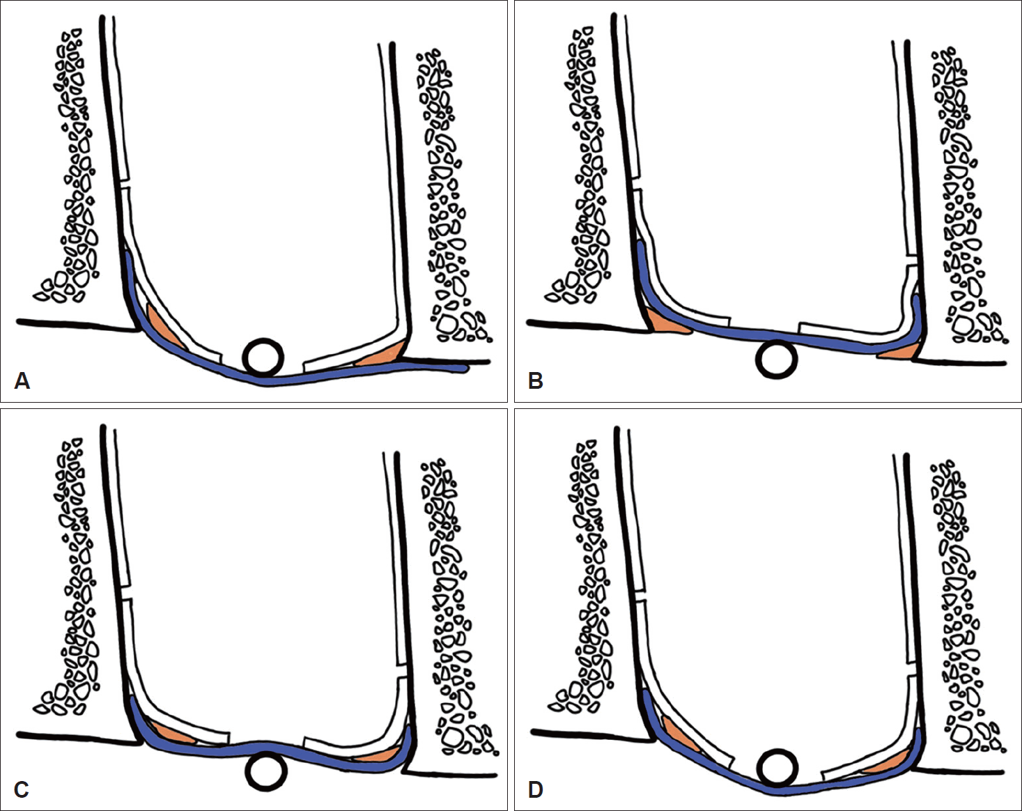 | Fig. 3.Various graft techniques in tympanoplasty. A: Underlay graft. This graft was placed medially on the posterior half of the fibrous annulus and was medial to the anterior half of the fibrous and bony annulus. B: Overlay graft. This graft was placed lateral to the fibrous annulus and malleus. C: Circumferential subannular graft. This graft was placed medial to the fibrous annulus, and lateral to the bony annulus and malleus. D: Modified circumferential subannular graft. This graft was placed medial to the fibrous annulus and lateral to the bony annulus. The risk for lateralization was minimized by inserting the graft into the medial side of the handle of the malleus, and the attachment to the handle of the malleus was stabilized.
|
Circumferential subannular tympanoplasty is an efficient technique that covers the shortcomings and takes advantage of underlay and overlay tympanoplasty. The previously reported procedure is to elevate the fibrous annulus circumferentially and place the graft to the lateral side of the handle of the malleus and the bony annulus (Fig. 3C) [15]. This procedure is relatively easier than the overlay technique because the pseudosulcus is not needed, and the supportive area of the graft is as wide as that of the overlay technique, so the graft remains stable and the middle-ear space is secured. However, the conventional microscopic technique still requires a postauricular incision and canaloplasty and has a risk of lateralization and anterior blunting similar to the overlay technique [19]. To compensate for these shortcomings, we modified the previous method and supported the medial and lateral sides by placing the graft on the medial side of the malleus or by inserting the graft into the malleus. The risk of side effects, such as lateralization, was reduced with this technique. In addition, a postauricular incision was not required using an endoscopic approach, and sufficient exposure of the entire TM was possible without canaloplasty. Grafts were taken from the perichondrial layer of the tragus in all patients including the MCST group. Perichondrium was harvested as wide as possible, and there were no problems in covering the whole bony annulus (Fig. 3D).
The results of MCST in this study, such as operating times, hearing improvement, and graft success rates, were comparable with the underlay technique, and the incidence of reperforation was rather low in patients with anterior perforations. In particular, as MCST was performed in patients with relatively severe anterior perforation, the stability of MCST in anterior perforation could be significant. Moreover, no cases of anterior blunting or lateralization were observed after surgery. These results were not different from previous studies that used other graft techniques, and thus, it is considered a reasonable option for the operator depending on the location of the perforation. In cases with anterior or large perforations, better stability can be achieved by using MCST with the wide supportive area of the graft.
Several factors should be considered when performing MCST. The procedure is easier using an endoscope than the conventional microscopic overlay technique or the circumferential subannular graft technique because no canaloplasty is required. Moreover, the process of making a pseudosulcus or removing the epithelial layer is not needed, so it is relatively easier than the endoscopic overlay technique. However, despite these advantages, it still requires expertise and skill compared to the underlay technique. In addition, the handle of the malleus may need to be denuded to position the graft properly on the handle of the malleus; thus, the risk of manipulating the ossicles and causing subsequent hearing damage is higher than in the underlay technique. Finally, it is important to determine the extent of the incision of the tympanomeatal flap. A wide meatal incision that extends to the anterior recess is essential to elevate the anterior flap smoothly. However, an excessive incision can affect the blood supply to the flap, which is associated with flap viability and the graft success rate. In this study, as in most previous studies, the incision was made from 11 to 1 o’clock, but further studies related to the correlation between the extent of the incision and flap viability are needed. In addition, as the sample size of our study was relatively small and the follow-up period of at least 6 months with an average of 9 months was not sufficient, further studies are still required to examine the risk of recurrence and long-term complications.
Graft success is the most important factor in tympanoplasty. In this study, MCST was successfully performed during endoscopic transcanal tympanoplasty, and the difference in operating time was not significant. The postoperative results were similar to those of the underlay technique. In addition, unlike the microscopic approach, no postauricular incision or canaloplasty was required. Hence, this modified technique is more suitable for an endoscopic approach, and addresses the shortcomings of conventional techniques to achieve a stable graft for anterior and subtotal perforations. In conclusion, MCST with an endoscope is a feasible and effective technique to improve the tympanoplasty results depending on the location of the perforation or the operator’s preference.
Go to :
 XML Download