

 PDF
PDF Citation
 Print
Print


Introduction
Exostosis is a rare condition, characterized by a benign bony growth extending outwards from the surface of a bone. External auditory canal exostosis (EACE) is benign hyperplasia of the lamellar bone secondary to chronic thermal, physical, or chemical irritation. It is well known to have a high prevalence in frequent cold water exposurers such as surfers or divers [1]. EACE, or surfer’s ear, has broad base and is multiple, unlike osteoma which is a pedunculated solitary bony tumor. It is usually asymptomatic at the beginning, but as it grows, external auditory canal (EAC) obstruction, conductive hearing loss, or even cholesteatoma might occur. Symptomatic EACE can be managed surgically. The surgery consists of complete EACE removal and the restoration of EAC skin.
External auditory canal cholesteatoma (EACC) is a rare condition in which keratin materials accumulate in the EAC and form a bone-destructive cystic mass. Small lesions can be managed conservatively, whereas larger lesions with symptoms such as otalgia and otorrhea need surgery, which consists of removing the cholesteatoma and reconstructing the defect site [2].
Until now, bony structures in otologic surgery have been cut by motorized drills with customized tips for efficient bone cutting or grinding. However, motorized instruments are prone to produce large amounts of heat which results in burning and consequential delayed bone healing [3]. During the drilling, the neighboring soft tissues may get caught in the rotating burr. Resultant skin loss and bare bone exposure delay healing and may require additional procedures such as skin graft.
To reduce the heat production, rotational speed should be controlled. However, lowering the speed hinders the cutting action. To compensate for the low-rotation speed, the surgeon should increase manual pressure, which consequently results in augmented macrovibrations and low surgical sensitivity.
The complications of drilling also include noise induced hearing loss, perforation of the tympanic membrane (TM) and EAC stenosis. The aforementioned problems promoted the development of the new instruments that are capable of cutting the bone with minimal heat production without adjacent tissue injury.
After considering all of the innovative items, we decided to perform transcanal endoscopic ear surgeries (EES) with piezoelectric device (PZD) for EACE or EACC. This relatively new device is based on ultrasonic microvibrations 4]. The passage of electric currents across certain ceramics transforms the structures of the device to produce microvibration.
The main advantage of the PZD was the absence of macrovibration so that the accurate and safe bone dissection would be possible. The absence of any rotating parts in the device also allowed us to use the cottonoid for protecting the EAC skin without annoying spooling and even to contact directly between the PZD tip and EAC skin.
Recently, EES has been extensively used in the field of otology as a minimally invasive procedure, allowing the entire view and complete resection of the EAC lesions. The use of a PZD in the treatment of EACE has been reported in only one English literature [5]. In this report, we present case series of EACE and EACC that were safely treated by EES with PZD.
Case
Case 1
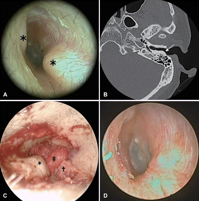
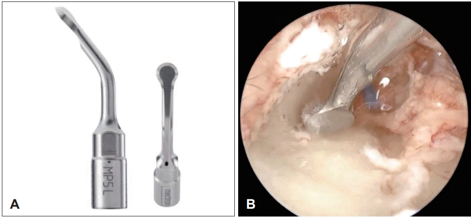
A 41-year-old male patient presented with a several-year history of slow growing masses in the left EAC. He had been operated for right EACE 5 years ago by postauricular approach under the view of microscope. At that time, he had decided to remove the larger right EACE first. He re-visited the clinic for the purpose of removing the remaining left EACE. Painless, even-surfaced hard masses were observed in the left bony EAC (Fig. 1A). Although the surgery was not indicated as the patient was asymptomatic, he strongly wanted to have the surgery before going abroad for years, where proper management of EACE was not expected. The computed tomography of temporal bone (TBCT) revealed about 4 mm-sized bony protrusions at anterosuperior wall of left EAC (Fig. 1B). The broad based bony proliferations were consistent with exostosis. During the following operation, the skin of EAC was infiltrated with 2 percent lidocaine and 1:100000 epinephrine. After making several circumferential incisions along the masses, skin flaps were elevated using round knife and duckbill elevator as laterally based flaps for the lateral surface of the each mass and medially based flaps for the medial side [5]. During the procedure, EAC skin was protected with the cottonoid (Fig. 1C). After obtaining the sample for histologic work up, a PZD (Piezosurgery®, Mectron S.p.A., Genoa, Italy) and the long osteoplasty circular insert tip (MP5 L, Mectron S.p.A.) (Fig. 2A) were used to remove the eroded surface, layer by layer with continuous saline irrigation. The use of this PZD provided a smooth cut surface (Fig. 2B). The EAC was widened for the full visualization of the TM. After repositioning the skin flaps, the EAC was packed with several pieces of antibiotics soaked absorbable gelatin compressed sponge (Gelfoam ®, Pfizer, New York, NY, USA) (Supplementary Video 1). The histologic analysis confirmed a diagnosis of EACE. At a 3-month follow-up after surgery, the patient was asymptomatic and free from any complications or relapse (Fig. 1D).
Case 2
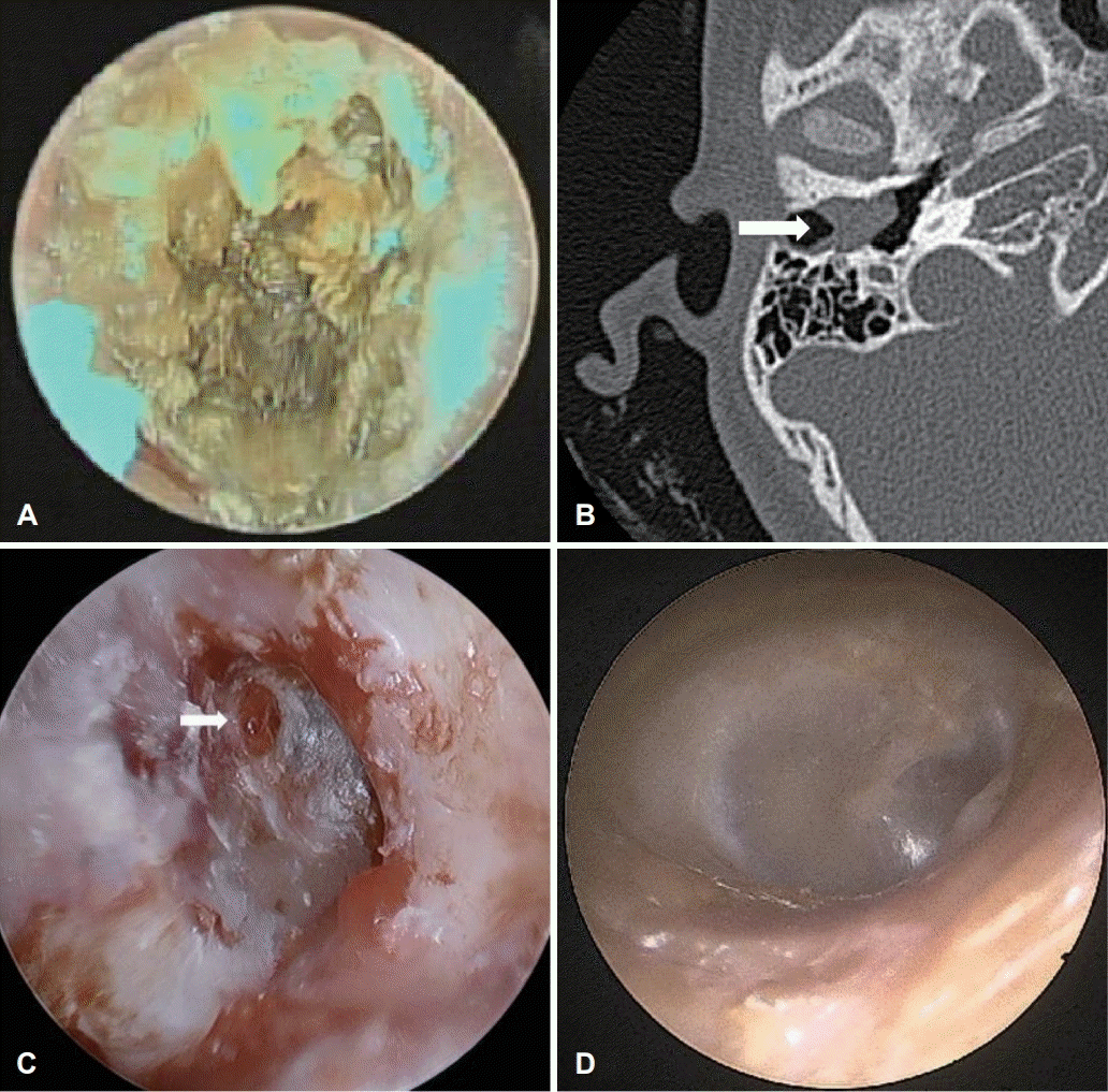
A 12-year-old female visited our clinic with hearing difficulty in her right ear. The endoscopy showed an irregularshaped EAC mass, of which all margins were firmly attached to the skin (Fig. 3A). The following TBCT revealed a 1.3 cm sized expansile mass-like lesion confined to the right bony EAC with subtle adjacent bony erosion (Fig. 3B). Suspecting an EACC, the EES using PZD was planned to remove the mass. Under general anesthesia, the chole sac around the destructed EAC was extirpated. The remaining granulation at the TM was also removed (Fig. 4C). The burring off the destructed EAC and the excavation of tissue using PZD to form a shallow shelving depression were performed. The perforated TM and the bare bone were reconstructed with acellular dermal matrix (MegaDerm®, L&C Bio Inc., Seongnam, South Korea). The EAC was packed with antibiotics soaked absorbable gelatin compressed sponge (Gelfoam®) and fibrin sealant (Tisseel®, Baxter Healthcare Corp., Deerfield, IL, USA) (Supplementary Video 2). The histologic analysis confirmed a diagnosis of EACC. At a 3-month follow-up, the patient was free from any complications or relapse (Fig. 3D).
Case 3
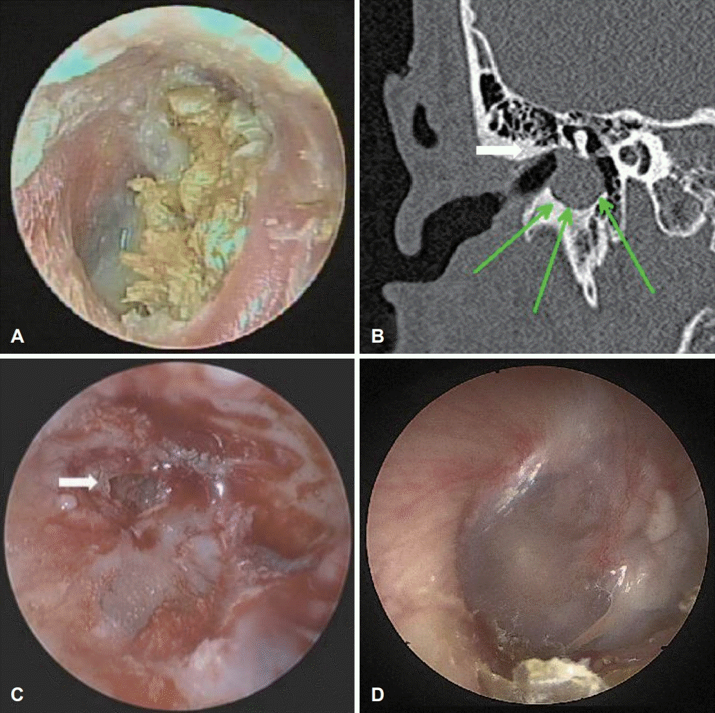
A 15-year-old male patient visited our clinic with right hearing disturbance. The endoscopy revealed a yellowish protruded irregular-shaped mass attached to the TM (Fig. 4A). The TBCT showed a homogenous soft tissue mass at the medial end of the right EAC with bone erosion (Fig. 4B). As in case 2, the operation was performed with above-mentioned materials in the same manner except that the defect was reconstructed with sliced cartilage and perichondrium (Fig. 4C). The pathology report confirmed the mass to be an EACC. At a 2-month follow-up, the endoscopy showed that the TM and EAC were all in well healing status (Fig. 4D).
Discussion
In the 20th century, technology of otology has been advanced in terms of instrumentation, visualization and intraoperative nerve monitoring. The developments of the microscope, endoscope and surgical drills are representative exemples [6]. After dentists had applied ultrasound technology as a cutting tool, thoracic surgery was the first department in medicine to use the ultrasound device in 1972. However, insufficient power of the original ultrasonic instrument inevitably led to the macrovibration and excessive heat production causing bony necrosis [7]. To overcome such problems, the PZD, a system with microvibrations, was developed. This relatively low frequency (24-36 kHz) device exclusively acts on the mineralized tissue, not influencing the surrounding soft tissues such as skin and nerves.
The term of piezo derives from Greek, meaning ‘to squeeze or press [8].’ Certain ceramics and crystals contract or expand when an electric current is passed across them, causing oscillation [8]. The microvibrations make the device easy to handle and enhance the tactile control, making it possible to remove the mineralized tissue layer by layer with precision [4,8]. When the device loses contact with the bone accidentally, the cutting head is programmed to stop immediately [4]. PZD converts just 30% of energy into heat, so remaining 70% is transformed into cutting power [9]. The integral peristaltic saline pump enables cooling of the tip, simultaneously removing debris from the operation field.
Many studies have also revealed that bone healing is improved after piezoelectric cutting at an early stage [10]. In animal studies of PZD, initial healing was faster and caused less epineural damage, compared to the conventional bur [11]. By avoiding coagulative necrosis at the bony margin [7], there is less chance of vascular occlusion or blood flow disruption [12,13].
The microvibrations also make it possible to avoid inner ear damage. The frequency of ultrasound is greater than the upper limit of normal human hearing. The audiologic results verify the safety of the PZD in regard to hearing [4,8,10,14].
Nevertheless, our impression was that the PZD removed the bone slightly slower than the conventional bur. This is consistent with others’ studies [14. The operative time itself was 35% longer with PZD compared to a conventional bur [15]. However, when considering the effort of a surgeon to protect skin or soft tissue, the time required for addition skin graft, and the delayed healing by traditional drill, it is likely that there is in fact more merits in PZD.
EACE and EACC can be managed surgically if EAC obstruction or symptoms arise. In our case series, removing symptomatic EACE and EACC with the PZD was safe and effective approach. The minimal bleeding, lack of damage to the EAC skin and reduction of postoperative complications highlight the safety of EES with PZD. Such advantages make PZD an ideal tool to use especially at the borders between soft and hard tissues in cases of EACE or EACC.

 XML Download