Introduction
Surgical phase of virtual surgical planning (VSP) and 3D bio-printing technique
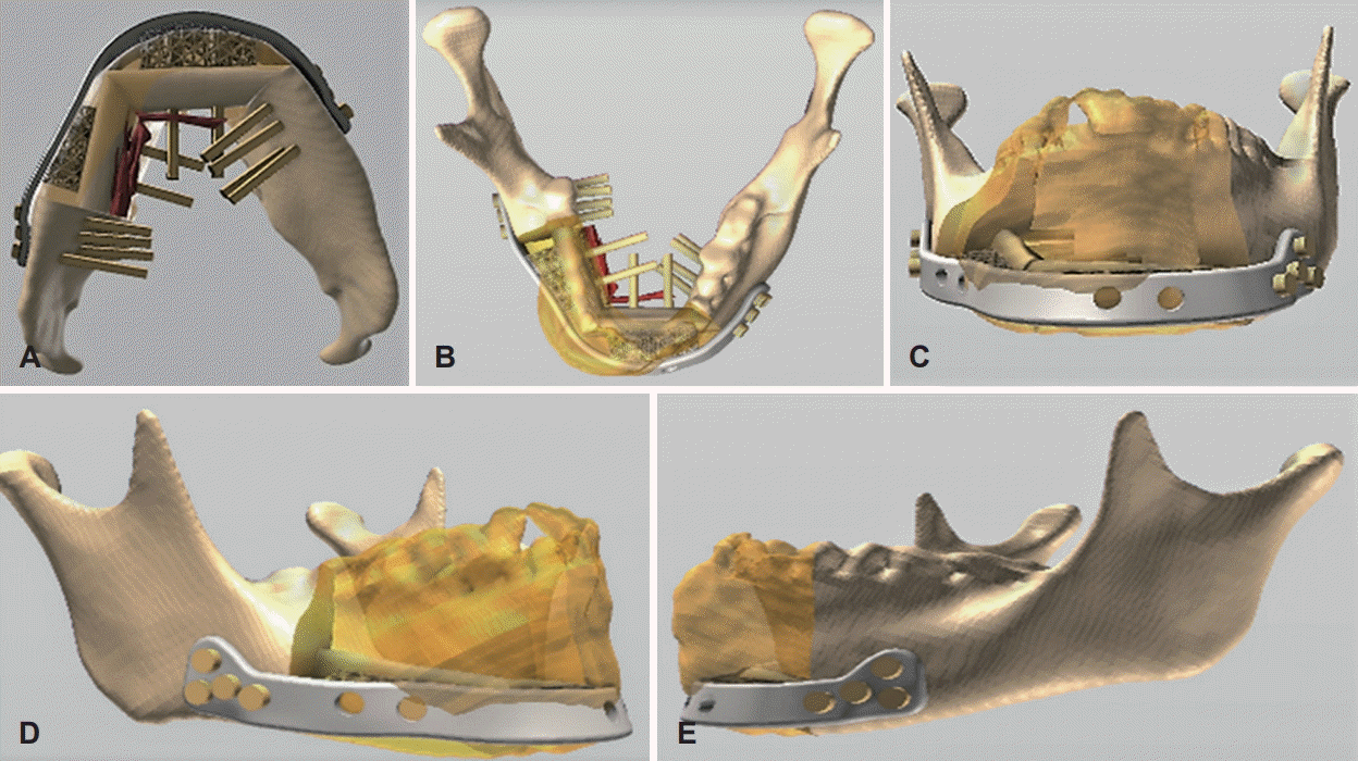
 | Fig. 3.Showed the step of 3D model printing of the mandible with the fixed prebent reconstruction titanium and screw. Virtual planning and prepared 3D model printing of the mandible with the fixed prebent reconstruction titanium and screw (A, B). Overlay of the virtually planned neomandible with pre-bent customized plate reconstruction with a pre-bent customized plate and screw evaluated by 3D CT (C-E). This is a sample case with diagnosis of benign lesion ‘Desmoplastic Ameloblastoma’ confirmed by the incisional biopsy at the Department of Oral and Maxillofacial Surgery, Mahidol University, Bangkok. 3D: three-dimensional. |
Advantages and disadvantages of VSP and 3D bio-printing technique
Previous studies
Table 1.
| Author & year | Diagnosis | Surgical intervention and evaluation of parameter | Sample size | ||
|---|---|---|---|---|---|
| Succo, et al. [15] 2015 | Both benign and malignant cases | Computer-assisted reconstruction of mandible with a microvascular fibular free flap using pre-operative virtual planning, customized cutting guides, along with laser pre-bent titanium plates. To demonstrate the overall reconstructive outcome, precise re-establishment of oro-facial functions, and esthetic outlines. | 5 cases | ||
| Rodby, et al. [16] 2014 | Maxilla and mandibular oncology (84%) | Computer-assisted head-neck surgery according to VSP, stereolithographic models along with specific cutting guides. | A total of 220 cases | ||
| ORN (16%) | Reconstructions performed by free fibula osteocutaneous flap 177 (80%), iliac crest flap 31 (14%), scapular flap 12 (6%) cases. A systematic review conducted to find-out the conveniences of the VSP technology-based reconstruction, identify its utilization and evaluate the advantages, and made comparison of surgical outcomes with conventional surgery on head neck oncology. | Reconstructions of mandible (193 cases, 88%) and reconstructions of maxilla (22 cases, 10%) | |||
| Toto, et al. [17] 2015 | SCC | Free fibula osteocutaneous flaps reconstruction after segmental mandibulectomies. To evaluate the influences of CT-directed preoperative planning on post-operative outcomes. | Sample size: 57 | ||
| 3 groups | |||||
| Group A: 12 patients | |||||
| Reconstructive surgeon based bended plates on acrylic model. Osteotomies went-through on back-table. | |||||
| Group B: 20 patients | |||||
| Reconstructive surgeon based bended plates on acrylic model. Osteotomies went-through in situ osteotomies. | |||||
| Both A & B groups without the use of preoperative planning or osteotomy guides. | |||||
| Group C: | |||||
| 25 patients went-through in situ osteotomies along with prefabricated osteotomy guides created from the preoperative CT-guided computerized planning session. | |||||
| Wang, et al. [18] 2016 | ORN, intraosseous carcinoma of the mandible, lower gingival carcinoma. Carcinoma in the oral floor, osteosarcoma, mandibular osteo-fibroma. | Virtual planning surgery group: | 2 group | ||
| Resection and reconstruction of mandible using vascularized fibular flap, utilizing VSP software, stereolithographic models, specific cutting guides, and specific reconstruction pre-bent plates. | 56 patients | ||||
| Conventional surgery group: | 21 patients went through mandibular reconstruction with vascularized fibula grafts using the VSP. | ||||
| Fibular flap harvest, resection and reconstruction of mandible by manual surgeon’s basis effort. To evaluate the preciseness and determine the clinical benefits in both virtual planning and conventional surgery patients. | 35 patients went through conventional surgery. | ||||
| Bosc, et al. [19] 2017 | SCC | Segmental mandibulectomy | Sample size:18 | ||
| Reconstruction by osteocutaneus free flap. To evaluate the potentiality and accuracy of an in-house technique that contains all the procedures of CAD and rapid prototype modeling that not needed any outside laboratory insertion. | |||||
| Chang, et al. [20] 2016 | SCC | Mandibular reconstruction with osteocutaneous free flaps. To determine the essentiality and proficiency of CT guided preoperative VSP on post-operative outcomes. | Sample size: 92 | ||
| ORN | Group A: 43 patients had a STL model to serve as a template, 18 of them (41.9 percent) having a prebent plate. Osteotomies performed on the back table without the use of pre-fabricated cutting guides. | ||||
| Group B: 49 patients | |||||
| Underwent VSP with STL models, prebent plates, and osteotomy guides based upon the preoperative, CT-guided planning session. | |||||
| De Maesschalck, et al. [21] 2017 | SCC | Reconstruction of mandible with fibular free flap. | Sample size: 18 | ||
| ORN | To compare morphological mandibular reconstruction outcome between CAD/CAM custom patient-specific plates and surgical cutting guides versus conventional freehand technique and to assess the accuracy and duplicability of VSP technology. | Group A: 7 patients. The CAS group experienced virtual planning with CAD/CAM customised patient-specific plates and surgical cutting guides. | |||
| Group B: 11 patients. The control group went through traditional freehand surgery. | |||||
| Tarsitano, et al. [22] 2017 | SCC, KCOT, OGS, odontogenic fibromyxoma | Disarticulation resection surgery following the preservation of articular meniscus and set a CAD/CAM reconstructive plate with fibular microvascular free flap. | 9 patients went through disarticulation resection surgery for benign and malignant mandibular tumors at the condylar region. | ||
| To assess the clinical benefits and complications by a series of oncological patients. | |||||
| Ren, et al. [23] 2018 | Ameloblastoma | Vascularized fibula flap mandibular reconstruction utilizing virtual planning and 3D printing modeling along with cutting guide osteotomies and pre-bent plates to find-out its capabilities and to compare the operation time and surgical results of this technique with the traditional method. | 30 patients | ||
| Ossifying fibroma | Study group: 15 patients went through vascularized fibula flap mandibular reconstruction utilizing virtual planning and 3D printing modeling, pre-bent titanium plates, and cutting guides. | ||||
| KCOT | |||||
| SCC | Control group: 15 patients experienced mandibular reconstruction utilizing fibula flap without the help of virtual planning and 3D printing models. | ||||
| Jacek, et al. [24] 2018 | SCC advanced stage | Mandible reconstruction surgery with the scapula and fibula free flaps. A comparison made between two types of mandible reconstructive operations either 3D models from thermoplastic materials or conventional planning surgeries using both scapula and fibula free flaps. | Sample size: 8 | ||
| 4 patients treated with free fibular flap with skin island. | |||||
| Another 4 patients treated with scapular flap with skin island. | |||||
| Among the 8 patients, 4 are treated with conventional technique and another 4 are by 3D model. | |||||
| Dell’Aversana Orabona, et al. [25] 2018 | SCC | Oncological mandibular reconstruction using fibular flap by VSP and 3D bio-printing in a home-made way. | Sample size:4 | ||
| To evaluate the viability and effectiveness of a protocol to produced cutting guides in a “homemade” way. | |||||
| Goormans, et al. [26] 2019 | SCC, ORN, RMS, ES, OS | Reconstruction of the mandible with free fibula flap, using VSP and 3D model tech, fixation tray, and cutting guides’ osteotomies, manually bended reconstruction plate according to 3D model. | Sample size: 26 | ||
| To assesses the more preciseness of computer-assisted mandibular reconstructions. | Patients divided by condylar involvement and subdivided by the number of fibular segments used for reconstruction. | ||||
VSP: virtual surgical planning, 3D: three-dimensional, ORN: osteoradionecrosis, SCC: squamous cell carcinoma, STL: stereolithography, CAD/CAM: computer-aided design/computer-aided modeling, CAS: computer-assisted surgery, KCOT: kerato-cystic odontogenic tumor, OGS: osteogenic sarcoma, RMS: rhabdomyosarcoma, ES: ewing sarcoma, OS: osteosarcoma
Discussion
Table 2.
| Author & year | Follow-up | Result |
|---|---|---|
| Succo, et al. [15] 2015 | 8-12 month | Outstanding accuracy of cutting guides, perfectly fit pre-bent plates, precise bone contact, and position found in mandible and fibula graft. |
| The average difference between the programed segment and CT control SL 0.098±0.077 cm. | ||
| Significantly shorter ischemia times an average of 75±8 min; Average post-operative hospital stay 18±3 days. | ||
| Rodby, et al. [16] 2014 | Mean follow-up month | Total: 33 articles, 220 cases |
| Functional outcomes reported in 99 cases (45%). | ||
| 14 months | 2 patients (2%) categorized as ‘ideal,’ 12 patients (12%) as ‘outstanding,’ 23 patients (23%) as good, 34 patients (35%) as ‘restored’ and comments from patient and physician-expressed satisfaction with function for 7 (7%) and 21 (21%) patients respectively. | |
| Esthetical outcomes reported in 84 patients (38%) and catagorized as ‘outstanding’ for 34 patients (41%) and ‘good’ in 17 patients (20%); ‘successful’ for 8 patients (10%); ‘symmetry achieved’ for 6 patients (7%); ‘near normal’ results in one patient; and patient and physician- satisfaction revealed with the esthetic outcome for 7 (8%) and 11 (13%) of patients respectively. | ||
| Precise accuracy of the reconstruction found on 93% of cases. | ||
| Shortened intraoperative time found in 175 cases (80%). | ||
| Especially described reduced flap ischemic time for 67 cases (30%). | ||
| User convenience especially reported for 53 cases (24%). | ||
| Negative margin found as resection cutting guides in 12 cases (5%). | ||
| Refined predictability of outcomes especially documented in 16% cases. | ||
| 22% of the patients improved patient satisfaction. | ||
| 6% of patients decreased complications. | ||
| Quantitative study data showed with an average score of 88 for patients from VSP and score 68 for traditionally reconstructed patients. | ||
| Quantitative results utilizing pre-operative and post-operative CT scan comparisons shown for 30% of cases. | ||
| Mean surface deviation in 1 case. | ||
| Repetition of segment placement found in 7 patients, and osteotomy site difference by plate overlap found in 11 patients. The position, the difference from the actual postoperative and projected CT images, all patients (50 cases total) were within 2.5 mm deviation/difference and overlap in the actual vs. projected reconstructive plates was seen in 11 patients, 58% (±8.96%). | ||
| Toto, et al. [17] 2015 | N/D | 3 groups, as mentioned in Table: 1 |
| Mean ischemia time 177.8 min, 86.69 min, and 76.92 min for group A, B, and C respectively. Mean OR time 706.9 min, 659.8 min, 534.2 min for group A, B and C respectively. Mean operating room values $20332.50, $19002.60, $15450.48 for group A, B, C respectively. Cost of plate $4200.00, $4200.00, $5500.00 for group A, B and C groups respectively. | ||
| Total costs $24532.50, $23202.60, $20950.48 for group A, B and C respectively. | ||
| Wang, et al. [18] 2016 | 6 months | Ischemia period (mean±SD) found 45±13 min in VSP group where 63±15 min in CS group, Total operation duration (mean±SD) 4.5±0.9 hr. in VSP group and 5.8±1.3 hr in CS group. Normal condyle position achieved 100% in the VSP group and 54.3% in the CS group. Sharp bone to bone contact found 95.2% in the VSP group and 71.4% in the CS group. Accurate position among plate in mandible and fibula segment achieved 95.2% in the VSP group and 62.9% in the CS group. Good facial appearance achieved 95.2% in the VSP group and 77.1% in the CS group. 100% of patients have their regular diet in the VSP group where CS group 85.7%. 100% of patients have their intelligible speech in the VSP group and 97.1% in the CS group. |
| Bosc, et al. [19] 2017 | 19 months | All reconstruction outcomes successfully matched between the pre-operative model and final surgery outcome both the fibula bone segments and angle measurements. |
| No major modification needed for any patient. The mean variation in angle value between the preoperative models to postoperative outcomes 4°. | ||
| Chang, et al. [20] 2016 | 12 months | Preoperative CT-guided surgery resulted less burring, lesser osteotomy revisions, and little bone grafting, decreased operative time (666 minutes for VSP group 545 minutes for conventional surgery group; p<0.005), decreased bony nonunion. |
| De Maesschalck, et al. [21] 2017 | Morphometric accuracy, from pre- and postoperative linear and angular measurements found no statistically significant variences in both groups but only for the axial angle on the non-affected side (1.0 in the CAS group versus 2.9 in the control group; p=0.03). | |
| In the CAS group, a mean distance deviation of 2.3±1.0 mm for mandibular osteotomies and 1.9±1.1 mm for fibular osteotomies. | ||
| Tarsitano, et al. [22] 2017 | 2-72 months, mean 24.8 month | Only 1 patient had condylar displacement anterior to the glenoid tubercle, other patients had usual mouth opening. |
| All patients developed low-grade mandibular deviation during maximum mouth opening. | ||
| Chewing ability, speech style, and esthetical facial contour equivalent to or better than baseline for all patients. | ||
| The average postoperative condylar deviation from the preoperative location about 3.8 mm (range: 1.3 to 6.7 mm). | ||
| Ren, et al. [23] 2018 | Reconstructive time hours and operative time hours on CAS group about 1.60±0.38 hrs and 5.54±0.50 hrs respectively where control group reconstructive times hours and operative times hours about 2.58±0.45 and 6.54±0.70 respectively and the p-value is <0.001. CAS group showed Intercondylar distance, intergonial angle distance, anteroposterior distance, gonial angles 2.92±1.15, 2.93±1.19, 4.31±1.24, 3.85±1.68 respectively and in control group 4.48±1.41, 4.79±1.48, 5.61±1.41, 5.88±2.12 respectively. | |
| Jacek, et al. [24] 2018 | Stability reported at conventional technology 84% and model 3D printing group 100%, mouth opening 2.5 cm for conventional technology group 3 cm for model 3D printing group. | |
| Chewing function restored results 72% on the conventional group and 90% on model 3D printing group. Cosmetically acceptable and operation time 60% and 8.5 hr respectively for the conventional group and 100% and 6.5 hrs respectively for Model 3D printing group. Mandibular counter symmetrical angle different 10.0±12.5 mm for the conventional group and 7.3±9.1 mm for Model 3D printing group. | ||
| Dell’Aversana Orabona, et al. [25] 2018 | All mandibular reconstructions were fruitful with a perfect match from the digitally planned 3D models to the final outcomes. No big adjustment of the mandibular bone resection needed in intra-operatively as suggested to the planned 3D simulation. The total period taken for the virtual planning design was about 3 h. 6 h were required for the printing process and sterilization. An average distance of 1.631 mm (range 0.594 to 4.067) and a SD of 5.496 mm (range 1.966 to 8.024). | |
| Goormans, et al. [26] 2019 | 50 fibular segments evaluation | |
| The mean deviation in fibular SL is 1.74 mm (range, 0.02-6.10 mm), the angular deviation of the osteotomy planes is 1.98° (range, 0.04-5.86°). | ||
| Mean values for each measure were intercoronoid distance deviation, 3.86 mm (range, 0.20-11.21 mm); interregional distance deviation, 3.14 mm (range, 0.05-8.28 mm); anteroposterior distance deviation, 2.92 mm (range, 0.03-8.49 mm); and intersegmental plane shift, 11.00° (range, 2.76-24.15°). These results illustrated dissimilarities in a large scale in final postoperative accuracy. | ||
| In the group with preserved condyles, mean intercoronoid and interregional deviations differed significantly (5.02 mm and 4.88 mm, respectively; both p<0.05) for one-segmented and three-segmented fibular reconstructions, respectively. Final postoperative accuracy lessens when more fibular segments are utilized for reconstruction. For intersegmental plane shift, the preserved and non-preserved condyle groups vary significantly (7.18°; p<0.05). | ||
| General ischemia period found <4.00 h where most ischemia times found <2.00 h. | ||
| The all-inclusive survival rate of fibular flaps 96.15%. |
Table 3.
| Authors & year |
The outcome of VSP |
|||||||
|---|---|---|---|---|---|---|---|---|
| No of cases | Decreased odds ratio time | Decreased ischemia time | Increased reconstruction accuracy | No major complications | Functional outcome | Decreased cost | ||
| Succo, et al. [15] 2015 | 5 cases | Yes | Yes | Yes | Yes | Not mentioned | Not mentioned | |
| 75±8 min | Difference at programed and CT control segment lengths 0.098±0.077 cm | |||||||
| Rodby, et al. [16] 2014 | 193 case | Yes | Yes | Yes | Yes | Yes | Yes | |
| Toto, et al. [17] 2015 | 25 cases | Yes | Yes | Yes | Yes | Not mentioned | Yes | |
| 534.2 min | 76.92 min | Excellent accuracy | ||||||
| Wang, et al. [18] 2016 | 21 cases | 4.5±13 hr | 45±0.9 min | Yes | Yes | Not mentioned | Not mentioned | |
| Excellent accuracy | ||||||||
| Bosc, et al. [19] 2017 | 18 cases | Mean | Yes | Yes | Artery anastomosis thrombosis: 1 | Not mentioned | Not mentioned | |
| 7-hour 2 min | Pulmonary embolism: 1 | |||||||
| Acute myocardial infarctio: 1 | ||||||||
| Chang, et al. [20] 2016 | 49 cases | 545±12.6 min | 120±19.8 min | Yes | Yes | Not mentioned | Not mentioned | |
| Excellent | ||||||||
| De Maesschalck, et al. [21] 2017 | 7 cases | Not mentioned | Not mentioned | Yes | Not mentioned | Not mentioned | Not mentioned | |
| Tarsitano, et al. [22] 2017 | 9 cases | Not mentioned | Not mentioned | Yes | Yes | Not mentioned | Not mentioned | |
| Mean difference between postoperative and preoperative condylar deviation by CT 3.8 mm | ||||||||
| Ren, et al. [23] 2018 | 15 cases | Yes | Not mentioned | Yes | Not mentioned | Not mentioned | Not mentioned | |
| Jacek, et al. [24] 2018 | 4 cases | Yes | Not mentioned | Yes | Not mentioned | Yes | Not mentioned | |
| 6.5 hour | Significantly improved accuracy | Significant improved functional outcome | ||||||
| Dell’Aversana Orabona, et al. [25] 2018 | 4 cases | Yes | Not mentioned | Yes | Yes | Not mentioned | Yes | |
| Mean distance of 1.631 mm with a standard deviation of 5.496 mm by CloudCompare software overlay analysis | ||||||||
| Goormans, et al. [26] 2019 | 26 cases | Yes | Yes | Not mentioned | Acute complications (<6 weeks postoperatively) | Not mentioned | Not mentioned | |
| All ischemia times were <4.00 h, most ischemia times <2.00 h | - Hemorrhage: 2 | |||||||
| - Venous congestion: 1 | ||||||||
| Late complications (>6 weeks postoperatively) | ||||||||
| - Removal of bony plate and reconstruction: 1 | ||||||||
| - Low-grade osteoradionecrosis: 2 | ||||||||
| - Trismus: 3 | ||||||||


 PDF
PDF Citation
 Print
Print


 XML Download