Abstract
Schwannoma, also known as neurilemmoma, is a benign neoplasm that originates from any nerves wrapped with a sheath made of Schwann cells. Schwannoma occurring in the head and neck region is not rare, but schwannomas of the anterior neck, especially ansa cervicalis, are extremely rare that only 7 cases have been reported to date worldwide. Although rare, it should be considered in differential diagnosis of anterior cervical mass and may be confused with other cervical and thyroid mass. We report a case of intramuscular schwannoma in the sternohyoid muscle. Preoperative diagnosis was established with an ultrasound-guided core needle biopsy. Although it was removed entirely without connection to any other nerves identified or any complication, clinically, the mass was thought to be derived from the nerve. To our knowledge, this is the first case of the intramuscular schwannoma occurring from ansa cervicalis reported in the literature.
신경초종은 신경초세포(Schwann cell)에서 기원하는 양성 종양으로 시신경과 후각신경을 제외한 신체 내 어느 신경 분지에도 생길 수 있다[1]. 전체 신경초종의 25~45%가 두경부에서 발생하며, 두경부 내에서는 부인두공간이 37.5%로 가장 흔하며, 악하 및 이하선 공간에 호발하는 것으로 보고되었으며[2], 전경부에 발생하는 경우는 매우 드물다. 전경부에 발생한 경우에는 갑상설관낭, 유피낭, 흉선 낭종, 지방종 등과 감별이 필요하며 해부학적 위치와 영상학적 검사를 토대로 진단을 추정할 수 있다[3]. 경신경고리에서 기원한 신경초종은 매우 드물어 현재까지 전 세계적으로 문헌상 보고된 사례는 7예로 확인되며 피대근의 근육 내부에서 신경초종이 발견된 사례는 현재까지 국내외에 보고된 바 없다. 저자들은 서서히 커지는 전경부 종괴로 내원한 43세 환자에서 피대근 내부에 발생한 경신경고리 기원의 신경초종을 완전절제하였고 술후 3년간 재발 및 후유증 없이 양호한 결과를 보였기에 드문 증례로서 문헌고찰과 함께 보고하는 바이다.
평소 건강하던 43세 남성이 내원 3개월 전부터 좌측 전경부에 만져지는 종물을 주소로 내원하였다. 계통문진에서 음성변화나 연하곤란 등의 특이증상은 없었다. 신체검사에서 좌측 갑상선엽 전방에 단단한 무통성 종괴가 촉지되었으며 삼킴 시 후두의 움직임을 따라 함께 움직이는 모습이 보였다. 경부 전산화단층촬영에서 좌측 상갑상선의 전방 경계로 흉골설골근 내부에 약 1.5 cm 크기의 주변과 경계가 뚜렷하고 내부에 약간의 조영증강을 보이는 저음영의 종괴가 확인되었다. 이외 양측 경부에 뚜렷한 이상 소견은 관찰되지 않았다(Fig. 1). 3개월 후 추적 초음파검사에서 종괴의 크기나 양상, 환자의 증상에는 큰 변화가 없었으며 조직학적 진단을 위해 초음파유도 하 중심바늘생검을 시행하였다(Fig. 2). 조직검사 결과 방추형의 신경초세포를 확인하여 신경초종으로 진단할 수 있었고 초음파와 전산화단층촬영에서는 뚜렷한 신경 연결 부위를 확인할 수는 없었다. 보다 세밀한 확인을 위해 MRI를 권유하였으나 환자 원치 않아 시행하지는 못하였다. 다만 종괴의 해부학적 위치상 갑상선이나 후두신경에서 기원하였을 가능성은 희박하고, 경신경고리에서 유래한 것일 가능성이 가장 크고, 근육 내부에 생겼으므로, 경신경고리의 원위 말단부의 신경 분지에서 유래하였을 것으로 생각하고, 수술로 인한 신경 손상의 가능성을 충분히 설명하고 수술을 진행하였다.
먼저 좌측 윤상연골의 외측으로 피부 주름을 따라 3 cm 절개하여 위아래로 광경근하 피판을 들어 피대근 내부의 종괴를 확인하고 피막하박리를 진행하였다. 육안 소견상 종괴는 일부 근섬유에 유착이 되어 있었으나, 비교적 근육과 구분되는 피막으로 잘 싸여 있었으나 신경을 포함하여 다른 조직과의 연결은 발견할 수 없었다(Fig. 3). 따라서 임상적으로 피대근에 분포하는 경신경고리의 근육 내 최종 원위말단부에서 유래하였을 것으로 생각하였고, 경신경고리가 기능적으로 임상적 의의가 크지 않고, 근육 내 분지의 경우 손상으로 인한 기능 소실은 극히 미미할 것으로 생각되어 종괴를 완전적출한 후, 근육을 수복하고 배액관 유치 없이 수술을 종료하였다.
육안적으로 종양은 잘 피막화 된 견고한 종물로 조직검사상 heamtoxylin and eosin(H&E) 염색에서 방추형의 신경세포 핵이 나란히 배열하는 책상배열(palisading) 모양을 볼 수 있었으며 베로카이 소체(Verocay body)를 포함하는 Antoni A 영역과 불규칙한 배열의 저세포성 Antoni B 영역이 혼재되어 관찰되었다. 또한 초음파에서 흉골설골근과 종괴의 피막이 유착되어 있던 부분의 경우, 조직염색에서 피막과 함께 절제된 근섬유를 확인할 수 있었다(Fig. 4). H&E 염색에서 전형적인 신경초종으로 확인되었기 때문에 병리과 전문의의 판단에 따라 S-100 등 추가 면역조직화학염색은 하지 않았다. 수술 후 환자는 특별한 불편감이나 합병증 없이 3일째 퇴원하였다. 이후 환자는 3년간 정기적인 추적관찰 중에도 수술과 관련하여 후유증이나 재발의 증거 없이 지내고 있다.
신경섬유초종(neurilemmoma)으로 불리기도 하는 신경초종은 신경초세포로 덮여있는 신경이라면 어떤 신경에서든 발생할 수 있는 양성종양으로 1910년 처음 소개되었다[3]. 대개 단일병변으로 발생하며 신경초종의 25~45%가 두경부 영역에 발생한다[4]. 두경부 신경초종이 주로 발견되는 위치는 측경부이며, 이는 부인두공간에서 가장 흔한 종양으로도 알려져 있다[1]. 두경부 영역 신경초종의 호발 부위는 측경부 중에서도 부인두공간, 악하공간 및 이하선의 순서로 알려져 있으며[2], 미주신경[1], 경부교감신경 및 상완신경총에서도 비교적 흔하다[5]. 드물게 구강, 비부비강, 안면신경이나 갑상선[6]에서 기원하는 경우도 보고되었으나, 경신경고리에서 신경초종이 발생하는 사례는 특히 드물다(Table 1). 또한, 저작근 내 및 사지의 골격근 내에서 발생한 신경초종과 같이 다양한 근육 내 신경초종이 보고되었으나[7], 본 증례와 같이 피대근 내에서 신경초종이 발견된 사례는 아직까지 전세계적으로 보고된 바는 없다.
두경부 영역에서 발생한 신경초종은 대부분 통증 없이 서서히 자라나는 경부 종괴의 형태로 발견되며 증상과 임상양상이 비특이적이다. 이러한 특징 때문에 경동맥체 종양, 림프절 비대, 새열 낭종, 혹은 갑상선 병변 등으로 오인되어 진단하기 어렵다[8]. 경신경고리에서 발생한 신경초종의 경우, 특징적으로 전경부 종괴의 형태로 나타나 갑상선 종양이나 갑상설관낭, 유피낭, 흉선 낭종, 지방종 등과 감별진단이 필요하다[4]. 진단을 위한 방법으로는 초음파, 전산화단층촬영, 자기공명영상 등 영상학적 검사를 사용할 수 있다. 이 중에서도 자기공명영상검사의 경우 T1 강조영상에서는 동등신호강도나 고신호강도를 나타내며 특징적인 “split fat sign”을 보이기도 한다. T2 강조영상에서는 고신호강도를 나타내며 “fascicular sign”, “target sign”, “thin high intense rim” 등 특징적인 소견을 보여 진단에 유용하다[7]. 세침흡인검사를 시행하여 세포학적 진단을 시도해볼 수 있으나 낭성변성 혹은 유리질화된 기질로 인해 검체에 포함된 세포성분이 진단에 불충분한 경우가 흔하다[9]. 본 증례에서는 중심바늘생검을 통해 조직학적인 진단을 내림으로써 세포 검사의 한계를 극복하였다.
경신경고리는 설골하 근육에 분포하는 원심성 신경으로 흉골설골근, 흉골갑상근, 견갑설골근의 운동을 담당한다. 일반적으로 첫 3개의 척추신경(C1-3)에서 기원한 신경고리와 상근(superior root), 하근(inferior root)로 이루어져 있고 거의 항상 내경정맥의 전방에 위치하며 경동맥초를 따라 하방으로 주행한다[10]. Park 등[9]과 Rath 등[11]에 따르면 경동맥초를 따라 종축으로 발생한 신경초종을 제거하는 과정에서 원위부와 근위신경을 자극하였을 때 수술 중 피대근의 수축을 관찰함으로써 경신경고리에서 기원한 신경초종임을 진단하였다고 보고하고 있다. Hirabayashi 등[12]의 보고에서도 기원 신경을 확인하지 못한 경부 신경초종의 일부는 경신경고리에서 유래했을 가능성을 제시하고 있다.
본 증례에서는 수술실 육안 소견상 종괴가 피대근 내부에 국한되어 있었고, 피막하박리 후 근육섬유와 유착 외에 신경을 포함한 다른 조직과의 연결을 확인할 수 없었다. 따라서 해부학적으로 신경기원을 유추한다면, 피대근에 분포하는 경신경고리의 근육 내 원위 말단부 신경분지에서 유래하였을 것으로 생각하였다(Fig. 5). 본 증례와 유사하게 신경의 원위 말단부이자 근육 내에 발생하는 신경초종의 예로 안와 내 신경초종이 보고된 바 있다. 외직근에서 발생한 안와 내 신경초종을 최초로 보고한 Irace 등[13]에 따르면, 수술 중 신경의 연결부위를 확인하지 못하고 전절제를 시행하였으나 종양의 기원을 외전신경으로 추측하였으며, 이유에 대해 다음과 같이 설명하였다. 1) 처음으로 나타난 증상이 외직근의 침범으로 인한 가측 복시였으며 검사에서 상당량의 근육결핍이 있었다; 2) MRI에서 외직근에서 기시하는 종양임을 강력하게 시사하고 있었다; 3) 수술 중 관찰 소견으로 종양의 발생이 특히 외전신경섬유의 진입 지점부터 외직근의 안구측 표면을 따라 발생했음을 확인했다. 이후 Rato 등[14]의 증례에서 외직근에 분포하는 외전신경의 원위 말단부와 외직근 내 신경초종의 연결부위를 확인함으로써 기원을 밝힌 바 있다. 본 증례에서도 종양이 발생한 혹은 종양과 인접한 근육에 분지하는 신경을 토대로 기원신경을 추정하였으며 구체적인 근거는 다음과 같다. 1) 환자가 해당 근육의 운동능력과 관련한 특별한 증상을 호소하지는 않았으나; 2) 초음파검사 및 수술 중 육안 소견을 통해 흉골설골근 내부에서 발생한 종양을 확인하였고; 3) 흉골설골근에 분지하는 신경이 경신경고리의 원위 말단부위라는 점으로 미루어보아 피대근 내 신경초종의 기원을 경신경고리로 유추할 수 있었다(Fig. 5).
경신경고리는 신경절단의 낮은 이환률로 인해 되돌이후두신경의 손상 시 직접 신경이식의 공여자로 사용되며, 특히 흉골쇄골근 분지는 호흡의 주기 운동에 영향을 미치지 않아 가장 적합한 후보로 생각된다[10,15]. 이와 같이 임상에서 경신경고리의 기능적 의의가 크지 않고 이환률이 낮다는 특성을 고려하여 본 증례에서는 종괴를 완전적출하였다.
문헌으로 보고된 경신경고리 기원 신경초종은 기존 증례와 본 증례를 포함하여 총 8예에 불과하며, 종양이 발생하는 장소는 경동맥공간, 전척추공간, 악하공간 및 전경부로 다양하다(Table 1). 따라서 전경부종괴에 대해 감별진단 시, 신경초종 중 기원이 확인되지 않은 경우, 경신경고리 유래일 가능성이 있으며 경신경고리가 주행하는 해부학적 경로를 따라 매우 다양한 위치에서 신경초종이 발생할 수 있다는 것을 염두에 두어야 한다(Fig. 5). 본 증례는 피대근 내에 발생한 두경부 영역 신경초종에 대한 첫 증례이며, 저자들은 본 증례를 통하여 일반적으로 경부의 신경초종이 측/후경부 부위에 잘 생긴다는 고정관념을 가지지 말아야 하며, 전경부의 종괴의 감별진단에서도 드물지만 신경초종을 염두에 두어야 한다는 교훈을 얻어, 이를 문헌고찰 및 신경기원에 대한 가설과 함께 보고하는 바이다.
ACKNOWLEDGMENTS
National Research Foundation of Korea (NRF) grants funded by the Korean government (MEST) (grant numbers: NRF2016R1D1A1B04932112), Chungnam National University Hospital Research Fund (2018-CF-031).
Notes
Author Contribution
Conceptualization: Jae Won Chang. Data curation: Seul Gi Lee. Investigation: Seul Gi Lee, Jae Won Chang. Methodology: Jae Won Chang. Project administration: Ho-Ryun Won. Supervision: Bon Seok Koo, Jae Won Chang. Writing—original draft: Seul Gi Lee. Writing—review & editing: Seul Gi Lee, Jae Won Chang.
REFERENCES
1. Hawkins DB, Luxford WM. Schwannomas of the head and neck in children. Laryngoscope. 1980; 90(12):1921–6.

2. Sharma P, Zaheer S, Goyal S, Ahluwalia C, Goyal A, Bhuyan G, et al. Clinicopathological analysis of extracranial head and neck schwannoma: A case series. J Cancer Res Ther. 2019; 15(3):659–64.
3. Kang GC, Soo KC, Lim DT. Extracranial non-vestibular head and neck schwannomas: A ten-year experience. Ann Acad Med Singapore. 2007; 36(4):233–8.
4. Okonkwo O, Doshi J, Minhas S. Schwannoma of the ansa cervicalis. J Surg Case Rep. 2011; 2011(8):3.

5. Nikte H, Virmani N, Dabholkar J. Cervical root schwannoma: A case series. Int J Otorhinolaryngol Head Neck Surg. 2016; 2(1):43–6.

6. Lee YS, Kim JS, Chung AM, Park WC, Kim TJ. Primary neurilemmoma of the thyroid gland clinically mimicking malignant thyroid nodule. J Pathol Transl Med. 2016; 50(2):168–71.

7. Lee SK, Kim JY, Lee YS, Jeong HS. Intramuscular peripheral nerve sheath tumors: Schwannoma, ancient schwannoma, and neurofibroma. Skeletal Radiol. 2020; 49(6):967–75.

8. Wang B, Yuan J, Chen X, Xu H, Zhou Y, Dong P. Extracranial non-vestibular head and neck schwannomas. Saudi Med J. 2015; 36(11):1363–6.

9. Park JH, Ahn D, Hwang KH, Jeong JY. Schwannoma of ansa cervicalis in the submandibular space. Korean J Otorhinolaryngol-Head Neck Surg. 2014; 57(9):616–9.

10. Kikuta S, Jenkins S, Kusukawa J, Iwanaga J, Loukas M, Tubbs RS. Ansa cervicalis: A comprehensive review of its anatomy, variations, pathology, and surgical applications. Anat Cell Biol. 2019; 52(3):221–5.

11. Rath S, Sasmal PK, Saha K, Deep N, Mishra P, Mishra TS, et al. Ancient schwannoma of ansa cervicalis: A rare clinical entity and review of the literature. Case Rep Surg. 2015; 2015:578467.

12. Hirabayashi S, Sakurai A, Fukuda O. Neurilemoma of the ansa cervicalis. Plast Reconstr Surg. 1987; 79(5):809–11.

13. Irace C, Davì G, Corona C, Candino M, Usai S, Gambacorta M. Isolated intraorbital schwannoma arising from the abducens nerve. Acta Neurochir (Wien). 2008; 150(11):1209–10.

14. Rato RM, Correia M, Cunha JP, Roque PS. Intraorbital abducens nerve schwannoma. World Neurosurg. 2012; 78(3-4):375.e1–4.

16. Tahri N, Marsot-Dupuch K, Chabolle F, Tubiana JM, Josset P. [An unusual cause of dysphagia: Cervical schwannoma of the prevertebral space. Radioclinical correlations]. Ann Radiol (Paris). 1994; 37(7-8):547–51.
17. de Diego Sastre JI, Melcón Díez E, Prim Espada MP. [Neurilemmoma of the ansa cervicalis: A case report]. Acta Otorrinolaringol Esp. 1996; 47(1):83–4.
18. Righini CA, Motto E, Faure C, Karkas A, Lefournier V, Reyt E. [Schwannomas of the neck. About 3 cases, and literature review]. Rev Laryngol Otol Rhinol (Bord). 2007; 128(1-2):109–15.
Fig. 1.
Preoperative CT scan. Axial CT scan shows about 1.5×1.3 cm sized solitary, well-defined low density mass (arrowheads) with slight enhancement (A). Coronal CT scan also shows well defined, low density lesion (arrowheads) in the anterior aspect of left upper thyroid gland. Beam hardening artifact appears as multiple streaking bands over the lesion (B).
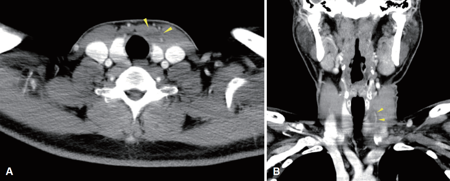
Fig. 2.
Neck ultrasound showed 1.5×1.2 cm sized well-capsulated hypoechoic nodule with heterogeneous internal echotexture in the strap muscle. The medial margin of the mass is intermingled with the surrounding SH muscle (asterisk), while the lateral margin is relatively well-demarcated from ST muscle. CCA: common carotid artery, OH: omohyoid, SCM: sternocleidomastoid muscle, SH: sternohyoid, Thy: thyroid, ST: sternothyroid.
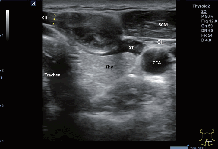
Fig. 3.
Intraoperative findings. The well-encapsulated solid mass was identified in the sternohyoid muscle through a longitudinal incision and dissection of the muscle.
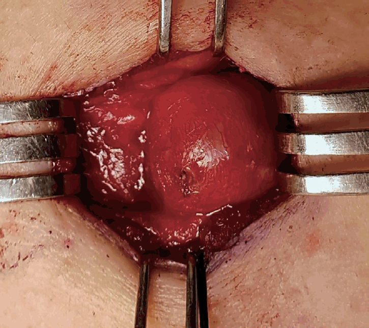
Fig. 4.
Histologic findings. Low magnification view of a spindle cell lesion (arrowheads) well-demarcated by fibrous capsule (white asterisk) and adjacent muscle fibers (thick arrows) (A: H&E original magnification ×40). High magnification view of verocay body with nuclear palisading (asterisk) and fascicles of spindle shaped cells consistent with diagnosis of schwannoma (arrows) (B: H&E original magnification ×200). H&E: hematoxylin and eosin stain.
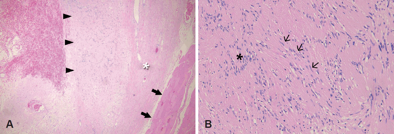
Fig. 5.
Schematic illustration suggesting the conceptual relationship between tumor and possible nerve origin (the fascicle of a distal branch of ansa cervicalis innervating in the sternohyoid muscle).
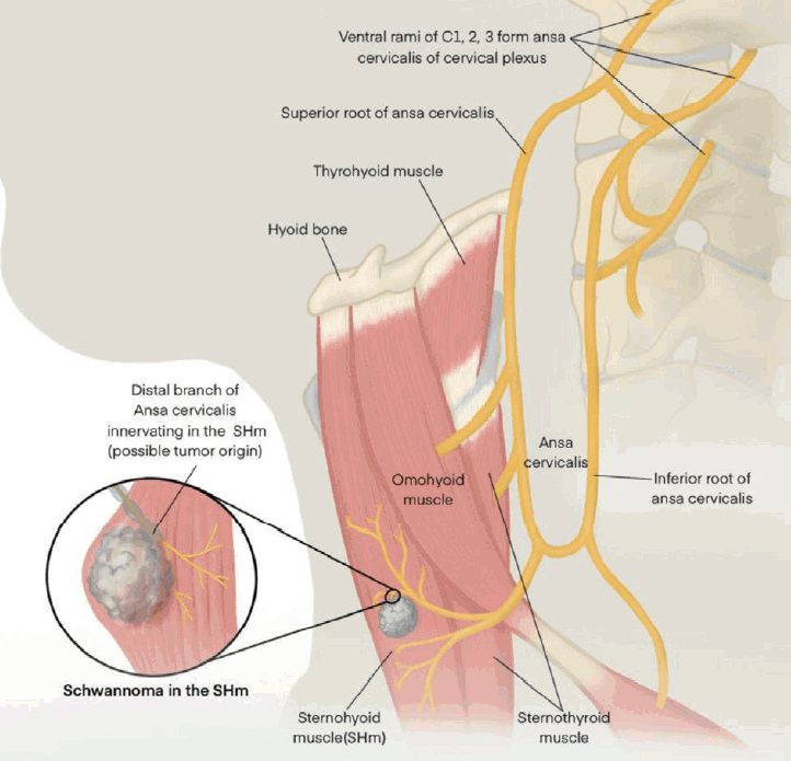
Table 1.
Review of the literature of schwannoma originated from ansa cervicalis
| Group | Year | Sex | Age | Site | Presenting signs and symptoms | Initial diagnosis/modality | Nerve origin | Nerve preservation | Language |
|---|---|---|---|---|---|---|---|---|---|
| Hirabayashi, et al [12] | 1987 | F | 14 | Carotid space | Asymptomatic neck mass | Schwannoma/CT | Intraoperatively confirmed | Sacrificed | English |
| Nerve stimulation | |||||||||
| Tahri, et al [16] | 1994 | M | Middle age | Prevertebral space | Dysphagia | Neurofibroma/MRI | Intraoperatively confirmed | Sacrificed | French |
| Identifying nerve continuity | |||||||||
| de Diego Sastre, et al [17] | 1996 | M | Adolescent | Anterior neck: N/A | Asymptomatic neck mass | Thyroid malignancy/N/A | N/A | Sacrificed | Spanish |
| Righini, et al [18] | 2007 | N/A | N/A | N/A | Asymptomatic neck mass | Schwannoma/N/A | N/A | Sacrificed | French |
| Okonkwo, et al [4] | 2011 | M | 25 | Anterior neck: between SH and TH muscle | Asymptomatic neck mass | Thyroid malignancy/US, FNA | Clinical expectation | Sacrificed | English |
| No continuity, but anatomical correlation | |||||||||
| Park, et al [9] | 2013 | M | 54 | Submandibular space: anterior to carotid sheath | Asymptomatic neck mass | Schwannoma/MRI | Intraoperatively confirmed | Sacrificed | English |
| Strap muscle contraction during operation | |||||||||
| Rath, et al [11] | 2015 | M | 62 | Submandibular space: between ICA and IJV | Asymptomatic neck mass | Schwannoma/US, FNA | Intraoperatively confirmed | Sacrificed | English |
| Nerve stimulation |


 PDF
PDF Citation
 Print
Print


 XML Download