함기화에 대하여
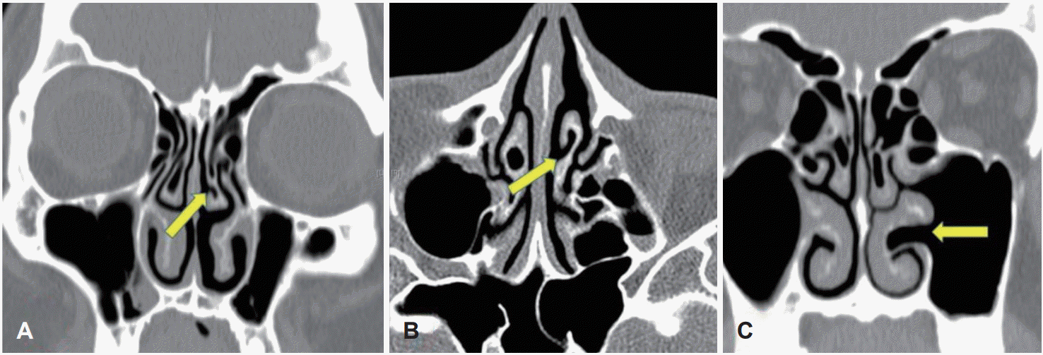
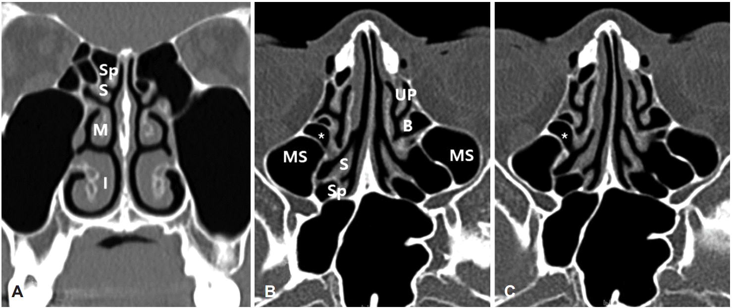 | Fig. 1.The inferior (I), middle (M), superior (S) and supreme turbinate (Sp) are visible in one coronal image (A). The uncinate process
(UP), ethmoid bulla (B), middle turbinate, superior turbinate and supreme turbinate were seen in axial view (B, C). The space (*) behind the ethmoid bulla is connected to superior meatus and located in posterior ethmoid sinus. MS: maxillary sinus. |
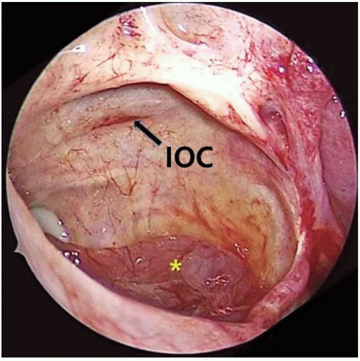
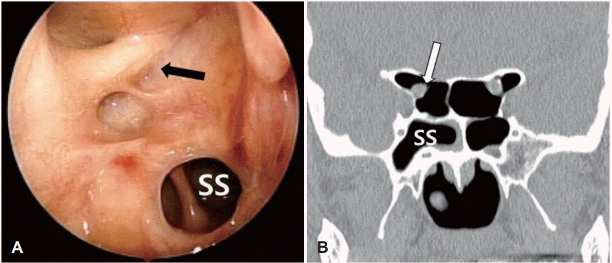 | Fig. 2.Post-op endoscopic finding of optic canal (black arrow) in right Onodi cell (A). Surgical opening of sphenoid sinus (SS) is also seen. Pre-op coronal CT finding showing optic canal (white arrow) in Onodi cell (B). |
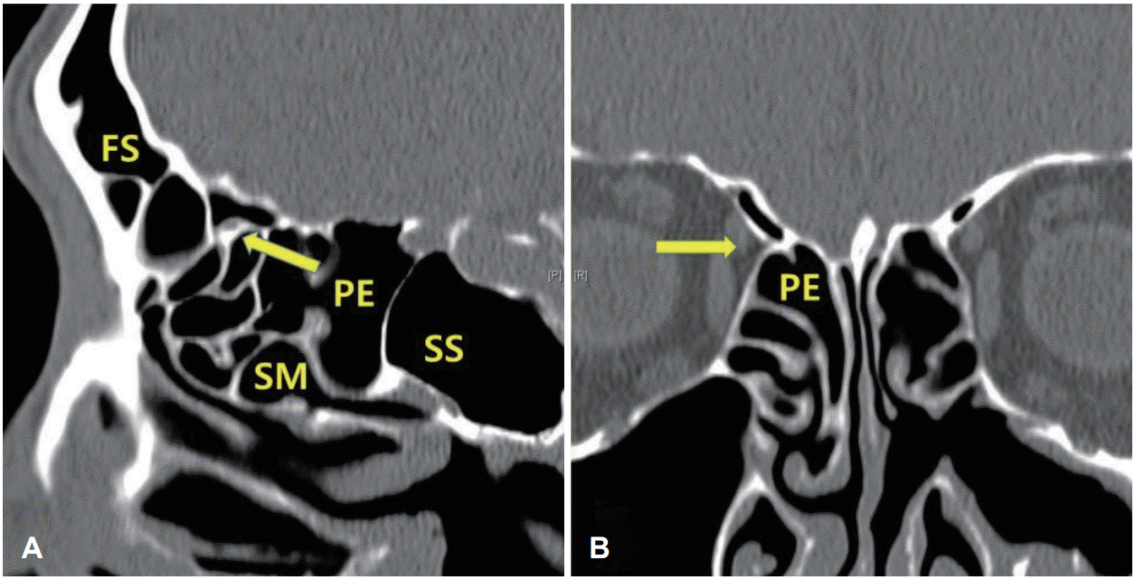
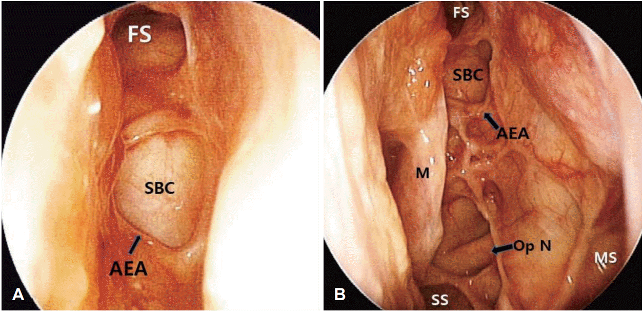 | Fig. 3.Post-op endoscopic finding showing the right-side roof of anterior ethmoid sinus (A). Pneumatization to the frontal bone, anterior to the anterior ethmoid canal (AEA, arrow) is divided with frontal sinus (FS) and suprabullar cell (SBC). Post-op endoscopic finding of left ethmoid roof (B). From posterior to the anterior direction, sphenoid sinus (SS), optic nerve (Op N, arrow) in Onodi cell, suprabullar cell (SBC) and frontal sinus (FS) are seen. MS: maxillary sinus, M: middle turbinate. |
부위별 수술 해부
사골동 하부로 진입하기
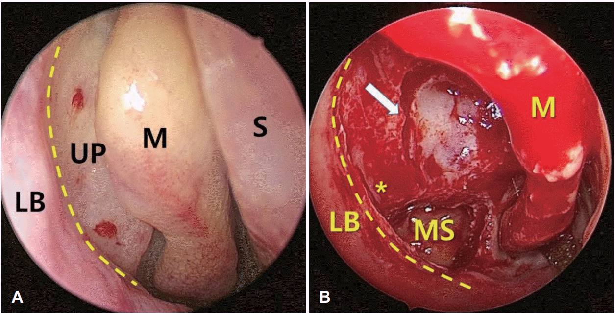 | Fig. 5.Endoscopic finding of lateral nasal wall anterior to the middle turbinate (M) of right nasal cavity (A). The bulge of lacrimal structure (LB) is noted just anterior to the attachment potion (yellow dotted line) of the uncinated process (UP). Finding after removal of uncinated process, anterior wall of ethmoid bulla and partial removal of middle turbinate (B). The opened Infundibulum (*) is noted between cutting edges of uncinated process and cutting margin of anterior wall of ethmoid bulla (white arrow). The inside of the maxillary sinus (MS) is also noted. |
후사골동 진입
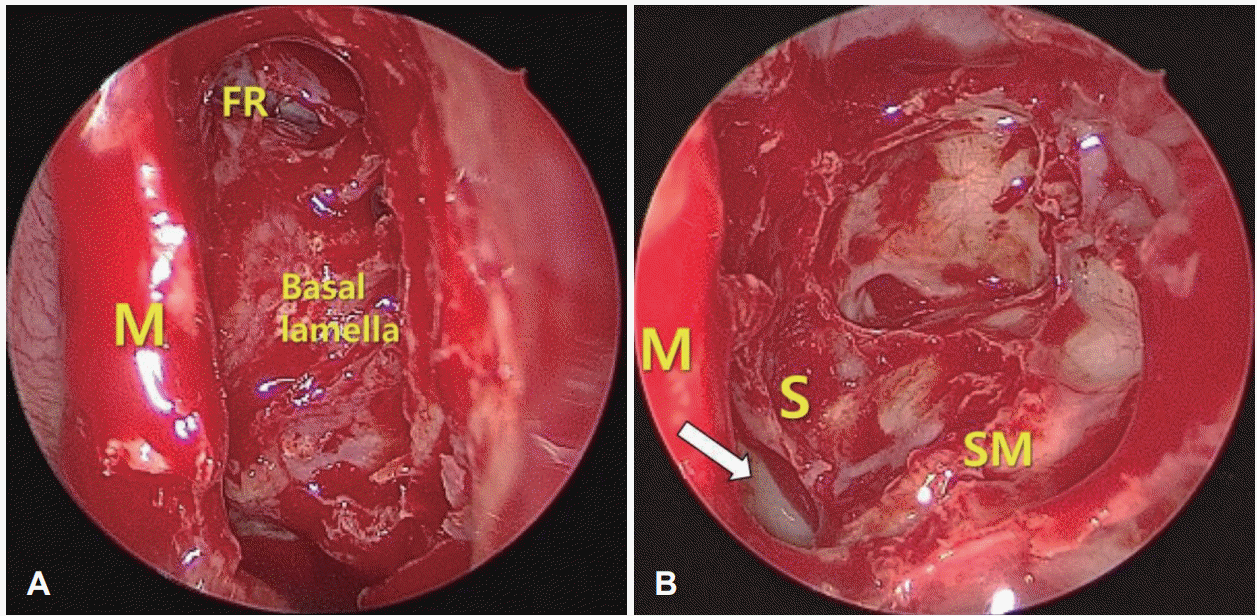 | Fig. 6.Endoscopic finding of the basal lamella of middle turbinate which is the posterior wall of anterior ethmoid sinus after removal of uncinate process and ethmoid bulla (A). Frontal recess (FR) is seen at the anterior portion. The anterior portion of the middle turbinate (M) is removed partially. Endoscopic finding of posterior ethmoid sinus after removal of the basal lamella of middle turbinate (B). The anterior inferior part of superior turbinate (S) was removed and septal mucosa (arrow) is seen through the superior meatus (SM). |
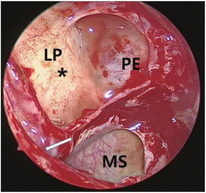 | Fig. 7.Endoscopic finding of right ethmoid sinus after removal of the uncinated process, anterior wall of ethmoid bulla and widening of fontanelle. The mucosa in the lateral wall of ethmoid sinus (*) looks white compare to the bone septa. Arrow depicts the exposed infundibulum. LP: lamina, PE: posterior ethmoid sinus, MS: maxillary sinus. |
접형동 접근
사골동 후방에서 앞으로
전사골동의 외측 상부에서 전두동까지
상악동으로의 접근
하안와부위와 중비갑개로의 함기화
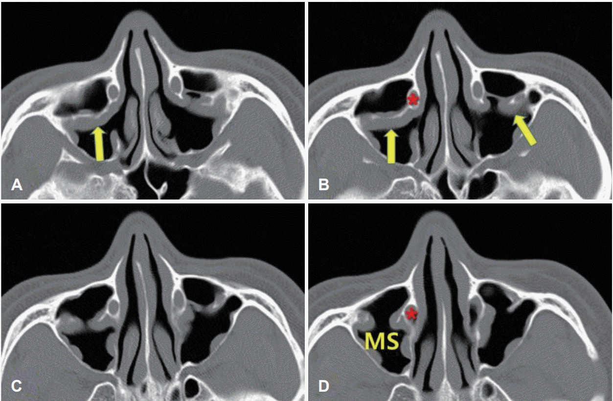
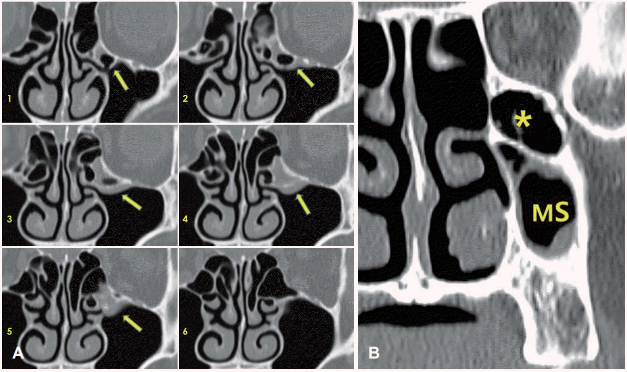 | Fig. 10.Serial coronary images showing infraorbital cell (arrow) (A). The opening of the cell is noted in the middle meatus in image 1 and the cell is filled with secretion partially. The posterior margin of this cell is noted in image 5. Another case of infraorbital cell (*) pneumatized from superior meatus over the maxillary sinus (MS) (B). |
수술시 제거 범위와 안전한 기구 사용
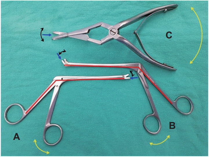 | Fig. 12.The movement pattern of surgical instruments. In the Grunwald cutting fcorceps (A, B), fixed portion is depicted with red line. Fixed portion and mobile portion are present in the cutting portion and handles. The range of movement in the cutting portion is depicted with black curved lines with tip in both side and that in the handle is depicted with yellow curved line with tip in both side. The blue arrow depicts the planes of cutting area. In the Jansen Middleton septum forceps (C), the cutting portion and handles move simultaneously. The plane of cutting area is the center line of instrument. |


 PDF
PDF Citation
 Print
Print


 XML Download