서 론
증 례
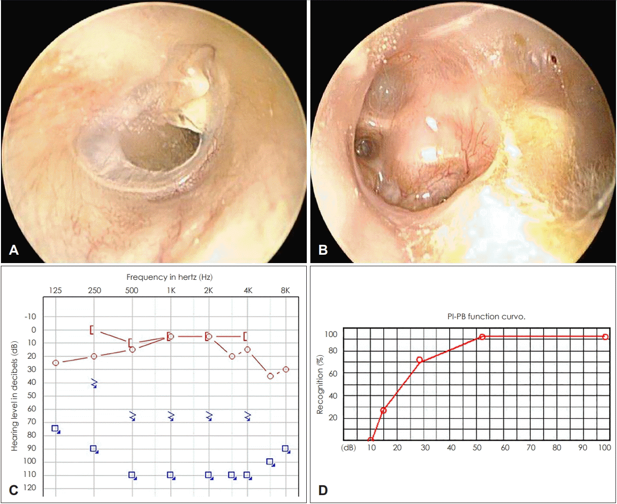
 | Fig. 1.Preoperative evaluation: oto-endoscopic images show a healed state of tympanic membrane on the right ear (A) and a postoperative state of canal wall down mastoidectomy, concomitant with adhesive otitis media with otorrhea on the left ear (B). Pure tone audiometry and speech audiogram show a completely deaf left ear and relatively normal hearing on the right ear (C, D). |
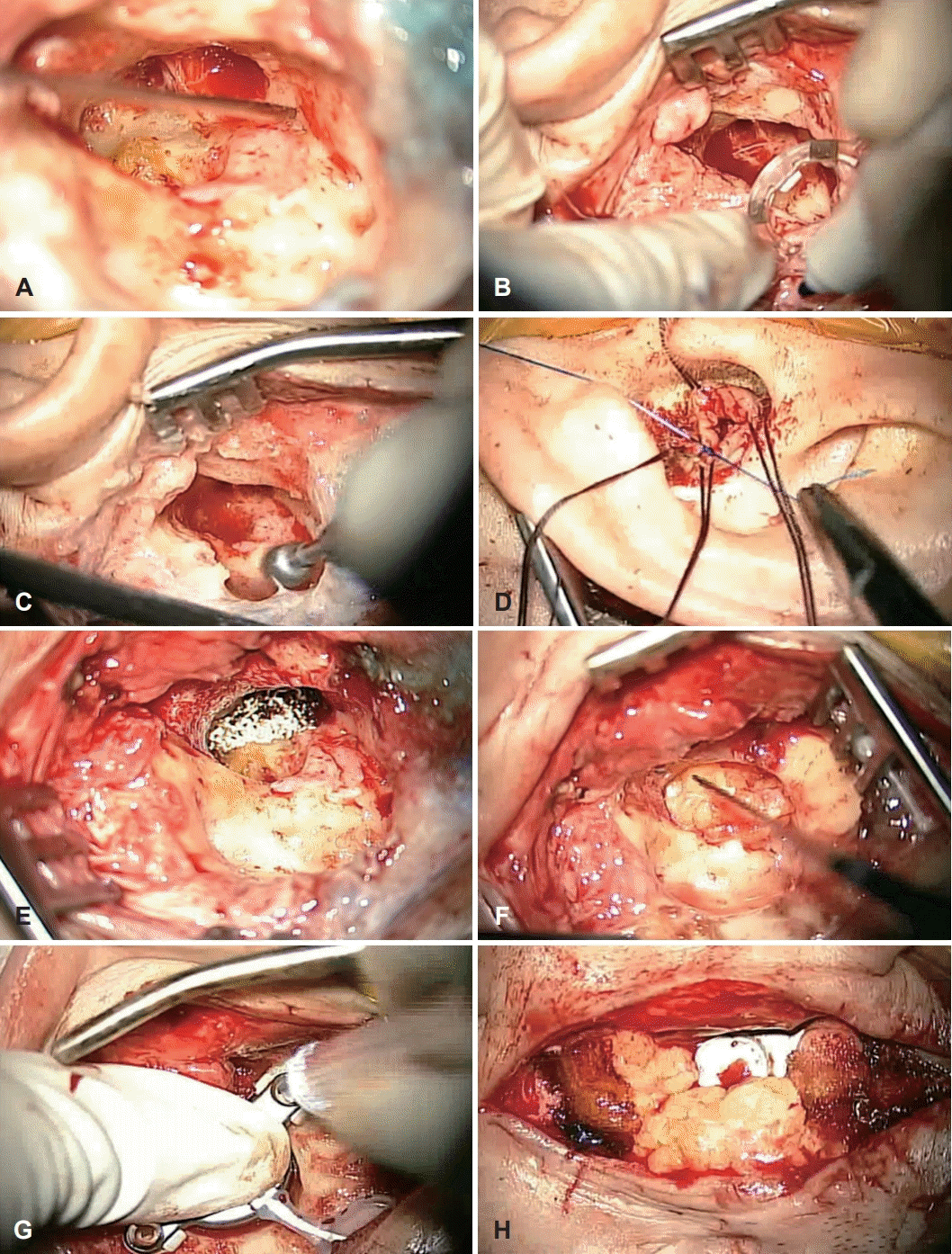
 | Fig. 3.Surgical procedure of single-staged operation of BonebridgeTM and subtotal petrosectomy. Removal of the air cells and mucosal tissues in the middle ear and mastoid cavity (A). Using a Sizer to mark the BC-FMT space (B). Additional drilling of mastoid cortical bone for BC-FMT (C). Closure of the external auditory canal (D). Obliteration of the Eustachian tube using harvested conchal cartilage, fat and muscle (E). Obliteration of tympanomastoid cavity using abdominal fat (F). Insertion of the BC-FMT. The level of the BC-FMT was adjusted by implant lifts (anterior side; 1 mm lift, posterior side; 2 mm lift) (G). Obliteration of the remaining space with abdominal fat (H). BC-FMT: bone conduction floating mass. |
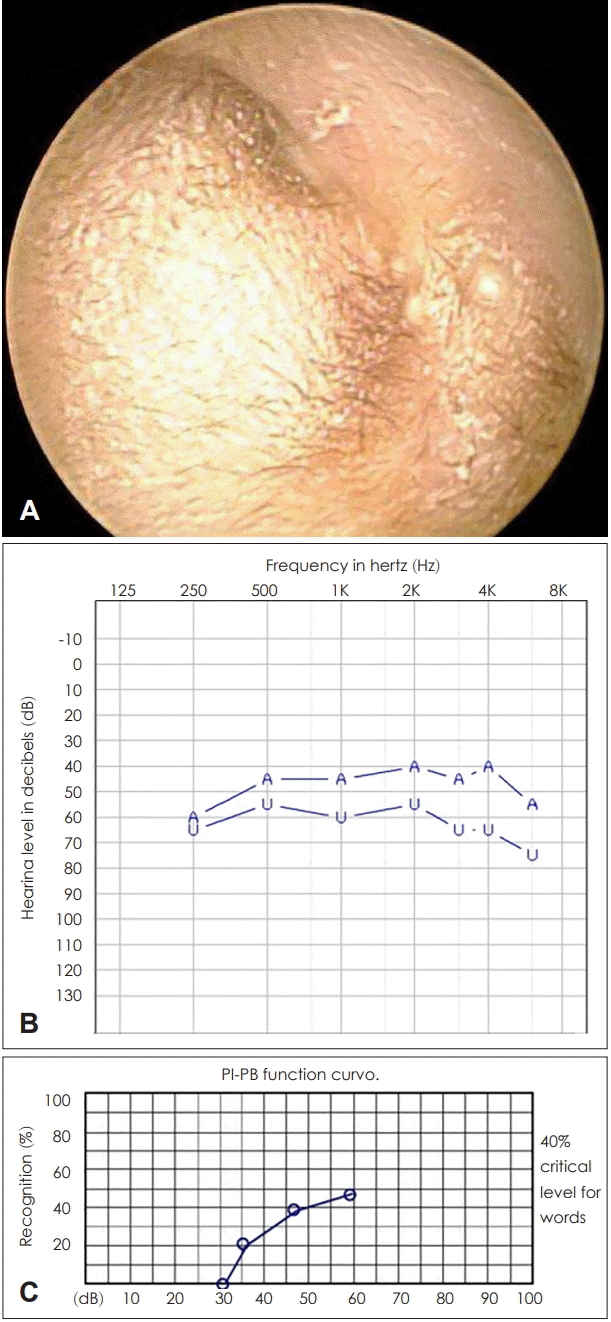
 | Fig. 4.Postoperative states at 6 months follow-up visit. Oto-endoscopic image shows the state of the obliterated external auditory canal without any complications (A). The aided sound field test shows 42.5 dB threshold in the pure tone audiometry test (B) and 48% of word recognition score (C), which indicate sufficient hearing gain on the left ear. A: aided, U: un-aided. |


 PDF
PDF Citation
 Print
Print


 XML Download