

 PDF
PDF Citation
 Print
Print


서 론
갑상선 유두암(papillary thyroid carcinoma, PTC)의 한 아형 중 cribriform-morular variant(CMV)는 매우 드문 조직학적 아형으로, 조직검사상 사상체 및 방추체 모양의 세포들이 관찰된다[1,2]. CMV-PTC의 임상 양상은 아직 뚜렷하게 보고되는 것은 없으나, 50% 이상에서 가족성 선종성 용종증(familial adenomatous polyposis, FAP)이 동반된다[3].
FAP는 대장암의 전암성 병변으로 adenomatous polyposis coli(APC) 유전자의 5q21 변이와 연관되어 있으며, APC 유전자 변형은 어린 나이에서 대장암 발병 위험성을 높이므로, CMV-PTC 환자들은 대장 내시경검사를 통한 대장 및 소장의 용종 여부 및 APC 유전자의 변이 여부 검사가 필요하다[4]. 갑상선암이 FAP를 가진 환자의 첫 증상으로 발현될 수 있으며, 4~12세 어린 환자들의 약 30%에서는 대장용종의 발현이 있기 전에 갑상선암이 먼저 발견되기도 한다[5].
저자들은 갑상선의 다발성 결절을 주소로 내원한 16세 환자의 CMV-PTC와 APC 유전자 돌연변이를 보이는 FAP가 동반된 증례에서, 이전에 보고되지 않은 APC 유전자 돌연변이가 관찰되어 보고하고자 한다.
Go to : 
증 례
16세 여자 환자는 2016년 5월에 전경부에 만져지는 종물로 내원하여, 시행한 갑상선 초음파 및 컴퓨터 전산화단층촬영상 갑상선 협부의 3.5 cm 크기의 종양과 양측 갑상선에 다양한 크기의 결절이 여러 개 관찰되었다(Fig. 1). 양전자 방출 컴퓨터단층촬영(positron emission tomography-computed tomography) 에서는 갑상선에 다발성 섭취 증가가 보였고, 경부림프절이나 전신 전이는 관찰되지 않았다(Fig. 2).
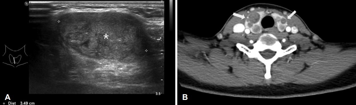 | Fig. 1.Representative images of US and CT. Initial US scan shows huge thyroid mass with isoechoic to hypoechoic heterogeneous attenuation about 3.5 cm sized- nodule in isthmus of thyroid (white asterisk) (A). Preoperative CT scan shows multiple indeterminate solitary nodules in both thyroid glands (white arrows). There is no evidence of enlarged lymph node which is suspicious for malignancy (B). US: ultrasonograpy, CT: computed tomography.
|

흡입천자세포검사 결과상 사상체 및 상실배 모양의 조직이 관찰되었고, 면역염색상 β-catenin 양성 소견의 갑상선암으로 진단되어, 이에 전신마취하 갑상선 전 절제술을 시행하였다. 육안적으로 갑상선 피막 외 침범은 보이지 않았다.
최종 병리조직검사 결과상, 갑상선 협부에 최대 3.5×3×2.5 cm의 종양과 함께 총 18개 이상의 다양한 크기의 종양이 갑상선 유두암으로 진단되었고, 현미경적으로 갑상선 피막 외 침범과 혈관 침범 소견은 관찰되지 않았다. 최종 조직검사 결과는 이전 세침흡인검사 결과와 마찬가지로 상실배 모양(morular morphology) 및 사상체 모양(cribriform morphology)의 조직과 함께 β-catenin 면역염색 양성으로 CMV-PTC에 합당한 소견이 보고되었다(Fig. 3). 수술 2개월 이후 방사선 요오드 치료를 50 mCi를 추가로 1차례 시행하였다.
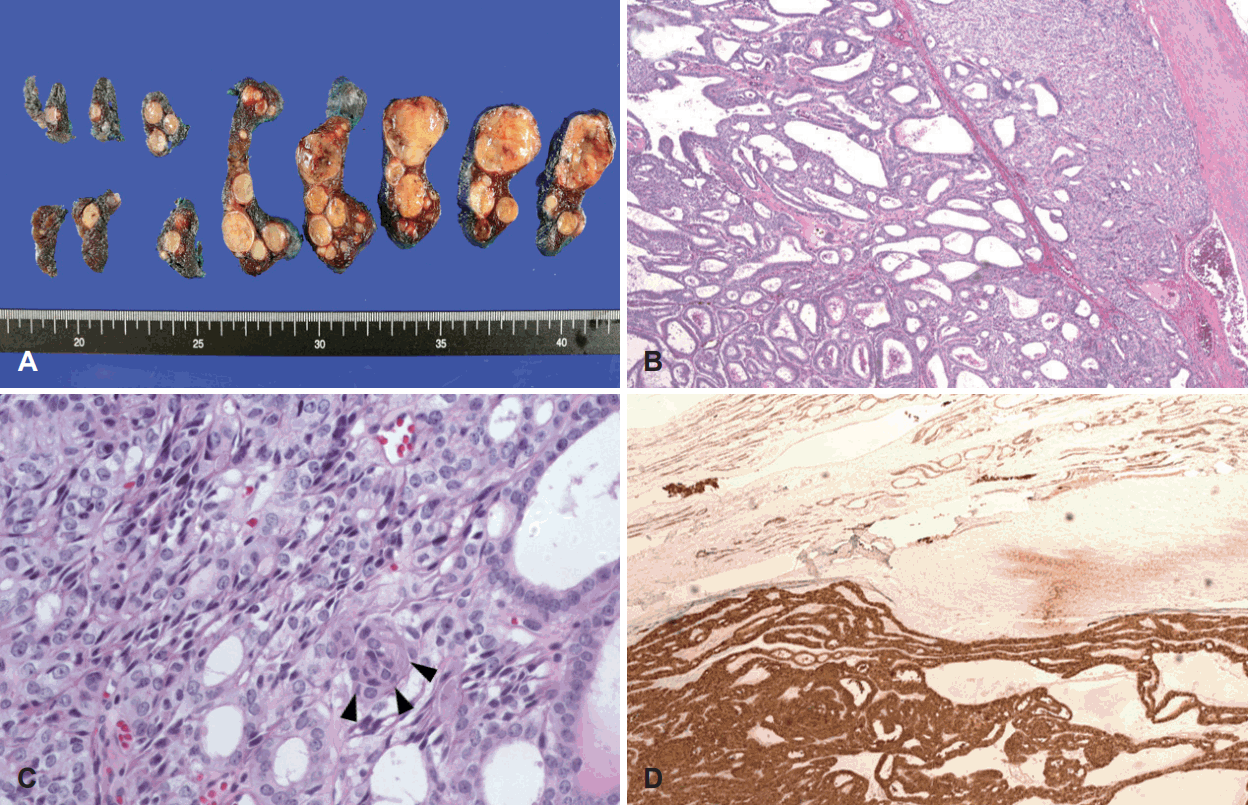 | Fig. 3.Thyroid gland cross section shows total 18 mass. Largest diameter of thyroid tumor was measured 3.5×3×2.5 cm at isthmus (A). Thyroid tumor with compact arrangement of cribriform, follicular, papillary, trabecular, and solid patterns by columnar cells thick. The tumor is surrounded by a fibrous capsule and colloid is not present (H&E, ×50) (B). Morular (cuboid) shaped cells shows (arrowheads) (H&E ×400) (C). The nuclei and cytoplasm was stained by β-catenin (×50) (D).
|
CMV-PTC는 가족성 선종성 용종증이 동반되는 경우가 많기 때문에, 병력 청취 및 유전자 분석을 시행하였다. 환자의 아버지가 대장의 용종증이 있었고, 대장암으로 10년 전 사망한 가족력이 있었다. 이에 위 및 대장 내시경검사와 APC 유전자 분석을 시행하였다. 위 내시경검사상, 위저 용종(stomach fundic polyp) 및 대장 내시경검사상 소장에서 1개, 대장에서 15개의 선종이 관찰되었다. APC 유전자에 대한 염기서열 분석 결과, 5번 염색체 q arm의 21번 유전자 4181~4188번째 염기서열이 결손(deletion)되어 1394번째 아미노산인 아스파트산(Aspartic acid)이 글리신(Glycine)으로 치환되면서, 조기 종결(premature termination)되는 프레임시프트 돌연변이(frameshift mutation)가 관찰되었다(Table 1, Fig. 4). 이 돌연변이는 이전에 보고된 적 없는 새로운 서열 변화이다[6].
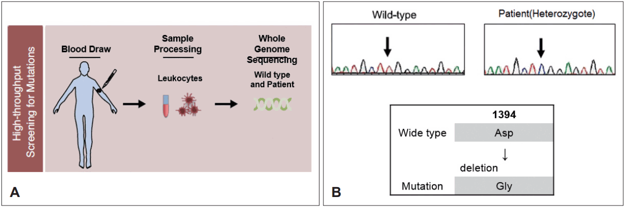 | Fig. 4.APC gene analysis diagram (A). Through direct sequencing, APC gene on chromosome 5q21-22 of the cribriform-morular variant papillary thyroid carcinoma patient was analyzed. A novel gene mutation, which deleted 4181-4188 nucleotides substitute Glycine for Aspartic acid, was discovered causing premature termination of the APC protein (B). APC: adenomatous polyposis coli, ASP: aspartic acid, Gly: glycine.
|
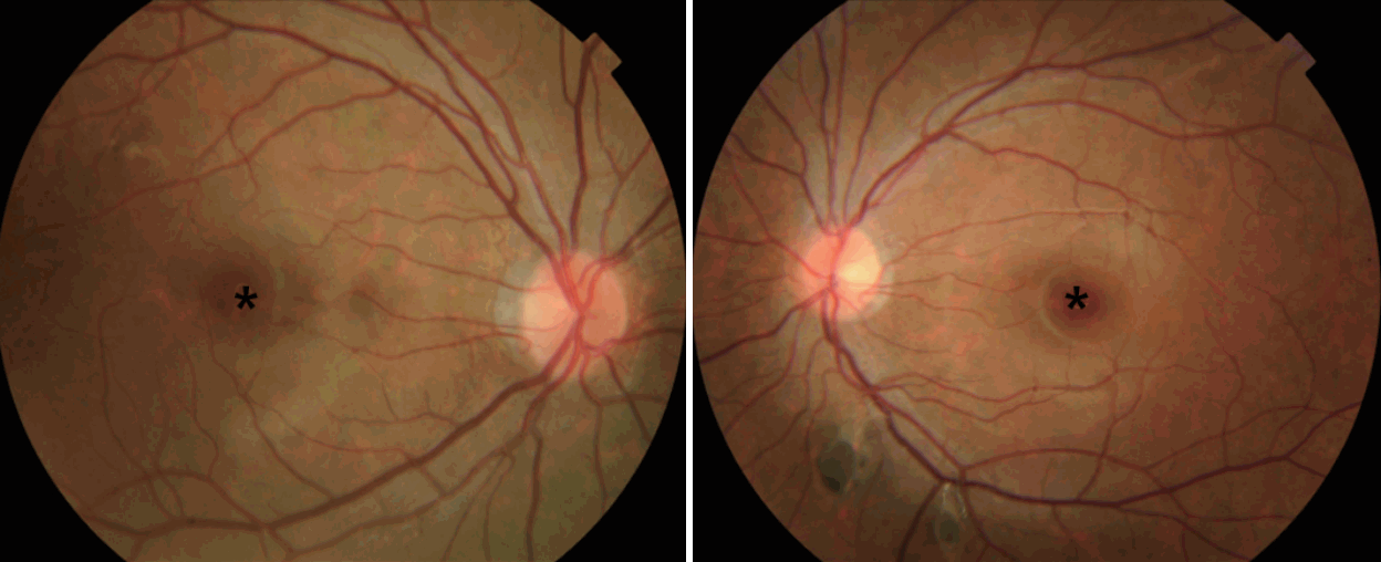
환아는 다발성 갑상선암 및 가족성 선종성 용종증과 더불어 시력 저하를 호소하여 시행한 안과 검진상 좌안 망막 하방으로 색소가 침착된 선천성 망막 색소 침착성 상피 비대증(congenital hypertrophy of the retinal pigment epithelium, CHRPE) 소견이 관찰되었고, 그 외 다른 피부 병변은 관찰되지 않았다(Fig. 5).
수술 후 1년째, 환자는 초음파검사 및 동위원소검사 등을 통해 정기검진을 시행하고 있으며, 재발은 관찰되지 않았다. 대장 선종에 대해서는 고등학교 졸업 이후에 수술적 치료를 고려하고 수술 전까지 1년마다 대장 내시경 및 위 식도 내시경을 시행하고, 좌측 눈 병변에 대해서도 1년마다 정기검진을 시행할 예정이다.
Go to :
고 찰
CMV-PTC는 갑상선 유두암의 아형으로 분류되지만, 전형적인 유두암과 구별되는 특징적인 임상 양상을 가지고 있다. 갑상선 유두암은 최근 World Health Organization 분류법에 의하여 15가지로 분류되며, 이 중 CMV-PTC는 0.2%를 차지하는 드문 아형이다[7].
갑상선 유두암이 40대에 호발하는 반면 CMV-PTC는 20대에 호발하며[3], 본 증례와 같이 10대에도 비교적 호발하는 것으로 알려져 있다. 또한 갑상선 유두암은 림프절 전이의 유병률이 높은 반면에, CMV-PTC는 약 10% 정도의 림프절 전이를 보이며 원격 전이의 유병률도 낮게 관찰되었다[3,8].
CMV-PTC가 임상적으로 가장 특징적인 것은 FAP와의 연관성이다. FAP는 상염색체 우성 질환으로, 대장의 다발성 용종과 APC 유전자의 5번 염색체 q arm의 22번 유전자에서 변이가 관찰된다. FAP 환자는 거의 대부분이 대장암으로 진행하며, 어린 나이에 다발성으로 발생할 수 있다. 기존의 메타분석에서는 53%의 CMV-PTC 환자에서 FAP가 동반되었으며, 이 중 약 50%에서 갑상선암이 먼저 진단되었다[3,9]. 본 환자의 경우 APC gene mutation 확인을 위하여, 말초혈액의 백혈구로부터 Genomic DNA를 추출하여 5번 염색체의 q arm의 21~22번째에 위치한 APC 유전자의 모든 coding exon 및 인접 intron 부위의 염기서열을 direct sequencing을 통한 염기서열 분석법으로 확인하였다. 총 15개 exon의 염색체 분석에서 APC 유전자의 5번 염색체 q arm의 21번 유전자 4181~4188번째 염기서열이 deletion 되면서 Aspartic acid가 Glycine으로 치환되는 변이가 관찰되면서, APC 유전자가 조기에 종결되었다. 대부분 밝혀진 APC gene mutation의 경우, 3927~3931 염기서열이 deletion 되면서 frameshift mutation이 발생하여 APC 유전자의 premature termination에 의해 변이가 된다고 밝혀져 있으나, 본 증례에서는 기존과는 달리 4181~4188번 염기서열의 deletion에 의한 APC 유전자 돌연변이로, 기존에 보고되지 않은 유전자 변이이다.
이와 더불어 FAP를 가진 환자의 경우 약 75%에서 CHRPE가 동반되고 있으며, 특히 양측 망막의 색소 침착 혹은 병변의 저색소증이 동반된 경우에는 FAP와 연관된 CHRPE의 지표라고 할 수 있어 CMV-PTC 환자의 경우, 정기적으로 망막검사가 필요하다[10].
CMV-PTC는 조직학적으로 cribriform의 구조를 가지고 있고, morule 형태의 세포 구조를 가지고 있으며[1,2], β-catenin 면역염색에서 핵과 세포질이 염색되는 것을 확인함으로써 진단을 할 수 있다[11].
CMV-PTC의 치료법은 유병률이 낮아 정립되지는 않았지만, 여타 갑상선 유두암의 치료 원칙을 따른다. 본 증례와 같이 다발성 갑상선암이 흔하기 때문에 갑상선 전 절제술을 시행하며, 림프절 전이가 확인이 되면 림프절 절제술을 추가하게 된다[3,9,12]. 본 증례에서는 갑상선의 다발성 종양에 대해 전 갑상선 절제술을 시행하였고, 대장 용종에 대해서는 경과관찰 후 FAP의 치료 원칙에 따라 환아 성장 이후 대장의 부분 절제술을 고려하고 있다.
본 증례에서는 드물게 보고되고 있는 CMV-PTC 환자에서 전형적인 임상 양상을 가졌으나, 이전에 보고되지 않았던 새로운 APC 유전자의 변형이 관찰되었고, FAP가 동반된 증례를 진단 및 치료하였기에 보고하였다. CMV-PTC는 갑상선암의 특징적인 임상 양상과 더불어, 대장암의 전암성 병변으로 알려진 FAP가 흔하게 동반되기 때문에 이에 대한 각별한 주의가 필요하겠다.
Go to :
 XML Download