서 론
증 례
증 례 1
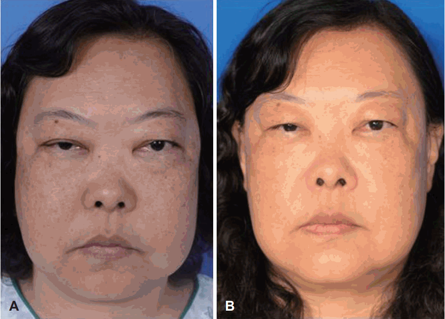 | Fig. 1.Preoperative photo (case 1) shows mild conjunctival chemosis and severe exophthalmos (25.5 mm/ 26.6 mm) in both eyes (A). Postoperatively, exophthalmos was improved by 4 mm in both eyes from 25.5 mm to 21.5 mm in the right eye and from 26.6 mm to 22.5 mm in the left eye, respectively (B). |
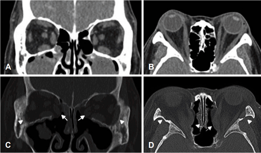 | Fig. 2.Preoperative coronal CT scan (case 1) shows severe enlargement of all rectus muscles (A). Axial view shows the distinct exophthalmos in both eyes (B). Postoperative coronal CT scan shows the effect of bilateral medial, inferomedial wall (arrows) and lateral wall removal (arrowheads). Medial rectus muscle and orbital fat tissue were extruded to nasal cavity, which decrease orbital volume and pressure (C). Axial view shows loss of medial (arrows) and lateral wall (arrowheads) of both orbit and improvement of exophthalmos in both eyes compared to preoperative one (D). |
증 례 2
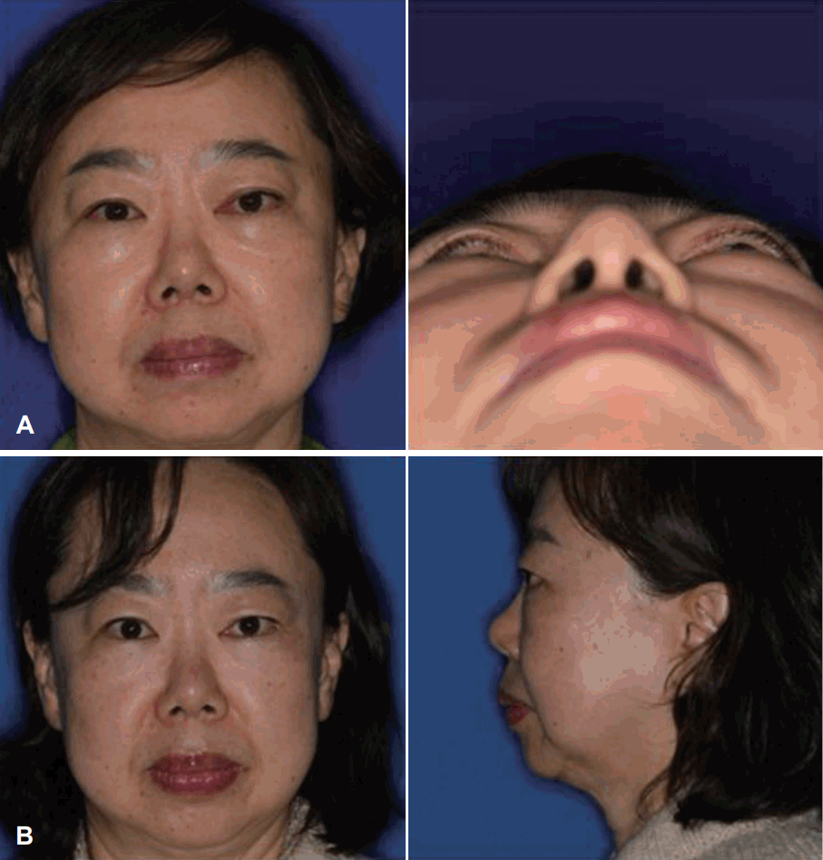 | Fig. 3.Preoperative photos (case 2) show severe exophthalmos of left orbit (A). Postoperatively, left exophthalmos was improved by 4 mm (B). |
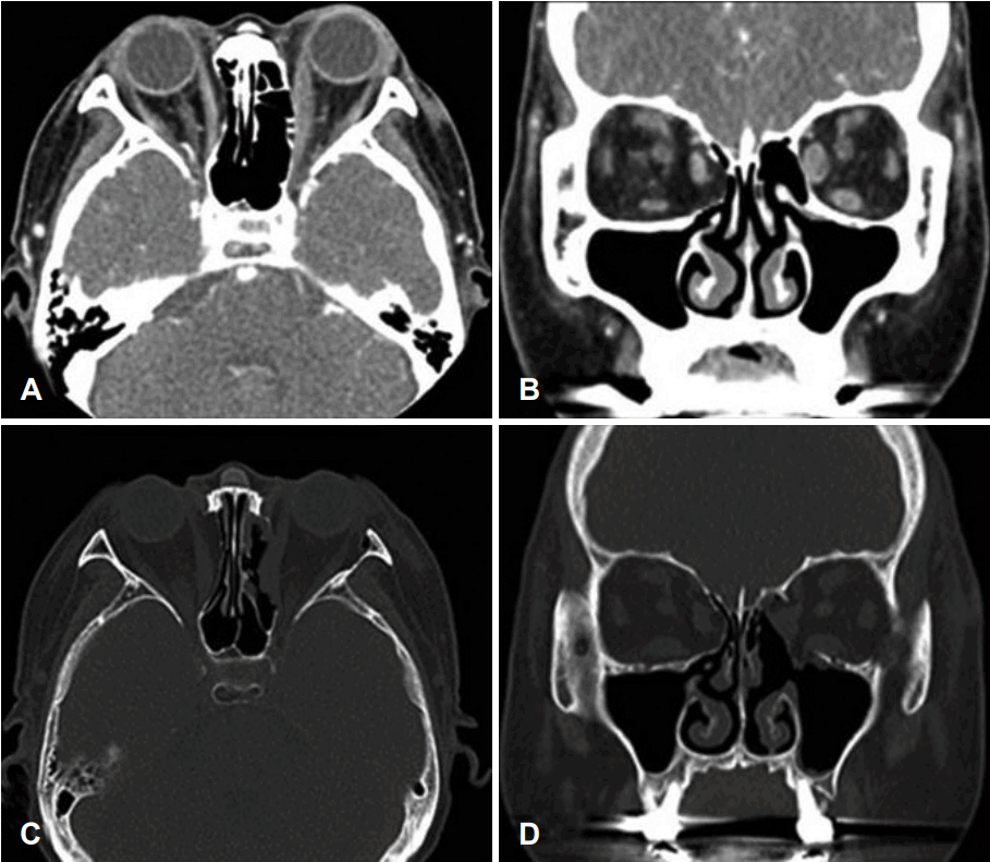 | Fig. 4.Preoperative Axial CT scan (case 2) shows the distinct exophthalmos in left eye (A). Coronal view shows severe enlargement of left rectus muscles (B). Postoperative axial view shows loss of medial and lateral wall of left orbit and improvement of exophthalmos in left eye (C). Coronal CT scan shows the effect of medial, inferomedial wall and lateral wall removal of left orbit (D). |
증 례 3
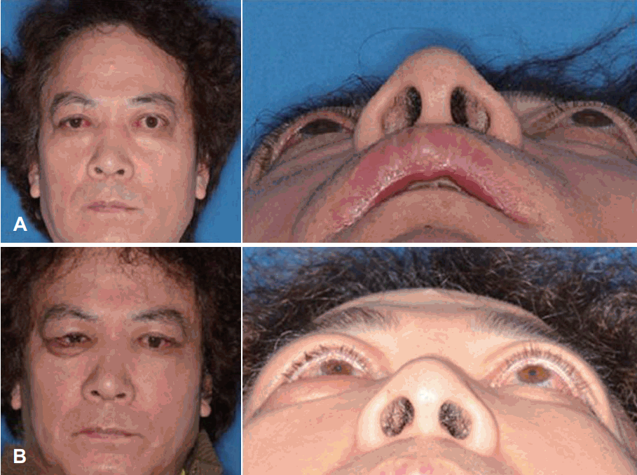 | Fig. 5.Preoperative photo (case 3) shows conjunctival chemosis and severe exophthalmos (25 mm/23 mm) in both eyes (A). Postoperatively, exophthalmos was improved from 25 mm to 19 mm in the right eye and from 23 mm to 18 mm in the left eye (B). |
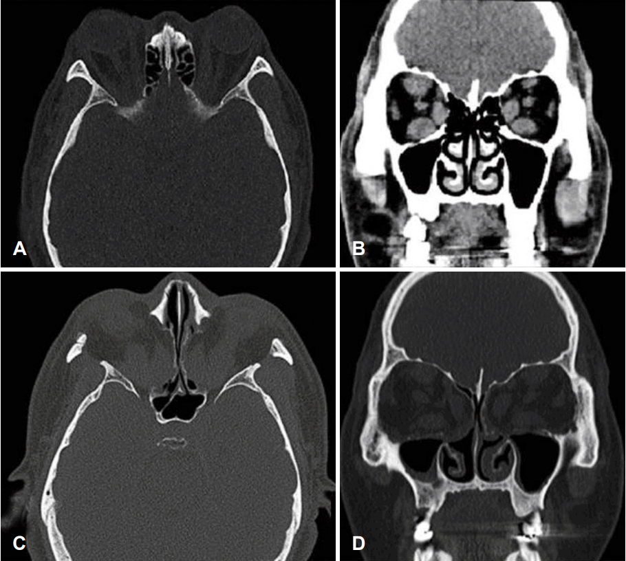 | Fig. 6.Preoperative Axial CT scan (case 3) shows the distinct exophthalmos in both eyes (A). Coronal view shows severe enlargement of all rectus muscles (B). Postoperative axial view shows loss of medial and lateral wall of orbit and improvement of exophthalmos in both eyes (C). Coronal view of facial bone CT scan shows the effect of medial, inferomedial wall and lateral wall removal (D). |


 PDF
PDF Citation
 Print
Print


 XML Download