서 론
증 례
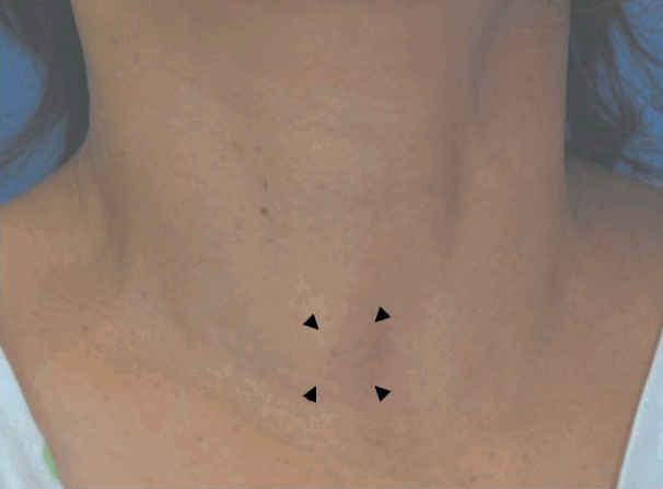
 | Fig. 1.External photograph. It shows 2×2 cm sized, soft, round, slightly movable and non-tender mass, which did not move with swallowing (arrowheads). |
 | Fig. 2.Transverse scan of ultrasonography shows 2.2×1.3 cm sized, well-defined heterogeneously hypoechoic mass (asterisk) above the right sternohyoid (arrows) (A). The contrast-enhanced axial image of computed tomography revealed that focal bulging of the strap muscle is observed without obvious shadow of mass (arrows) (B). Intraoperative findings. It shows well-margined, smooth and round mass slightly adherent to the sternohyoid (asterisk) (C). |
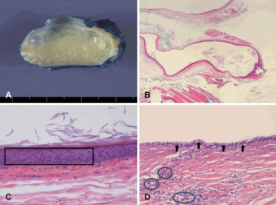
 | Fig. 3.Cut surface of the specimen showed that the uni-locular cyst measures 2.5×2.0×1.5 cm and the inner cystic surface contains yellowish white cheezy material (A). The thin cystic wall is observed without nodular excrescences in low power view (H&E, ×40) (B). Cystic lining epithelium consists of stratified squamous epithelium (square) (H&E, ×200) (C). Cystic lining epithelium consists of ciliated columnar epithelium (arrows) (D). In the fibrous cystic wall, a little lymphoid infiltrates (encircled in black) are multi-focally observed beneath the lining epithelium (H&E, ×200) (D). |


 PDF
PDF Citation
 Print
Print


 XML Download