Journal List > Korean J Otorhinolaryngol-Head Neck Surg > v.60(9) > 1157031

구강내 장치를 사용한 폐쇄성 수면무호흡증 환자에서 장기간 사용 시 나타나는 합병증 분석

Abstract

Background and Objectives

Recently, mandibular advancement device (MAD) has been used widely used for the treatment of obstructive sleep apnea (OSA) because it can effectively reduce the collapsibility of upper airway during sleep. Although MAD is widely prescribed by otorhinolaryngologists, several studies described its complications arising from the anterior placement of the mandible with long-term use. However, there is still a lack of studies on long-term complications in Korean patients.

Subjects and Method

Retrospectively, we included a total of 57 OSA patients in the study. In this study, all enrolled OSA patients had used MAD over two years with more than 4 hours/day. Dental consulting and cephalometric analysis were conducted to identify the change of dental and skeletal findings at two different times (baseline and after 2 year).

Results

The dental findings showed that the overbite and overjet were significantly decreased. Additionally, the proclination of the lower incisors were increased significantly, whereas there was no significant change on the retroclination of the upper incisors. Three patients showed a change of malocclusion type. On the analysis of skeletal findings, we found a significant increase in the sella turcica central point-nasion-supramental point angle and a decrease in the subspinal point-nasion-supramental point angle. Moreover, the anterior lower facial height and anterior facial height were significant increased.

Conclusion

Consistent with Western studies, our findings suggest that the long-term use of MAD could induce changes in dental and skeletal morphologies in Korean OSA patients. Therefore, clinicians should thoroughly evaluate potential changes in dental and skeletal morphologies when they prescribe MAD to patients with OSA.

서 론

폐쇄성 수면무호흡증(obstructive sleep apnea)은 수면 중에 발생하는 상기도의 반복적인 부분 폐색 혹은 완전 폐색에 의해서 야기되는 질환으로, 수면의 질을 떨어뜨리고 만성 저산소증을 유발하여 주간에 과도한 졸리움증, 기억력 장애, 업무능력 저하, 교통사고 위험 증가와 같은 문제 등을 야기할뿐만 아니라, 장기적으로 고혈압, 허혈성 심장질환, 뇌졸중 등과 같은 심혈관계 질환을 유발하거나 악화시킬 수 있다[1]. 이러한 폐쇄성 수면무호흡증 환자의 치료 방법으로 수술적인 방법과, 양압기 치료(positive airway pressure treatment), 구강내 장치(oral appliance) 등의 비수술적인 방법이 있는데, 수술적 치료는 출혈과 통증, 수술 후 반흔에 의한 불편감, 완치의 어려움 및 증상 재발 등의 이유로[2], 양압기 치료는 장비가 비싸고 이동성이 제한되며 소음과 마스크 착용 시의 불편함으로 인한 낮은 순응도 문제의 제한점을 지닌다[3].
반면에, 최근 들어 구강내 장치는 경증에서 중등도의 수면호흡장애 환자 중 양압기 치료를 거부하는 환자 혹은 양압기 치료에 실패한 환자에서 효과적인 대체 치료 방법으로 대두되고 있으며, 오늘날 약 30여 종이 미국 식품의약안전청의 승인을 받아서 사용되고 있다[4,5]. 폐쇄성 수면무호흡증에 주로 사용되는 구강내 장치는 연구개 거상 장치(soft palatal lifter), 혀 유지 장치(tongue retaining device), 하악 전진 장치(mandibular advancement device) 등 크게 3종류로 나뉘어지며, 오늘날 가장 많이 사용되는 구강내 장치는 하악 전진 장치이며, 통상적으로 이를 구강내 장치라 일컫는다. 하악 전진 장치의 원리는 치열에 장치를 장착하여 하악을 전방으로 위치시켜 간접적으로 혀의 기저부를 전진시켜 혀 뒤의 공간을 넓히고 인두벽을 안정화시켜 수면 중 상기도 폐쇄를 방지하는 것으로 여러 문헌을 통해 구강내 장치 요법 중 가장 효과적으로 증명되어 있다. 일반적으로 하악 전진 장치는 하악의 운동을 허용하지 않는 일체형(mono-bloc type)과 하악의 전방, 측방 및 상하운동을 다소 허용할 수 있는 분리형(bi-bloc type)으로 나누어지며, 가역적이고 비침습적이어서, 환자의 적응도가 높은 장점을 지닌다[5,6].
이러한 장점에도 불구하고 구강내 장치는 하악을 전방으로 위치시킴으로 인해 발생하는 합병증이 필연적으로 존재할 수 있는데, 저작 시 불편감, 타액분비 증가, 구강건조증, 턱 관절부위 통증 등이 있으며, 이러한 합병증은 일시적인 경우가 대부분이다[5,7]. 그러나 장기간 사용과 관련된 연구 결과를 살펴보면 일부에서 구강내 장치의 사용은 교합의 변화나 안면골 구조의 변화 등 영구적인 문제를 일으킨다고 주장하고 있다[7-10]. 하지만 아직까지 한국인에서 시행된 폐쇄성 수면무호흡증 환자의 장기간 구강내 장치 사용의 합병증에 관한 연구는 부족한 실정이다. 그러므로 저자들은 본 연구에서 구강내 장치(하악 전진 장치)를 사용한 폐쇄성 수면무호흡증 환자에서 발생한 합병증의 종류 및 경과 등을 분석하고자 하였다.

대상 및 방법

대상 환자

본원에서 수면 검사를 진행하고, 폐쇄성 수면무호흡증으로 진단되어 구강내 장치를 처방받아 사용한 환자를 대상으로 후향적 연구를 시행하였다. 본 연구에서는 폐쇄성 수면무호흡증으로 진단된 환자 중 1) 수면 검사 시 무호흡-저호흡지수(apnea-hypopnea index, AHI)가 15 미만이면서 양압기 사용을 거부하는 경우, 2) 구강내 장치를 고정할 치아가 충분히 있는 경우, 3) 턱관절 장애를 가지지 않은 경우 구강내 장치를 처방하였다. 수면 검사에서 AHI≥5이면서 수면무호흡증과 관련된 주간 증상이 있으면 수면무호흡증으로 진단하였다. 장기간 사용 시 나타나는 합병증을 분석하기 위하여 2년 이상 하루 평균 4시간 이상씩 지속적으로 사용하는 환자를 연구 대상으로 하였다. 본 연구는 한림대학교 춘천성심병원 기관윤리심사위원회의 승인(Institutional Review Board No. 201623)을 받아 진행되었다.

방 법

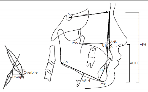
모든 환자들은 구강내 장치를 처방받기 전에 치과를 방문하여 환자의 치아가 충분히 건강한지와 측두하악관절의 이상 유무를 포함하여 환자가 하악을 충분히 전방 이동시킬 수 있는지를 평가하였다. 측두하악관절의 이상 유무는 개구량 및 하악의 전·측방 운동 양상, 턱관절음, 촉진 시 통증 유무 등으로 검사하였다. 구강내 장치 제작 시에는 안정상태에서 자연스럽게 반복적으로 재연할 수 있는 전방 편심위(protrusion bite)를 채득함을 원칙으로 하였고, 최대 전방 이동량의 60~75% 정도 전방 이동시킨 위치로 채득하였다. 구강내 장치의 장기간 사용으로 인한 합병증 평가를 위해 장치 제작 전과 사용 2년 후의 시점에서 시행한 구강 및 치열 검진과 두개계측영상을 통해 얻어진 정보로 대상환자의 수직 피개량(overbite), 수평 피개량(overjet), 상·하전치 경사도(incisor angulation) 및 안면 골격의 변화량을 비교하였다(Fig. 1). Angle’s classification(I, II, III)으로 교합관계를 평가하였으며[11], 이는 상하악의 첫 번째 어금니가 정상적인 맞물림 상태를 I, 하악의 첫 번째 어금니가 상대적으로 뒤쪽으로 들어가 있는 경우를 II, 이와는 반대로 하악의 첫 번째 어금니가 상대적으로 앞쪽으로 나와 있는 경우를 III으로 나타내는 분류법이다. 연구에서 모든 통계 분석은 SPSS Statistics version 21.0(IBM Corp., Armonk, NY, USA)를 사용하였고, p<0.05일 시 통계적으로 유의성을 가지는 것으로 판단하였다.

결 과

총 188명의 환자가 구강내 장치를 처방받았으며, 이들 중 57명의 환자가 2년 이상, 하루 평균 4시간 지속적으로 사용하였다. 성별 분포는 남자 38명, 여자 19명이었고, 연령분포는 21~69세이며 평균 연령은 44.3±12.3세였다. 환자의 병력, 신체검사 수치, 수면 설문지 및 수면 검사 결과는 Table 1과 같다. 대상 환자들에서 턱관절음 및 턱관절 통증 등 주관적인 측두하악관절의 이상은 없었으며, 본인의 치열 및 교합 등 치아 구조의 변화를 주관적으로는 인식하는 환자 역시 없었다.
치아 구조 변화 분석에서 환자의 수직 피개량은 3.53±2.1에서 2.87±2.4로, 수평 피개량은 2.54±2.2에서 2.01±2.0으로 통계학적으로 의미 있게 감소하였다(Table 2). 반면에 전치 경사도는 상전치의 경우 98.11±5.6°에서 97.82±3.9°로 감소하였으나 통계학적 의미가 없었고, 하전치의 경우 99.23±7.8°에서 101.89±4.3°로 통계학적으로 의미 있는 증가가 관찰되었다(Table 2). 교합의 변화는 총 5명의 환자에서 관찰되었는데, 이는 초기에 Class I이었던 14명의 환자 중 2명이 2년 후에 Class III이 되었고 초기에 Class II 환자 43명 중 3명이 2년 후에 Class III이 되었다(Table 2).
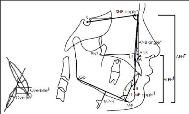
두개계측영상을 통한 안면 골격의 변화 정도를 살펴보았을 시에 sella turcica central point-nasion-subspinal point(SNA) angle은 의미 있는 변화가 관찰되지 않았으나, sella turcica central point-nasion-supramental point(SNB) angle은 77.5±4.1°에서 79.4±2.0°로, subspinal point-nasion-supramental point(ANB) angle은 3.8±1.6°에서 2.3±1.9°로 통계학적으로 의미 있는 변화가 관찰되었다(Table 3). 안면길이의 경우 anterior lower facial height(ALFH)와 anterior facial height 모두 의미 있게 증가하였으나, posterior airway space와 mandibular plane to the hyoid bone(MP-H)의 경우는 구강내 장치를 2년 사용한 경우에 평균값이 증가하는 양상이었으나 통계학적인 차이는 없었다(Table 3, Fig. 2). 추가적으로 시행한 성별에 따른 치아 구조와 안면 골격의 변화 정도는 의미 있는 차이점이 관찰되지 않았다.

고 찰

오늘날 구강내 장치는 폐쇄성 수면무호흡증 환자 치료에 있어서 치과뿐만 아니라 이비인후과에서도 많이 사용되고 있다. 이는 양압기 치료가 구강내 장치보다 수면 중 발생하는 상기도 폐쇄를 효과적으로 치료할 수 있음에도 불구하고, 고가의 장비이면서 소음, 마스크 사용, 호기 시의 불편감, 그리고 휴대가 어렵다는 이유로 환자 순응도 측면에서 구강내 장치가 보다 우수하기 때문이다[12,13]. 가장 흔히 사용하는 구강내 장치인 하악 전진 장치는 그 모양에 따라 일체형과 분리형으로 구별할 수 있는데, 이들 중 폐쇄성 수면무호흡증에 어느 것이 더 효과적인지는 여러 의견이 있으나[14,15], 분리형이 환자 순응도가 보다 좋은 것으로 알려져 있다[16]. 본 연구에서는 환자 순응도를 고려하여 모두 분리형을 사용하였다. 또한, 여러 연구를 통하여 하악의 전진이 클수록 구강내 장치의 치료 효과가 항상 높은 것이 아니라고 알려져 있기 때문에[17-19], 본 연구에서는 최대 전방 이동량의 60~75%에서 환자가 가장 편안하게 느끼는 만큼 하악을 전방 이동시켰다.
일반적으로 구강내 장치의 초기 부작용으로는 저작 시 불편감, 치은 자극, 과도한 타액배출, 구강건조증, 저작근 및 턱 관절 부위 통증 등이 있다. 그러나 이러한 초기 합병증들은 대게 단기간에 나타나며, 보통 그 강도가 경미하고 치료가 지속되면 대부분 사라지는 것들이다. 이에 비해 장기간 장착시 발생되는 합병증으로는 수직 및 수평 피개량의 감소, 전치 경사도의 변화, 안면길이의 증가, 하악평면각의 증가, 개구량의 증가 및 교합의 변화 등이 있다[20]. 그러나 아직까지 국내에서 이비인후과를 방문한 환자를 대상으로 구강내 장치의 장기간 사용 시 발생할 수 있는 합병증과 관련된 연구는 부족한 실정이다.
저자들의 본 연구 결과에서 2년 동안 구강내 장치를 충분한 시간 동안 사용한 환자들의 경우 하악이 다소간 전방 이동하면서 여러 계측치들의 변화가 나타나는 것을 관찰할 수 있었다. 대표적으로 수직 및 수평 피개량의 감소, 하전치 경사도의 증가를 관찰하였으며, 일부 환자(8.7%, 57명 중 5명)에서는 교합의 변화를 확인하였다. 두개계측영상에서는 SNB angle, ANB angle의 변화와 안면하부길이(ALFH)의 증가와 같은 하악골의 위치변화와 관련 있는 변수들의 의미 있는 변화가 관찰되었다. 그러나 하악평면각(MP-H)의 경우 2년 동안 구강내 장치를 사용한 환자들에서 평균적으로 증가하는 양상을 보였으나 이는 통계적으로 유의하지는 않았다. 본 연구에서 관찰된 결과들은 기존의 구강내 장치의 장기간 합병증을 연구한 국외 논문들의 결과와 매우 유사하였다[8-10,21,22].
그러나 일부 기존의 연구 결과에서는 하악평면각이 증가하였다고 보고하였으며[10,23], 이러한 차이점은 본 연구 결과가 2년으로 기존의 연구보다 관찰기간이 짧기 때문으로 판단된다. 게다가, 본 연구에서도 하악평면각의 평균값은 통계적으로 유의하지는 않았으나 증가하는 경향을 나타내었다. 그러므로 관찰기간이 길어지면 하악평면각에서 의미 있는 변화를 관찰할 수 있을 것으로 생각된다. 또한, 기존에 발표한 연구 결과에 따르면 수직 및 수평 피개량의 감소와 상·하전치 경사도의 변화를 대부분 환자들은 인식하지 못한다[9]. 이러한 모습은 본 연구에서도 관찰되는데, 수직 및 수평 피개량의 감소와 전치 경사도의 변화가 발생한 경우 이를 인식하고 있는 환자는 전혀 없었다. 이는 피개량의 변화량이 1 mm 미만이고, 하전치 경사도 변화 역시 2° 정도로 매우 적다는 점과 폐쇄성 수면무호흡증으로 인한 주간 졸림증 등 주관적 증상 해소로 인한 만족도가 높기 때문에 이러한 구조적인 변화를 환자들이 잘 인식하지 못한 것으로 생각된다.
본 연구의 한계점으로는 구강내 장치의 사용기간이 2년이라는 점과 하악 전방 이동량이 최대 전방 이동량의 60~75%로 일정한 전방 이동량을 환자에게 균등하게 제공하지 못한 점, 그리고 연단위 혹은 6개월 단위로 검진이 이루어지지 못해 어느 시점에서 하악골의 위치 변화가 발생하고, 이러한 변화가 점점 증가하는지 혹은 증가하다가 더 이상 변화되지 않았는지와 관련된 양상을 파악하기 어렵다는 점이 있다. 그러므로 추후에 더 많은 환자를 대상으로 더 장기간의 경과 관찰을 순차적으로 시행하면서 폐쇄성 수면무호흡증 환자에서 구강내 장치의 장기간 사용과 관련된 합병증 연구가 이루어져야 할 것으로 생각된다. 또한 추후 연구에는 구강내 장치 장기간 사용 후에 수면 검사를 시행하여 구강내 장치 치료로 인한 AHI 수치 등 수면 검사 결과의 변화를 비교 분석한 내용이 필요할 것으로 생각된다.

REFERENCES

1. Epstein LJ, Kristo D, Strollo PJ Jr, Friedman N, Malhotra A, Patil SP, et al. Clinical guideline for the evaluation, management and longterm care of obstructive sleep apnea in adults. J Clin Sleep Med. 2009; 5(3):263–76.
2. Kim DK, Lee JW, Lee JH, Lee JS, Na YS, Kim MJ, et al. Drug induced sleep endoscopy for poor-responder to uvulopalatopharyngoplasty in patient with obstructive sleep apnea patients. Korean J Otorhinolaryngol-Head Neck Surg. 2014; 57(2):96–102.
3. Kim HY, Jang MS. Improving compliance for continuous positive airway pressure compliance and possible influencing factors. Korean J Otorhinolaryngol-Head Neck Surg. 2014; 57(1):7–14.
crossref
4. Kushida CA, Morgenthaler TI, Littner MR, Alessi CA, Bailey D, Coleman J Jr, et al. Practice parameters for the treatment of snoring and obstructive sleep apnea with oral appliances: an update for 2005. Sleep. 2006; 29(2):240–3.
crossref
5. Ramar K, Dort LC, Katz SG, Lettieri CJ, Harrod CG, Thomas SM, et al. Clinical practice guideline for the treatment of obstructive sleep apnea and snoring with oral appliance therapy: an update for 2015. J Clin Sleep Med. 2015; 11(7):773–827.
crossref
6. Kim YH, Park DS, Son DH, Cho JH. Polysomnographic parameters related to the successful treatment of oral appliance in patients with sleep-disordered breathing. Korean J Otorhinolaryngol-Head Neck Surg. 2012; 55(12):771–6.
crossref
7. Spencer J, Patel M, Mehta N, Simmons HC 3rd, Bennett T, Bailey JK, et al. Special consideration regarding the assessment and management of patients being treated with mandibular advancement oral appliance therapy for snoring and obstructive sleep apnea. Cranio. 2013; 31(1):10–3.
crossref
8. Rose EC, Staats R, Virchow C Jr, Jonas IE. Occlusal and skeletal effects of an oral appliance in the treatment of obstructive sleep apnea. Chest. 2002; 122(3):871–7.
crossref
9. Robertson C, Herbison P, Harkness M. Dental and occlusal changes during mandibular advancement splint therapy in sleep disordered patients. Eur J Orthod. 2003; 25(4):371–6.
crossref
10. Wang X, Gong X, Yu Z, Gao X, Zhao Y. Follow-up study of dental and skeletal changes in patients with obstructive sleep apnea and hypopnea syndrome with long-term treatment with the Silensor appliance. Am J Orthod Dentofacial Orthop. 2015; 147(5):559–65.
crossref
11. Peck S. The contributions of Edward H. Angle to dental public health. Community Dent Health. 2009; 26(3):130–1.
12. Clark GT, Blumenfeld I, Yoffe N, Peled E, Lavie P. A crossover study comparing the efficacy of continuous positive airway pressure with anterior mandibular positioning devices on patients with obstructive sleep apnea. Chest. 1996; 109(6):1477–83.
crossref
13. Tan YK, L’Estrange PR, Luo YM, Smith C, Grant HR, Simonds AK, et al. Mandibular advancement splints and continuous positive airway pressure in patients with obstructive sleep apnoea: a randomized cross-over trial. Eur J Orthod. 2002; 24(3):239–49.
crossref
14. Bloch KE, Iseli A, Zhang JN, Xie X, Kaplan V, Stoeckli PW, et al. A randomized, controlled crossover trial of two oral appliances for sleep apnea treatment. Am J Respir Crit Care Med. 2000; 162(1):246–51.
crossref
15. Rose E, Staats R, Virchow C, Jonas IE. A comparative study of two mandibular advancement appliances for the treatment of obstructive sleep apnoea. Eur J Orthod. 2002; 24(2):191–8.
crossref
16. Lee WH, Wee JH, Lee CH, Kim MS, Rhee CS, Yun PY, et al. Comparison between mono-bloc and bi-bloc mandibular advancement devices for obstructive sleep apnea. Eur Arch Otorhinolaryngol. 2013; 270(11):2909–13.
crossref
17. Lowe AA, Sjöholm TT, Ryan CF, Fleetham JA, Ferguson KA, Remmers JE. Treatment, airway and compliance effects of a titratable oral appliance. Sleep. 2000; 23 Suppl 4:S172–8.
18. Mehta A, Qian J, Petocz P, Darendeliler MA, Cistulli PA. A randomized, controlled study of a mandibular advancement splint for obstructive sleep apnea. Am J Respir Crit Care Med. 2001; 163(6):1457–61.
crossref
19. Henke KG, Frantz DE, Kuna ST. An oral elastic mandibular advancement device for obstructive sleep apnea. Am J Respir Crit Care Med. 2000; 161(2 Pt 1):420–5.
crossref
20. Hoffstein V. Review of oral appliances for treatment of sleep-disordered breathing. Sleep Breath. 2007; 11(1):1–22.
crossref
21. Doff MH, Finnema KJ, Hoekema A, Wijkstra PJ, de Bont LG, Stegenga B. Long-term oral appliance therapy in obstructive sleep apnea syndrome: a controlled study on dental side effects. Clin Oral Investig. 2013; 17(2):475–82.
crossref
22. Ghazal A, Jonas IE, Rose EC. Dental side effects of mandibular advancement appliances - a 2-year follow-up. J Orofac Orthop. 2008; 69(6):437–47.
crossref
23. Doff MH, Hoekema A, Pruim GJ, Huddleston Slater JJ, Stegenga B. Long-term oral-appliance therapy in obstructive sleep apnea: a cephalometric study of craniofacial changes. J Dent. 2010; 38(12):1010–8.
crossref

Fig. 1.
Analysis parameters. PNS: posterior nasal spine, MP-H: mandibular plane to the hyoid bone, S: sella, Go: gonion, Me: menton, L1: mandibular incisor, B: innermost point on the anterior portion of mandible, U1: maxillary incisor, A: innermost point on the anterior portion of maxilla, ANS: anterior nasal spine, N: nasion, ALFH: anterior lower facial height, AFH: anterior facial height.
kjorl-hns-2017-00178f1.tif
Fig. 2.
Dental and skeletal change after 2 years. *p<0.05, †p<0.010, ‡p<0.001. PNS: posterior nasal spine, MP-H: mandibular plane to the hyoid bone, S: sella, Go: gonion, Me: menton, L1: mandibular incisor, B: innermost point on the anterior portion of mandible, U1: maxillary incisor, A: innermost point on the anterior portion of maxilla, ANS: anterior nasal spine, ALFH: anterior lower facial height, AFH: anterior facial height, SNB: sella turcica central point-nasion-supramental point, ANB: subspinal point-nasion-supramental point, L1-MP angle: mandibular incisor angle related to mandibular plane.
kjorl-hns-2017-00178f2.tif
Table 1.
Baseline characteristics of the study subjects
Variables (n=57) Values
Age (years) 44.3±12.3
Sex (male) 38 (66.7%)
Cardiovascular diseases 38 (66.7%)
Cerebrovascular diseases 6 (10.5%)
Chronic respiratory diseases 16 (27.6%)
Diabetes mellitus 14 (24.6%)
Body mass index (kg/m2) 25.4±2.9
Neck circumference (cm) 37.2±4.3
Epworth Sleepiness Scale 8.0±4.4
Pittsburgh sleep quality index 7.3±4.8
Beck depression inventory 6.0±3.3
Respiratory disturbance index 16.0±13.3
Apnea-hypopnea index 12.2±9.3
Lowest O2 saturation 92.4±11.2

Data are presented as mean±standard deviation or number of subjects. Cardiovascular diseases include the history of hypertension, angina, and acute myocardial infarction. Cerebrovascular diseases include the history of ischemia and hemorrhage. Chronic respiratory diseases include the history of asthma and chronic obstructive pulmonary disease

Table 2.
Dental change after long-term use of oral appliance
Variables (n=57) Baseline 2-year p-value
Overbite (mm) 3.53±2.1 2.87±2.4 <0.001
Overjet (mm) 2.54±2.2 2.01±2.0 0.004
U1-SN angle (°) 98.11±5.6 97.82±3.9 0.054
L1-MP angle (°) 99.23±7.8 101.89±4.3 <0.001
Angle’s classification Class I Class II Class III
Baseline n=14 n=43 n=0
2-year n=12 n=40 n=5

U1-SN angle: maxillary incisor angle related to anterior cranial base, L1-MP angle: mandibular incisor angle related to mandibular plane

Table 3.
Facial skeletal change after long-term use of oral appliance
Variables (n=57) Baseline 2-year p-value
SNA angle (°) 81.3±6.5 81.7±7.4 0.731
SNB angle (°) 77.5±4.1 79.4±2.0 0.038
ANB angle (°) 3.8±1.6 2.3±1.9 0.017
ALFH (mm) 77.1±6.7 79.8±7.3 0.004
AFH (mm) 116.3±7.6 119.4±4.7 0.008
PAS (mm) 8.7±2.1 9.8±2.3 0.072
MP-H (mm) 16.4±5.4 18.1±6.7 0.054

SNA: sella turcica central point-nasion-subspinal point, SNB: sella turcica central point-nasion-supramental point, ANB: subspinal point-nasion-supramental point, ALFH: anterior lower facial height (anterior nasal spine to menton), AFH: anterior facial height (nasion to menton), PAS: posterior airway space, MP-H: mandibular plane to the hyoid bone

TOOLS
Similar articles