Abstract
Proficiency in ultrasound-guided regional anesthesia (UGRA) requires the practitioner to acquire cognitive and technical skills. For anesthesiology residents, an assortment of challenges has been identified in learning UGRA skills. Currently, a validated UGRA curriculum for residents does not exist, and the level of UGRA proficiency achieved during residency training can vary considerably. Simulated practice has been shown to enhance proficiency in UGRA, and a competency-based education with simulation training has been endorsed for anesthesiology residents. The objective of this review is to outline simulation-based training that can be implemented in a UGRA curriculum and to explore educational tools like gamification to facilitate competency in regional anesthesiology.
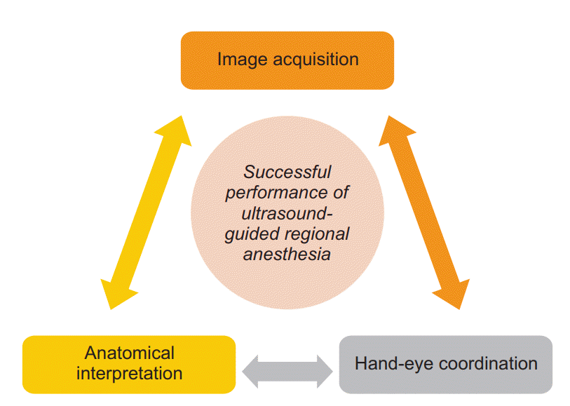
Anatomical-based ultrasound imaging has been one of the most invigorating innovations in the field of regional anesthesiology in recent years. For the first time in the history of regional anesthesia, a real-time visual representation of the internal anatomy is available to clinicians. This applied technology has ignited enthusiasm among anesthesiologists to perform ultrasound-guided regional anesthesia (UGRA). Compared to other nerve localization techniques, benefits of UGRA include increased nerve block success rate, faster onset time, decreased volume of local anesthetic, and reduced risk of local anesthetic complications [1,2]. Most experts now consider ultrasound-guidance to be the standard of care for peripheral nerve blocks [3]. To successfully perform a UGRA procedure, a triad of three distinct but interrelated skills are required: image acquisition, anatomical interpretation, and hand-eye coordination (Fig. 1). First, the clinician must successfully acquire an image and then correctly interpret the sonoanatomy. Next, the needle must be visualized and guided to the desired target. This dynamic process often requires constant needle manipulation with simultaneous adjustments of the ultrasound transducer.
While some clinicians can quickly assimilate ultrasound use into clinical practice, the majority face an extensive learning curve when initially acquiring UGRA skills [4,5]. Considering that each anesthesiology resident inherently possesses different levels of hand-eye coordination and can have varying degrees of ultrasound experience prior to their anesthesiology training, there can be wide variability in the rate at which UGRA skills are learned [5–8]. To provide a framework for UGRA teaching, the American Society of Regional Anesthesia and Pain Medicine and the European Society of Regional Anaesthesia and Pain Therapy (ASRA-ESRA) jointly identified core competencies and skill sets for UGRA and proposed that residency programs implement a UGRA curriculum with simulation training [9]. However, a standardized UGRA curriculum currently does not exist and residents may be expected to perform UGRA in a clinical setting without adequate preparation and training. The objective of this review is to examine the merit of simulation-based UGRA education when designing a resident curriculum and to explore educational tools such as gamification to augment UGRA training.
The traditional “apprenticeship model” or “see one, do one” method of learning while concurrently providing clinical care can result in inconsistent learning experiences, variability in case numbers, and compromise in patient safety [10,11]. In these models, the educational content can focus on what is being taught by the instructor instead of what is being learned by the resident [10], and time pressure during clinical care can limit teaching opportunities. Lack of a standardized curriculum may result in teaching individual preferences in UGRA techniques, equipment, and ergonomics [11]. Regional anesthesia rotations in which residents have dedicated time to perform nerve blocks can increase the overall number of completed procedures [12]; however, merely performing a high volume of nerve blocks does not necessarily equate with UGRA proficiency [13,14]. In the United States, the Accreditation Council for Graduate Medical Education (ACGME) requires anesthesiology residents to complete a regional anesthesia rotation (minimum of four weeks) and perform, before graduation, at least 40 epidural anesthetics, 40 spinal anesthetics, and 40 peripheral nerve blocks [15]. Yet, even with formal recommendations and standardized requirements, the number and types of UGRA procedures performed by residents can vary among training programs [16] and residents may not feel confident in performing UGRA upon completion of their training [17]. Subsequently, it has been accepted that residents will require more than the established minimum number to become proficient in regional anesthesia [16]. Furthermore, since the ACGME does not specify types of nerve blocks, residents may fulfill their requirements by repeatedly performing the same few nerve blocks while being insufficiently trained in others [17]. Not surprisingly, residency training programs are globally undergoing a transition from time- or volume-based requirements to a competency-based educational model [18].
Other challenges of learning UGRA include retention of knowledge and technical skills. For novices, skill attrition can occur as fast as 24 hours after initial learning of UGRA skilled tasks [19] but it may be difficult for residents to find time to incorporate multiple regional anesthesia rotations during residency training due to the growing number of requirements prior to graduation and other time constraints. Additionally, lack of trained or experienced faculty in UGRA may limit exposure to advanced nerve block sites [11]. Inconsistent availability of ultrasound equipment for training and clinical use can also be obstacles to learning UGRA [20].
The UGRA core competencies outlined by the ASRA-ESRA joint committee are encompassed into six domains: patient care, medical knowledge, system-based practice, practice-based learning and improvement, interpersonal and communication skills, and professionalism [9]. This competency-based learning model with simulation-based education is the recommended residency pathway for UGRA training [9,14]. Simulation has been utilized for training in a wide range of medical fields [21,22] and is an effective educational tool for acquiring knowledge and skills, gaining hands-on experience through repetitive practice without harm to patients, and receiving individualized feedback [23]. For UGRA, various simulation modalities have been used for resident education, from phantom gels and part-task trainers to virtual reality and full-scale mannequins [23,24]. Advantages of UGRA simulation include shortening the learning curve [3], allowing residents to learn at their individual rate [7], and improving block success [25]. Additional benefits of simulation training include learning non-technical skills (e.g., communication, situational awareness) and creation of a low-stress environment for learning [26]. This latter point may be critical for residents since anxiety can negatively affect novice performance of UGRA [27]. The majority of anesthesiology residents support simulation as a method for learning and assessment [28] and feel a simulation-based curriculum would be helpful in their education of UGRA [17].
In-depth knowledge of anatomy is a prerequisite for performing UGRA. Identification of key anatomical landmarks (e.g., muscles, fascias, bones, and blood vessels) is the first task outlined in the joint ASRA-ESRA recommendations for UGRA [9], and detailed knowledge of relevant anatomy is essential to identify sonoanatomy [29]. Without an understanding of relevant anatomy, novice learners cannot progress into “advanced sonoanatomy” (i.e., using ultrasound to identify anatomical landmarks surrounding target nerve) but instead can fall into a habit of “pattern recognition” (i.e., scanning until the ultrasound image closely resembles sonoanatomy they have seen previously) [8,30]. Furthermore, without knowledge of anatomy and correct interpretation of sonoanatomy, novices can fail to recognize abnormal anatomy and may endanger patients with inadvertent needle puncture of tissues or vessels [31].
Simulation-based education can be a time-efficient modality to learn UGRA-related anatomy. Ramlogan et al. [32] enrolled anesthesia residents and fellows in a training session using an online virtual-reality simulator that presented interactive 3-dimensional images and allowed virtual scanning. After a 1-hour self-study session, participants scored higher than their baseline on a multiple-choice exam and reported that the simulator tool was easy to use and beneficial to their learning. In a study by Woodworth et al. [33], a 25-minute video about anatomy complemented by a 5-minute interactive simulation module employing a virtual transducer (i.e., scanning produced both ultrasound and corresponding magnetic resonance images) led to improved scores on a written exam compared to a control group that only watched a sham video. However, the intervention group did not show improvement in image acquisition while scanning on live models, implying that virtual reality scanning may have limited clinical applications.
A few studies endorse the inclusion of real-time ultrasound images to learn anatomy and sonoanatomy. In a study by VanderWielen et al. [34], participants who viewed a brief instructional video (viewing session did not exceed ten minutes) that partly reviewed basic anatomy and demonstrated ultrasound scanning techniques improved their ability to acquire ultrasound images and identify anatomical landmarks on a live model. However, a group randomized to a hands-on training session with an anatomy-based gel phantom (scanning session did not exceed ten minutes) was more successful than the video group in identifying anatomical landmarks when both were compared to a control group, reinforcing the value of hands-on experience and dynamic image interpretation to learn sonoanatomy. During a 4-week regional anesthesia rotation, Orebaugh et al. [31] introduced a UGRA training curriculum to residents prior to performing nerve block procedures. Didactics covered numerous UGRA-related topics, including anatomy, and teaching of sonoanatomy involved focused scanning of patients under supervision. Compared to their pre-rotation standardized exam scores, residents at the end of the rotation demonstrated improvement in acquiring ultrasound images and identifying anatomical and neural structures at four different block sites (interscalene, supraclavicular, femoral, popliteal fossa). Likewise, Barrington et al. [35] demonstrated that a combination of standardized didactics followed by supervised hands-on scanning sessions on live models allowed residents to become proficient in acquiring ultrasound images and identifying relevant anatomical landmarks. This group specifically focused on axillary brachial plexus blocks and determined residents needed eight to ten training sessions with feedback from expert faculty to achieve proficiency. For residents who may not have access to experienced trainers but seek additional knowledge of sonoanatomy, scanning live models or patients with the aid of an electronic tutorial may enhance learning. In a study by Wegener et al. [29], novice clinicians who practiced with an ultrasound machine that simultaneously displayed anatomical tutorials alongside real-time images identified correctly more anatomical structures on a live model exam than the control group that practiced without the tutorial.
Ultrasound knowledge is a core competency outlined by the joint ASRA-ESRA committee and includes skills like understanding basic ultrasound physics (e.g., frequency, attenuation, refraction), equipment functions (e.g., depth, gain), and artifacts (e.g., reverberations, acoustic shadowing) [9]. Prior to performing simulated UGRA, it is recommended that through didactics residents become familiarized with concepts like appropriate transducer selection, transducer orientation to target (transverse vs. longitudinal), scanning techniques (rotation, alignment, tilt, pressure), needle visualization (in- vs. out-of-plane), and correct local anesthetic spread around the nerve [9]. Appropriate preparatory teaching can help residents understand the technical skills required to successfully perform UGRA and deconstruct procedures into individual tasks to facilitate learning [7]. Then, residents can proceed to hands-on practice to acquire proficiency in those individual UGRA tasks until sufficient skills are obtained to perform a UGRA procedure in its entirety [7].
For practice of UGRA technical skills, numerous simulated models have been introduced, including self-assembled phantoms (gelatin, meat, tofu) [34,36,37], commercially available trainers [25,38], hybrid simulators [39,40], and cadavers [6,41,42]. Simulation-based training in needle visualization has been shown to improve hand-eye coordination [43], decrease technical errors [44], and reduce number of needle passes [45]. A group of residents that underwent a 1-hour hands-on training session on a simulation model, focusing on needle visualization and hand-eye coordination, had higher clinical success rate of UGRA compared to a control group that did not receive simulation training [25].
Although recommendations cannot be made on the ideal simulation modality for learning UGRA-related technical skills, comparative studies suggest that low-fidelity phantom models can be cost-effective and accessible [46,47]. Chuan et al. [46] randomized novices to practice on either a porcine meat model with embedded bovine tendon (low-fidelity) or a fresh-frozen cadaveric upper limb (high-fidelity). Each participant underwent a deliberate practice session of 45 minutes during which time 30 practice trials were allowed with individualized feedback. The post-training examination consisted of identifying the sciatic nerve on a cadaver and injecting saline around the nerve. Between the low- and high-fidelity groups, there were no differences in time to complete the simulated sciatic nerve block, errors committed, and ultrasound image quality of the nerve. In another study, Friedman et al. [47] investigated the effects of a high- versus low-fidelity simulator on performance of epidural catheter placement in actual patients. In the high-fidelity group, anesthesiology residents practiced epidural needle insertion on a mannequin simulator with a “virtual-reality display of needle progression,” while the low-fidelity group practiced inserting needles into a banana (“greengrocer’s” model) to simulate loss of resistance. Each group was allowed sixty minutes of supervised training followed by live procedural observations. Throughout a six-month period, the two groups did not show a difference in skill acquisition when assessed by a checklist and global rating scale (GRS). For educators, the selection of a simulation modality will depend on availability of financial resources and time commitment; however, when devising a simulated environment, the priority should rest in supporting resident skill acquisition and assessment rather than simulation fidelity [8,48]. Practice sessions should have defined objectives that provide exposure to different types of transducers, superficial and deep targets, inand out-of-plane needle visualization, and short- and long-axis target views. Residents should also become familiarized with ultrasound artifacts, which may not be replicated on simulated models, but can confuse novices during UGRA procedures on actual patients [49,50].
When initially learning UGRA, it is common for residents to focus primarily on the hands-on aspects of UGRA [13]; however, technical competency alone does not result in UGRA proficiency [51]. Table 1 outlines examples of non-technical knowledge and skills necessary to progress toward independent practice. Specific topics like clinical pharmacology and risks and contraindications of regional anesthesia can be covered through didactics. Other tasks and skills, like aseptic technique, monitoring of vital signs and patient comfort, management of complex patients or complications, and coordination of care with other health care providers, can be learned through clinical experience. Although simulation training has not been studied extensively for non-technical UGRA skills, residents can practice providing informed consent, explaining post-procedural care, and discussing management of complications as part of a simulated UGRA procedure [52]. Additionally, simulation can be used to teach certain skills like situational awareness, multi-tasking, and teamwork during crisis management [21,24], and standardized simulated scenarios can allow residents to develop an approach to managing UGRA-related complications [53]. For example, during simulation-based training for local anesthetic systemic toxicity (LAST), trainees who used a checklist of therapeutic interventions demonstrated superior management of LAST events and exhibited better decision-making skills compared to trainees who relied on their memory [53]. Other scenarios for simulation that have been described include high spinal anesthesia and postoperative nerve injury [54].
There is a well-known adage about performance assessment: “When performance is measured, performance improves. When performance is measured and reported, the rate of improvement accelerates.”1) The principle of performance measurement is equally applicable to learning UGRA. Objective assessments and feedback are critical components of simulation training that can reduce learning curves [4] and enhance UGRA education [3,8,14]. For UGRA, a few validated assessment tools can be considered during training. Woodworth et al. [55] demonstrated reliability, content validity, and construct validity of a multiple-choice exam that specifically assesses knowledge of sonoanatomy. The strengths of this exam include accessibility (online format), applicability to residents at various levels of UGRA experience, and immediate feedback that is not dependent on an evaluator. However, this exam was designed to assess knowledge of UGRA and does not necessarily correlate with technical skills. Other assessment tools that have been used for regional anesthesia include checklists and GRS [47,56]. A checklist and GRS specifically for UGRA was first published in 2012 [57] and since has been validated through assessment of residents performing nerve block procedures on patients [58] and modified to allow evaluation of regional anesthesia performance irrespective of regional anesthesia type (neuraxial or peripheral) and nerve localization technique (ultrasound, nerve stimulation, or combined) [59]. Checklists can deconstruct a procedure into a discrete series of steps and allow educators to provide objective and specific feedback to residents at various stages of learning [59].
For competency-based models of training, a standardized assessment tool has been introduced. In the United States, the ACGME and the American Board of Anesthesiology jointly released the Regional Anesthesiology and Acute Pain Medicine Milestone Project to assess UGRA competencies [60]. Although this assessment was created for accredited regional anesthesiology fellowship programs, the milestones can be potentially modified and implemented as part of a UGRA curriculum to evaluate residents’ knowledge, interactions with patients and other health care providers, technical skills, and professionalism. Proficiency in each competency is measured by achievement of milestones that are categorized into five levels. Level 1 indicates performance expected of a fellow (or resident) at the beginning of their training and level 5 denotes performance of a clinician who has been in independent practice for several years. For technical skills, the primary measure of milestone achievement is the level of supervision required to successfully perform a procedure in a clinical setting. To standardize attainment of technical skill milestones (i.e., progression from one level to the next), educators can consider incorporating checklists and/or GRS to provide consistent and objective requirements for advancement.
Feedback is a critical learning tool for residents during all stages of UGRA skill acquisition [7]. With feedback, novice learners can improve performance time of UGRA tasks, decrease number of needle passes, and reduce errors [19]. Simulation-based training that combines deliberate practice with feedback has been shown to improve residents’ block performance and success rate in a clinical setting [25,61]. It also has been observed that learning of advanced or complex UGRA procedures requires feedback from experienced instructors [50]. The quality of feedback by educators may be even more effective than the fidelity of the simulation training in improving novice performance [46]. Conversely, simulation training can provide an optimal environment for residents who require time to assimilate new knowledge and learn from internal feedback [7] and self-reflection [51] (i.e., independent deliberate practice). Residents who underwent self-guided practice of UGRA in a hybrid simulator performed equally well when assessed by a checklist and GRS compared to residents who received coaching during simulation training [62]. This finding supports a prior observation that excessive feedback during task training or procedural performance can result in dependence on external feedback and decrease in skill retention [7,19].
The various types and timing of feedback and their application to UGRA have been previously described by Slater et al. [7]. Although further studies are needed to elucidate the optimal feedback model for resident learning of UGRA [3,19], educators should take an individualized approach since each resident will have different aptitudes, experiences, and learning preferences. Prior to training sessions, learning objectives should be clearly delineated and feedback should focus on specific tasks with emphasis placed on “constructive criticism, identification of weaknesses, problem-solving, and positive reinforcement of the resident’s technique” [7].
Gamification is the integration of game principles into nongame contexts and has been applied in numerous sectors from personal fitness to scientific endeavors [63,64]. Game design elements (Table 2) [65] that endorse competition, recognition of accomplishments, and social interactions can foster motivation and provide a positive reward system. When gamification principles are applied to an educational curriculum, the synergistic effect of training combined with elements of success, rewards, and social recognition can benefit learners by increasing engagement, productivity, content learning, knowledge retention, and collaboration [63]. For graduate medical education, game designs can create a “fun” learning environment, enhance resident training at various stages of learning, and serve as a method of assessing core competencies [66].
Elements of gamification have been incorporated into various residency training curricula (Table 3) [64,67–70], and gamification of simulation-based training has been shown to be a promising educational tool. In a study by Enter et al. [67], firstyear cardiothoracic surgery residents were invited to voluntarily participate in a 6-week simulation-based training (coronary anastomosis on a low-fidelity simulator) as part of a selection process to become a contestant in a live competition. On completion of their training, residents increased their competency in task performance and reported that training was a valuable investment of their time. Furthermore, the baseline disparity in skill level observed among residents was nonexistent after six weeks, suggesting that initial poor performers were able to catch up with more skilled residents by engaging in self-initiated training. In another study, Lobo et al. [68] integrated game designs (team competition) into a traditional simulation-based curriculum to teach point-of-care-ultrasound to emergency medicine residents. Resident participation resulted in improved knowledge of basic ultrasound technical skills, image interpretation, and patient management. Additional reported benefits of gamification included improved camaraderie and communication among residents. Although gamification is in its nascent stages of development for anesthesiology education, especially in UGRA, and may not be suitable for all learners [71], this innovative approach to spur motivation and engagement can be a useful supplement to enhance pre-existing teaching modules and enrich the resident learning experience.
To date, little is known about the design and implementation of UGRA curricula across residency programs. It is likely that each institution will incorporate a curriculum based on availability of resources. For example, the two-month regional anesthesia rotation established by Smith et al. [10] consists of a longitudinal care model in which residents are assigned daily to orthopedic cases and are expected to participate in pre-, intra-, and post-operative care plans. Outside of the clinical setting, residents are expected to immerse in self-learning activities that include an ultrasound curriculum (didactics, hands-on training, and simulated practice), simulation training, cadaveric dissections, and personal study (interactive DVDs, online learning modules, training videos). At the institution of Garcia-Tomas et al. [54], the regional anesthesia education consists of residents participating in a two-day teaching session every month that includes anatomy workshops, ultrasound scanning practice, simulated crisis management, and problem-based learning sessions. Moving forward, there is a need to identify best educational practices to teach UGRA during residency training; furthermore, the effects of different UGRA curricula on clinical performance need to be clarified. To facilitate academic and research collaboration among institutions, the development of a UGRA network has been proposed [3]. A forum that allows communication among UGRA coordinators [9] and/or rotation directors can promote sharing of curriculum design, implementation strategies, and educational interventions specific to resident learning. When designing or modifying a curriculum, UGRA coordinators at each institution can also incorporate feedback from recent graduates in independent practice to assess local expectations after residency training and identify deficiencies in UGRA teaching during residency [72].
In novices, skill attrition can occur rapidly after gaining initial proficiency in UGRA tasks [19,34], and frequent exposure has been identified as an essential element for learning UGRA [11]. For acquisition and retention of technical skills, two educational models have been investigated: massed practice in which residents participate in a consolidated training session and a distributed model that spaces training sessions over a period of time [73]. Although the effects of massed versus distributed practice can vary on the type and complexity of the UGRA task [7,35] and need to be further delineated in anesthesiology residents, these training models have been studied more extensively in surgery residents who also experience technical skill decay in a short period of time without continued practice [74,75]. Simulation-based training was adopted early by the surgery community to facilitate learning of laparoscopic skills [76] and continues to be used widely across surgical specialties [22]. In a recent systematic review comparing massed versus distributed practice for retention of surgery-related technical skills, novice learners who participated in spaced practice sessions performed better on retention tests than novices who underwent massed practice [77]. However, the effects of distributed learning on skill maintenance for novices can also be influenced by the quality of the practice sessions [78]. Simulation-based UGRA education has been shown to result in greater retention of knowledge compared to non-simulation teaching [25]; however, extrapolating from the surgical literature, anesthesiology residents will likely require periodic retraining through simulated practice to maintain technical proficiency. The ideal time interval between training sessions remains unknown at this time for UGRA and will likely vary for each resident and skill set. According to the surgery literature, however, technical skills may decline noticeably after three to six months [74,79].
Motivating residents to participate in regularly scheduled training can be challenging due to demands of residency and time constraints. Gamification of a UGRA curriculum can be an innovative modality to encourage residents to seek independent learning and simulated practice outside of required didactics and clinical rotations. A medical knowledge competition using a Web-based question-bank [64] or Twitter [69] to assign daily multiple-choice or open-ended questions resulted in increased academic reading, retention of knowledge, and standardized in-training exam scores. Similar gamification strategies can be considered to engage anesthesiology residents in UGRA-related knowledge questions even when they are not on a dedicated regional anesthesia rotation. Likewise, incentives offered by gamification can also compel residents to take advantage of available simulation equipment to engage in deliberate practice in their own time after completion of clinical responsibilities [80]. As part of resident training in point-of-care ultrasound, the Society for Academic Emergency Medicine hosts an annual, four-hour, interactive competition called the “SonoGames” [81] that has been shown to increase competency, enthusiasm, and clinical use of ultrasound in participating residents [70]. For cardiothoracic surgery residents, the implementation of an annual, national competition for technical and cognitive skills assessment motivated residents to engage in simulation training and increased their utilization of a standardized educational curriculum to prepare for the live contest [82]. Gamification of simulation training has yet to be explored for UGRA, but successful integration of game design elements can likely offer anesthesiology residents an interactive learning environment that can promote self-initiated learning and retention of skills.
Residency programs with limited resources can consider alternative learning opportunities for residents. Remote simulation teaching in collaboration with other institutions has been described and consists of tele-simulation workshops incorporating live and simulation model scanning, didactics, and self-directed practice sessions [52]. Additionally, residency programs can support resident participation in programs like the UGRA Education and Clinical Training Portfolio offered by ASRA and American Society of Anesthesiology (http://www.asahq.org) or the European Diploma for Regional Anesthesia and Acute Pain Management endorsed by ESRA (http://academy.esraeurope.org), both of which require completion of didactics and workshops, a minimum number of clinical procedures, and standardized examinations as part of their certification process.
In order to achieve a comprehensive UGRA education, a curriculum must define required skills, choose appropriate teaching models, develop relevant assessment methodology, provide timely feedback, and foster resident motivation and engagement. Educating residents to perform invasive UGRA procedures requires an ongoing balance between patient safety and frequent procedural training. A simulation-based UGRA curriculum can enhance learning of anatomy and sonoanatomy, facilitate acquisition of UGRA technical and non-technical skills, and provide recurrent training outside of the clinical setting. As the practice of UGRA advances, innovative methods are needed to enhance resident education. The application of gamification to augment simulation training can potentially transform the learning experience for residents, which in turn may also lead to increased proficiency and improved patient safety. Challenges in creating a UGRA curriculum with simulation and gamification are the cost of purchasing and maintenance of simulation equipment, investment of time, and implementation of a sustainable curriculum. Further investigations in resident education of UGRA should include identification of optimal simulation and gamification modalities for residents at various stages of learning, ideal time interval between simulation training, and clinical outcomes after simulation-based training.
References
1. Marhofer P, Harrop-Griffiths W, Kettner SC, Kirchmair L. Fifteen years of ultrasound guidance in regional anaesthesia: part 1. Br J Anaesth. 2010; 104:538–46.
2. Barrington MJ, Kluger R. Ultrasound guidance reduces the risk of local anesthetic systemic toxicity following peripheral nerve blockade. Reg Anesth Pain Med. 2013; 38:289–99.
3. Chen XX, Trivedi V, AlSaflan AA, Todd SC, Tricco AC, McCartney CJ, et al. Ultrasound-guided regional anesthesia simulation training: a systematic review. Reg Anesth Pain Med. 2017; 42:741–50.
4. Sites BD, Gallagher JD, Cravero J, Lundberg J, Blike G. The learning curve associated with a simulated ultrasound-guided interventional task by inexperienced anesthesia residents. Reg Anesth Pain Med. 2004; 29:544–8.
5. de Oliveira Filho GR, Helayel PE, da Conceição DB, Garzel IS, Pavei P, Ceccon MS. Learning curves and mathematical models for interventional ultrasound basic skills. Anesth Analg. 2008; 106:568–73.
6. Barrington MJ, Wong DM, Slater B, Ivanusic JJ, Ovens M. Ultrasound-guided regional anesthesia: how much practice do novices require before achieving competency in ultrasound needle visualization using a cadaver model. Reg Anesth Pain Med. 2012; 37:334–9.
7. Slater RJ, Castanelli DJ, Barrington MJ. Learning and teaching motor skills in regional anesthesia: a different perspective. Reg Anesth Pain Med. 2014; 39:230–9.
8. Nix CM, Margarido CB, Awad IT, Avila A, Cheung JJ, Dubrowski A, et al. A scoping review of the evidence for teaching ultrasound-guided regional anesthesia. Reg Anesth Pain Med. 2013; 38:471–80.
9. Sites BD, Chan VW, Neal JM, Weller R, Grau T, Koscielniak-Nielsen ZJ, et al. The American Society of Regional Anesthesia and Pain Medicine and the European Society of Regional Anaesthesia and Pain Therapy joint committee recommendations for education and training in ultrasound-guided regional anesthesia. Reg Anesth Pain Med. 2010; 35(2 Suppl):S74–80.
10. Smith HM, Kopp SL, Jacob AK, Torsher LC, Hebl JR. Designing and implementing a comprehensive learner-centered regional anesthesia curriculum. Reg Anesth Pain Med. 2009; 34:88–94.
11. O'Sullivan O, Shorten GD, Aboulafia A. Determinants of learning ultrasound-guided axillary brachial plexus blockade. Clin Teach. 2011; 8:236–40.
12. Martin G, Lineberger CK, MacLeod DB, El-Moalem HE, Breslin DS, Hardman D, et al. A new teaching model for resident training in regional anesthesia. Anesth Analg. 2002; 95:1423–7.
13. Tan JS, Chin KJ, Chan VW. Developing a training program for peripheral nerve blockade: the "nuts and bolts". Int Anesthesiol Clin. 2010; 48:1–11.
14. Niazi AU, Peng PW, Ho M, Tiwari A, Chan VW. The future of regional anesthesia education: lessons learned from the surgical specialty. Can J Anaesth. 2016; 63:966–72.
15. Accreditation Council for Graduate Medical Education. ACGME program requirements for graduate medical education in anesthesiology [Internet]. Chicago (IL): ACGME;2018. July 1 [cited 2018 Sep 29]. Available from https://www.acgme.org/Specialties/Program-Requirements-and-FAQs-and-Applications/pfcatid/6/Anesthesiology.
16. Neal JM, Gravel Sullivan A, Rosenquist RW, Kopacz DJ. Regional anesthesia and pain medicine: US anesthesiology residenttraining-the year 2015. Reg Anesth Pain Med. 2017; 42:437–41.
17. Moon TS, Lim E, Kinjo S. A survey of education and confidence level among graduating anesthesia residents with regard to selected peripheral nerve blocks. BMC Anesthesiol. 2013; 13:16.
18. Yamamoto S, Tanaka P, Madsen MV, Macario A. Comparing anesthesiology residency training structure and requirements in seven different countries on three continents. Cureus. 2017; 9:e1060.
19. Farjad Sultan S, Iohom G, Shorten G. Effect of feedback content on novices' learning ultrasound guided interventional procedures. Minerva Anestesiol. 2013; 79:1269–80.
20. Ramlogan R, Manickam B, Chan VW, Liang L, Adhikary SD, Liguori GA, et al. Challenges and training tools associated with the practice of ultrasound-guided regional anesthesia: a survey of the American society of regional anesthesia and pain medicine. Reg Anesth Pain Med. 2010; 35:224–6.
21. Boet S, Bould MD, Fung L, Qosa H, Perrier L, Tavares W, et al. Transfer of learning and patient outcome in simulated crisis resource management: a systematic review. Can J Anaesth. 2014; 61:571–82.
22. Atesok K, Satava RM, Van Heest A, Hogan MV, Pedowitz RA, Fu FH, et al. Retention of skills after simulation-based training in orthopaedic surgery. J Am Acad Orthop Surg. 2016; 24:505–14.
23. Udani AD, Kim TE, Howard SK, Mariano ER. Simulation in teaching regional anesthesia: current perspectives. Local Reg Anesth. 2015; 8:33–43.
24. Murray DJ. Progress in simulation education: developing an anesthesia curriculum. Curr Opin Anaesthesiol. 2014; 27:610–5.
25. Niazi AU, Haldipur N, Prasad AG, Chan VW. Ultrasound-guided regional anesthesia performance in the early learning period: effect of simulation training. Reg Anesth Pain Med. 2012; 37:51–4.
26. Yunoki K, Sakai T. The role of simulation training in anesthesiology resident education. J Anesth. 2018; 32:425–33.
27. Shafqat A, Ferguson E, Thanawala V, Bedforth NM, Hardman JG, McCahon RA. Visuospatial ability as a predictor of novice performance in ultrasound-guided regional anesthesia. Anesthesiology. 2015; 123:1188–97.
28. Chiu M, Tarshis J, Antoniou A, Bosma TL, Burjorjee JE, Cowie N, et al. Simulation-based assessment of anesthesiology residents' competence: development and implementation of the Canadian National Anesthesiology Simulation Curriculum (CanNASC). Can J Anaesth. 2016; 63:1357–63.
29. Wegener JT, van Doorn CT, Eshuis JH, Hollmann MW, Preckel B, Stevens MF. Value of an electronic tutorial for image interpretation in ultrasound-guided regional anesthesia. Reg Anesth Pain Med. 2013; 38:44–9.
30. Ihnatsenka B, Boezaart AP. Ultrasound: Basic understanding and learning the language. Int J Shoulder Surg. 2010; 4:55–62.
31. Orebaugh SL, Bigeleisen PE, Kentor ML. Impact of a regional anesthesia rotation on ultrasonographic identification of anatomic structures by anesthesiology residents. Acta Anaesthesiol Scand. 2009; 53:364–8.
32. Ramlogan R, Niazi AU, Jin R, Johnson J, Chan VW, Perlas A. A virtual reality simulation model of spinal ultrasound: role in teaching spinal sonoanatomy. Reg Anesth Pain Med. 2017; 42:217–22.
33. Woodworth GE, Chen EM, Horn JL, Aziz MF. Efficacy of computer-based video and simulation in ultrasound-guided regional anesthesia training. J Clin Anesth. 2014; 26:212–21.
34. VanderWielen BA, Harris R, Galgon RE, VanderWielen LM, Schroeder KM. Teaching sonoanatomy to anesthesia faculty and residents: utility of hands-on gel phantom and instructional video training models. J Clin Anesth. 2015; 27:188–94.
35. Barrington MJ, Viero LP, Kluger R, Clarke AL, Ivanusic JJ, Wong DM. Determining the learning curve for acquiring core sonographic skills for ultrasound-guided axillary brachial plexus block. Reg Anesth Pain Med. 2016; 41:667–70.
36. Xu D, Abbas S, Chan VW. Ultrasound phantom for hands-on practice. Reg Anesth Pain Med. 2005; 30:593–4.
37. Pollard BA. New model for learning ultrasound-guided needle to target localization. Reg Anesth Pain Med. 2008; 33:360–2.
38. Rosenberg AD, Popovic J, Albert DB, Altman RA, Marshall MH, Sommer RM, et al. Three partial-task simulators for teaching ultrasoundguided regional anesthesia. Reg Anesth Pain Med. 2012; 37:106–10.
39. Mariano ER, Harrison TK, Kim TE, Kan J, Shum C, Gaba DM, et al. Evaluation of a standardized program for training practicing anesthesiologists in ultrasound-guided regional anesthesia skills. J Ultrasound Med. 2015; 34:1883–93.
40. Kim TE, Ganaway T, Harrison TK, Howard SK, Shum C, Kuo A. Implementation of clinical practice changes by experienced anesthesiologists after simulation-based ultrasound-guided regional anesthesia training. Korean J Anesthesiol. 2017; 70:318–26.
41. Tsui B, Dillane D, Pillay J, Walji A. Ultrasound imaging in cadavers: training in imaging for regional blockade at the trunk. Can J Anaesth. 2008; 55:105–11.
42. Hocking G, Hebard S, Mitchell CH. A review of the benefits and pitfalls of phantoms in ultrasound-guided regional anesthesia. Reg Anesth Pain Med. 2011; 36:162–70.
43. Kim SC, Hauser S, Staniek A, Weber S. Learning curve of medical students in ultrasound-guided simulated nerve block. J Anesth. 2014; 28:76–80.
44. Baranauskas MB, Margarido CB, Panossian C, Silva ED, Campanella MA, Kimachi PP. Simulation of ultrasound-guided peripheral nerve block: learning curve of CET-SMA/HSL Anesthesiology residents. Rev Bras Anestesiol. 2008; 58:106–11.
45. Liu Y, Glass NL, Glover CD, Power RW, Watcha MF. Comparison of the development of performance skills in ultrasound-guided regional anesthesia simulations with different phantom models. Simul Healthc. 2013; 8:368–75.
46. Chuan A, Lim YC, Aneja H, Duce NA, Appleyard R, Forrest K, et al. A randomised controlled trial comparing meat-based with human cadaveric models for teaching ultrasound-guided regional anaesthesia. Anaesthesia. 2016; 71:921–9.
47. Friedman Z, Siddiqui N, Katznelson R, Devito I, Bould MD, Naik V. Clinical impact of epidural anesthesia simulation on short- and longterm learning curve: High- versus low-fidelity model training. Reg Anesth Pain Med. 2009; 34:229–32.
48. Norman G, Dore K, Grierson L. The minimal relationship between simulation fidelity and transfer of learning. Med Educ. 2012; 46:636–47.
49. Reusz G, Sarkany P, Gal J, Csomos A. Needle-related ultrasound artifacts and their importance in anaesthetic practice. Br J Anaesth. 2014; 112:794–802.
50. Kessler J, Wegener JT, Hollmann MW, Stevens MF. Teaching concepts in ultrasound-guided regional anesthesia. Curr Opin Anaesthesiol. 2016; 29:608–13.
51. Smith AF, Pope C, Goodwin D, Mort M. What defines expertise in regional anaesthesia? An observational analysis of practice. Br J Anaesth. 2006; 97:401–7.
52. Burckett-St Laurent DA, Cunningham MS, Abbas S, Chan VW, Okrainec A, Niazi AU. Teaching ultrasound-guided regional anesthesia remotely: a feasibility study. Acta Anaesthesiol Scand. 2016; 60:995–1002.
53. Neal JM, Hsiung RL, Mulroy MF, Halpern BB, Dragnich AD, Slee AE. ASRA checklist improves trainee performance during a simulated episode of local anesthetic systemic toxicity. Reg Anesth Pain Med. 2012; 37:8–15.
54. Garcia-Tomas V, Schwengel D, Ouanes JP, Hall S, Hanna MN. Improved residents' knowledge after an advanced regional anesthesia education program. Middle East J Anaesthesiol. 2014; 22:419–27.
55. Woodworth GE, Carney PA, Cohen JM, Kopp SL, Vokach-Brodsky LE, Horn JL, et al. Development and validation of an assessment of regional anesthesia ultrasound interpretation skills. Reg Anesth Pain Med. 2015; 40:306–14.
56. Naik VN, Perlas A, Chandra DB, Chung DY, Chan VW. An assessment tool for brachial plexus regional anesthesia performance: establishing construct validity and reliability. Reg Anesth Pain Med. 2007; 32:41–5.
57. Cheung JJ, Chen EW, Darani R, McCartney CJ, Dubrowski A, Awad IT. The creation of an objective assessment tool for ultrasound-guided regional anesthesia using the Delphi method. Reg Anesth Pain Med. 2012; 37:329–33.
58. Wong DM, Watson MJ, Kluger R, Chuan A, Herrick MD, Ng I, et al. Evaluation of a task-specific checklist and global rating scale for ultrasound-guided regional anesthesia. Reg Anesth Pain Med. 2014; 39:399–408.
59. Chuan A, Graham PL, Wong DM, Barrington MJ, Auyong DB, Cameron AJ, et al. Design and validation of the Regional Anaesthesia Procedural Skills Assessment Tool. Anaesthesia. 2015; 70:1401–11.
60. Edgar L, Elkassabany N, Maniker R, Mariano ER, Parra M, Rosenquist RW, et al. The regional anesthesiology and acute pain medicine milestone project. A joint initiative of the Accreditation Council for Graduate Medical Education and the American Board of Anesthesiology [Internet]. Chicago (IL): ACGME;2018. Feb [cited 2018 Sep 29]. Available from https://www.acgme.org/Specialties/Milestones/pfcatid/6/Anesthesiology.
61. Udani AD, Macario A, Nandagopal K, Tanaka MA, Tanaka PP. Simulation-based mastery learning with deliberate practice improves clinical performance in spinal anesthesia. Anesthesiol Res Pract. 2014; 2014:659160.
62. Udani AD, Harrison TK, Mariano ER, Derby R, Kan J, Ganaway T, et al. Comparative-effectiveness of simulation-based deliberate practice versus self-guided practice on resident anesthesiologists' acquisition of ultrasound-guided regional anesthesia skills. Reg Anesth Pain Med. 2016; 41:151–7.
63. Rutledge C, Walsh CM, Swinger N, Auerbach M, Castro D, Dewan M, et al. Gamification in action: theoretical and practical considerations for medical educators. Acad Med. 2018; 93:1014–20.
64. Nevin CR, Westfall AO, Rodriguez JM, Dempsey DM, Cherrington A, Roy B, et al. Gamification as a tool for enhancing graduate medical education. Postgrad Med J. 2014; 90:685–93.
65. Sailer M, Hense JU, Mayr SK, Mandl H. How gamification motivates: An experimental study of the effects of specific game design elements on psychological need satisfaction. Comput Human Behav. 2017; 69:371–80.
66. Yunyongying P. Gamification: implications for curricular design. J Grad Med Educ. 2014; 6:410–2.
67. Enter DH, Lee R, Fann JI, Hicks GL Jr, Verrier ED, Mark R, et al. "Top Gun" competition: motivation and practice narrows the technical skill gap among new cardiothoracic surgery residents. Ann Thorac Surg. 2015; 99:870–5.
68. Lobo V, Stromberg AQ, Rosston P. The sound games: introducing gamification into stanford's orientationon emergency ultrasound. Cureus. 2017; 9:e1699.
69. Lamb LC, DiFiori MM, Jayaraman V, Shames BD, Feeney JM. Gamified twitter microblogging to support resident preparation for the american board of surgery in-service training examination. J Surg Educ. 2017; 74:986–991.
70. Liteplo AS, Carmody K, Fields MJ, Liu RB, Lewiss RE. SonoGames: effect of an innovative competitive game on the education, perception, and use of point-of-care ultrasound. J Ultrasound Med. 2018; 37:2491–6.
71. Yuh DD. Gamification in thoracic surgery education: a slam dunk? J Thorac Cardiovasc Surg. 2015; 150:1038–9.
72. Corvetto MA, Echevarria GC, Espinoza AM, Altermatt FR. Which types of peripheral nerve blocks should be included in residency training programs? BMC Anesthesiol. 2015; 15:32.
73. Moulton CA, Dubrowski A, Macrae H, Graham B, Grober E, Reznick R. Teaching surgical skills: what kind of practice makes perfect?: a randomized, controlled trial. Ann Surg. 2006; 244:400–9.
74. Stefanidis D, Korndorffer JR Jr, Markley S, Sierra R, Scott DJ. Proficiency maintenance: impact of ongoing simulator training on laparoscopic skill retention. J Am Coll Surg. 2006; 202:599–603.
75. Scerbo MW, Britt RC, Montano M, Kennedy RA, Prytz E, Stefanidis D. Effects of a retention interval and refresher session on intracorporeal suturing and knot tying skill and mental workload. Surgery. 2017; 161:1209–14.
76. Derossis AM, Fried GM, Abrahamowicz M, Sigman HH, Barkun JS, Meakins JL. Development of a model for training and evaluation of laparoscopic skills. Am J Surg. 1998; 175:482–7.
77. Cecilio-Fernandes D, Cnossen F, Jaarsma DADC, Tio RA. Avoiding surgical skill decay: a systematic review on the spacing of training sessions. J Surg Educ. 2018; 75:471–80.
78. Van Bruwaene S, Schijven MP, Miserez M. Maintenance training for laparoscopic suturing: the quest for the perfect timing and training model: a randomized trial. Surg Endosc. 2013; 27:3823–9.
79. Mashaud LB, Castellvi AO, Hollett LA, Hogg DC, Tesfay ST, Scott DJ. Two-year skill retention and certification exam performance after fundamentals of laparoscopic skills training and proficiency maintenance. Surgery. 2010; 148:194–201.
80. Kerfoot BP, Kissane N. The use of gamification to boost residents' engagement in simulation training. JAMA Surg. 2014; 149:1208–9.
81. Lewiss RE, Hayden GE, Murray A, Liu YT, Panebianco N, Liteplo AS. SonoGames: an innovative approach to emergency medicine resident ultrasound education. J Ultrasound Med. 2014; 33:1843–9.
Table 1.
Table 2.
Table 3.
Authors | Residency program | Competency | Game design | Competition | Outcomes |
---|---|---|---|---|---|
Nevin et al. [64] | Internal medicine (multiple institutions) | Medical knowledge | Voluntary web-based competition, individually or in teams, with leaderboards and badges | Three rounds over an academic year; “Kaizen-IM” software generated daily multiple-choice questions (email and on program website) | Leaderboard provided motivation for participation; increased resident engagement and knowledge retention |
Enter et al. [67] | Cardiothoracic surgery (multiple institutions) | Coronary anastomosis using low-fidelity simulator | Voluntary six-week training to compete in a national competition | Five finalists compete in “Top Gun,” a live demonstration event at annual society meeting; best overall score wins “Resident Top Gun” award | Improved scores on standardized assessment tool; decreased time to complete task; resident support of simulation for skills acquisition |
Lobo et al. [68] | Emergency medicine (single institution) | Point-of-care ultrasound knowledge and clinical skills | Formation of teams to participate in a two-day interactive course | Teams compete in “Sound Games” consisting of three rounds of questions and one hands-on simulation session | Improved test scores; endorsement of competitive format to learn new content; increased rapport with coresidents |
Lamb et al. [69] | General surgery (single institution) | Medical knowledge | Voluntary social media-based competition with points | Daily open-ended question posted on Twitter for six months; residents microblogged responses | Improvement in in-service training examination (ITE) percentile rank |
Liteplo et al. [70] | Emergency medicine (multiple institutions) | Point-of-care ultrasound knowledge and clinical skills | Voluntary team participation in multiple elimination rounds | Four-hour interactive “SonoGames” hosted at annual society meeting; rounds of multiple-choice questions and live scanning | Self-reported increase in knowledge, competency, enthusiasm, and clinical application of ultrasound |